. 2018 Apr 1;314(4):G483-G493.
doi: 10.1152/ajpgi.00213.2017. Epub 2018 Feb 8.

CC chemokine receptor 2 promotes recruitment of myeloid cells associated with insulin resistance in nonalcoholic fatty liver disease

Affiliations

CC chemokine receptor 2 promotes recruitment of myeloid cells associated with insulin resistance in nonalcoholic fatty liver disease

Richard Parker et al. Am J Physiol Gastrointest Liver Physiol. .

Abstract

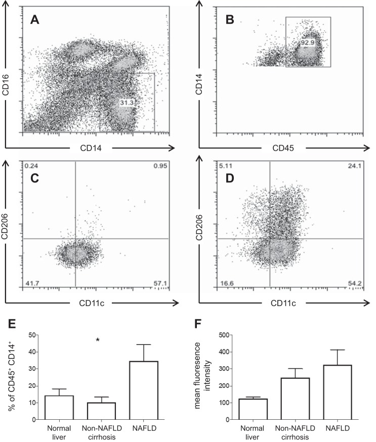
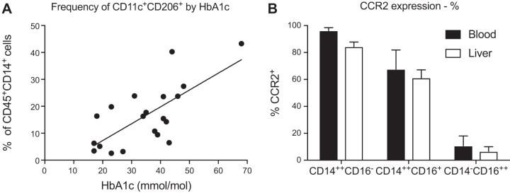
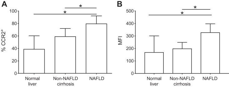
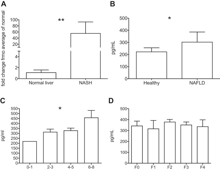
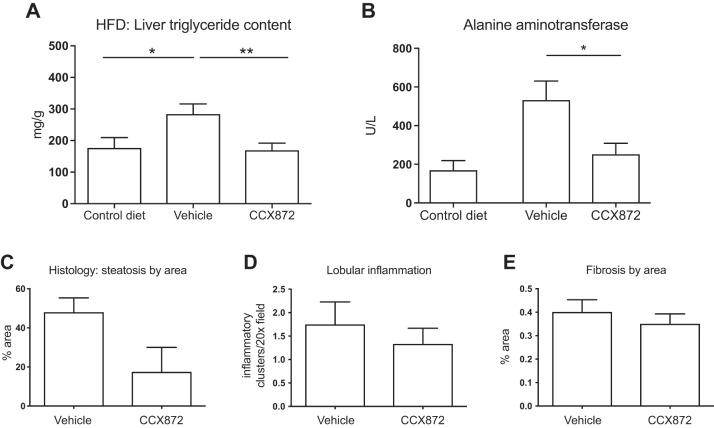
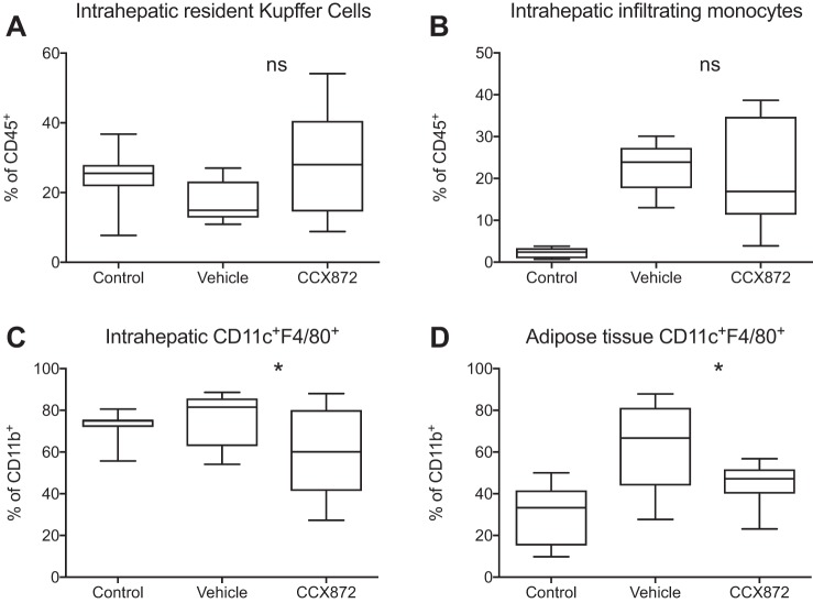
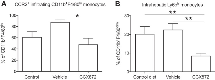
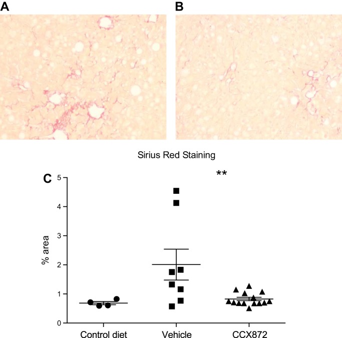
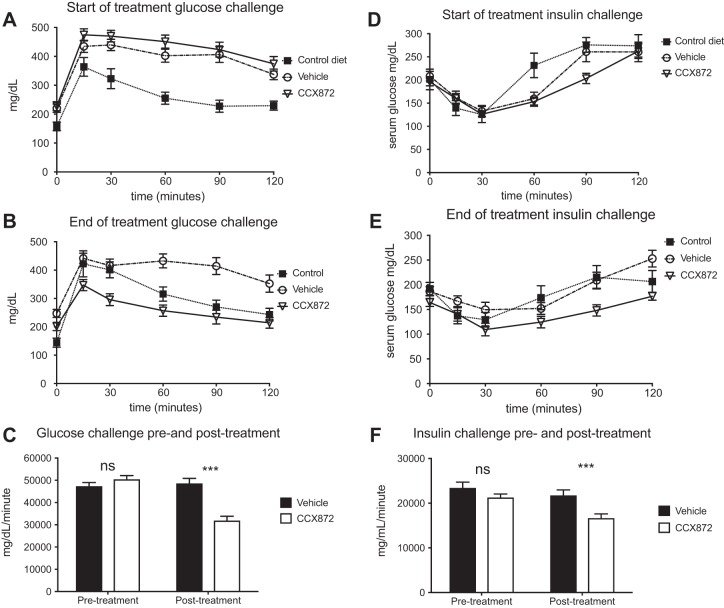
Nonalcoholic fatty liver disease (NAFLD) is a common disease, closely associated with obesity and insulin resistance. We investigated the presence of a subset of myeloid cells associated with metabolic disturbance in the liver of patients with NAFLD and a murine model of obesity-induced liver disease. Gene and protein expression in liver and serum was investigated with RT-PCR or ELISA and correlated to clinical disease. Liver-infiltrating immune cells were isolated from normal or diseased human liver for flow cytometric analysis. In animal experiments, mice were fed a high-fat diet (60% of calories from fat) for 16 wk, or high-fat diet with 30% fructose for 32 wk to induce steatohepatitis and fibrosis. A small molecule inhibitor of CC chemokine receptor 2 (CCR2), CCX872, was administered to some mice. A subset of CD11c+CD206+ immune cells was enriched in human liver tissue, and greater infiltration was observed in NAFLD. The presence of CD11c+CD206+ myeloid cells correlated with systemic insulin resistance. CD11c+CD206+ cells expressed high levels of CCR2, and liver CC chemokine ligand 2 (CCL2) expression was increased in nonalcoholic steatohepatitis and correlated with disease activity. In mice, CCR2 inhibition reduced infiltration of liver CD11b+CD11c+F4/80+ monocytes, which are functional homologs of human CD11c+CD206+ cells, and improved liver injury and glycemic control. A role for CCR2/CCL2 in human NAFLD has long been postulated. These data confirm a role for this chemokine/receptor axis, through mediating adipose and hepatic infiltration of myeloid cells. Inhibition of CCR2 improved hepatic inflammation and fibrosis in murine models of NAFLD. These data confirm the rationale for targeting CCR2 to treat NAFLD. NEW & NOTEWORTHY These data show for the first time that CD11c+CD206+ myeloid cells, previously associated with human adipose tissue inflammation, infiltrate into liver tissue in nonalcoholic fatty liver disease. These cells express CCR2. Inhibition of CCR2 in mice inhibits hepatic inflammation caused by a murine homolog of these myeloid cells and improves experimental liver disease.

Keywords: immunology; insulin resistance; nonalcoholic fatty liver disease; obesity.


Figures

Fig. 1.
Fig. 1.
CD11c+CD206+ monocytes are enriched in liver tissue. A and B: gating strategy to identify CD45+CD14+ monocytes. Representative samples of peripheral blood (C) and liver infiltrating monocytes (D) from the same individual (E) are shown. Liver tissue from patients with NAFLD (n = 8) showed a greater proportion of CD11c+CD206+ monocytes as a proportion of CD45+CD14+ monocytes, compared with other chronic liver disease (alcoholic liver disease, n = 4; primary sclerosing cholangitis, n = 3; primary biliary cholangitis, n = 2; hemochromatosis, n = 1; cryptogenic cirrhosis, n = 1) or normal liver (n = 5). *P < 0.05 by Kruskal-Wallis. F: mean fluorescence intensity of CD11c+CD206+ cells by liver disease (Kruskal-Wallis, P = 0.056).
Fig. 2.
Fig. 2.
Monocytes were isolated from liver tissue from patients with or without NAFLD and analyzed by flow cytometry. A: the frequency of CD11c+CD206+ monocytes in liver tissue correlated with insulin resistance, measured by HbA1c. n = 24; r2 = 0.499. B: CCR2 percent expression was greater on CD14++CD16 monocytes with a nonsignificant reduction of CCR2 expression on all intrahepatic monocytes.
Fig. 3.
Fig. 3.
Monocytes were isolated from liver tissue and analyzed by flow cytometry. CCR2 expression was higher on CD11c+CD206+ monocytes isolated from NAFLD liver tissue (n = 8) compared with non-NAFLD cirrhosis (n = 11) or normal liver tissue (n = 5) with regard to percentage of CCR2+ cells (normal, median 39.4%, IQR 40.1; non-NAFLD cirrhosis 59.7%, IQR 24.9; NAFLD 80.1%, IQR 24.7; A) and mean fluorescent intensity (normal 171, IQR 163.7; non-NAFLD cirrhosis 200.6, IQR 80.1; NAFLD 3,299, IQR 144.4; B). Data are shown as median and IQR; n = 23 in each case. *P < 0.05 by Mann-Whitney test.
Fig. 4.
Fig. 4.
A: RNA was isolated from liver tissue, and CCL2 gene expression was analyzed by semiquantitative PCR. CCL2 gene expression was significantly increased in liver tissue from patients with NASH (n = 6) compared with normal liver tissue (n = 6) (Mann-Whitney test, **P < 0.01). B: serum concentration of CCL2 measured by ELISA was higher in NAFLD (n = 20) compared with healthy volunteers (n = 10) (Mann-Whitney test, *P < 0.05). C: serum concentration of CCL2 increased with increasing disease activity as measured by the NAS score (one-way ANOVA, *P < 0.05). D: no relation was seen with fibrosis stage (one-way ANOVA, P > 0.05).
Fig. 5.
Fig. 5.
Improvements in steatohepatitis with inhibition of CCR2. Thirteen animals in each group were given HFD with daily administration of vehicle or CCX872, and a further eight animals were given a control diet for 16 wk. A: triglyceride content was measured with a colorimetric assay. CCR2 inhibition reduced triglyceride accumulation (*P < 0.05, **P < 0.01 by Student’s t-test). B: CCR2 inhibition reduced serum ALT (*P < 0.05 by Student’s t-test). Histological assessment of liver disease confirmed reduced steatosis, as assessed by area of staining (C), but there were no differences in histological inflammation (D) or histological fibrosis (E).
Fig. 6.
Fig. 6.
Myeloid cells from liver and adipose tissue from mice given a HFD were analyzed by flow cytometry. Treatment with a small molecule inhibitor of CCR2 did not affect proportions of intrahepatic Cd11b+F4/80hi Kupffer cells (A) or overall infiltrating CD11b+F4/80low monocytes (B) [Mann-Whitney test to compare vehicle and CCX872 groups, P > 0.05, nonsignificant (ns)]. CCR2 antagonism reduced infiltration of CD11c+F4/80+ cells into liver tissue (C) (Mann-Whitney test to compare vehicle and CCX872 groups, *P < 0.05) and adipose tissue (D) (Mann-Whitney test to compare vehicle and CCX872 groups, t-test, *P < 0.05). Data are shown as boxes to denote IQR, with line at median and whiskers showing maximum and minimum values.
Fig. 7.
Fig. 7.
Myeloid cells from liver and adipose tissue from mice given a HFD were analyzed by flow cytometry. Treatment with a small molecule inhibitor of CCR2 reduced hepatic infiltration with CCR2+CD11b+F4/80lo monocytes (A) (Student’s t-test, *P < 0.05) and infiltration of liver tissue by proinflammatory Ly6chi cells (B) (**P < 0.05 by Student’s t-test).
Fig. 8.
Fig. 8.
22 C57/Bl6 mice were fed HFD with 30% fructose in drinking water, or control diet without fructose, for 32 wk. CCR2 antagonism with a small molecule inhibitor, CCX872, reduced fibrosis compared with vehicle control. Representative pictures of liver sections from control (A) and CCX872-treated animals (B) are shown. C: fibrosis as assessed by percentage of collagen area by Sirius red staining of liver sections. Values are means and SE. **P < 0.05 by Student’s t-test.
Fig. 9.
Fig. 9.
CCR2 antagonism improved glycemic control in mice on a HFD with CCR2. Glycemic control was assessed at the beginning and end of the treatment period with glucose tolerance tests and insulin challenges. Mice in each group showed similar responses at the start of the treatment period [glucose (A) and insulin (D)]. At the time of death, mice treated with CCX872 showed significantly improved response to glucose and insulin (B and E, respectively). When assessed by measuring area under the curve, statistically significant changes were seen [glucose (C) and insulin (F)]. ***P < 0.001 by Student’s t-test.

References

    1. Angulo P, Kleiner DE, Dam-Larsen S, Adams LA, Bjornsson ES, Charatcharoenwitthaya P, Mills PR, Keach JC, Lafferty HD, Stahler A, Haflidadottir S, Bendtsen F. Liver fibrosis, but no other histologic features, is associated with long-term outcomes of patients with nonalcoholic fatty liver disease. Gastroenterology 149: 389–397.e10, 2015. doi:10.1053/j.gastro.2015.04.043. - DOI - PMC - PubMed
    1. Armstrong MJ, Barton D, Gaunt P, Hull D, Guo K, Stocken D, Gough SC, Tomlinson JW, Brown RM, Hübscher SG, Newsome PN; LEAN trial team . Liraglutide efficacy and action in non-alcoholic steatohepatitis (LEAN): study protocol for a phase II multicentre, double-blinded, randomised, controlled trial. BMJ Open 3: e003995, 2013. doi:10.1136/bmjopen-2013-003995. - DOI - PMC - PubMed
    1. Armstrong MJ, Hazlehurst JM, Hull D, Guo K, Borrows S, Yu J, Gough SC, Newsome PN, Tomlinson JW. Abdominal subcutaneous adipose tissue insulin resistance and lipolysis in patients with non-alcoholic steatohepatitis. Diabetes Obes Metab 16: 651–660, 2014. doi:10.1111/dom.12272. - DOI - PMC - PubMed
    1. Baeck C, Wehr A, Karlmark KR, Heymann F, Vucur M, Gassler N, Huss S, Klussmann S, Eulberg D, Luedde T, Trautwein C, Tacke F. Pharmacological inhibition of the chemokine CCL2 (MCP-1) diminishes liver macrophage infiltration and steatohepatitis in chronic hepatic injury. Gut 61: 416–426, 2012. doi:10.1136/gutjnl-2011-300304. - DOI - PubMed
    1. Boujedidi H, Bouchet-Delbos L, Cassard-Doulcier AM, Njiké-Nakseu M, Maitre S, Prévot S, Dagher I, Agostini H, Voican CS, Emilie D, Perlemuter G, Naveau S. Housekeeping gene variability in the liver of alcoholic patients. Alcohol Clin Exp Res 36: 258–266, 2012. doi:10.1111/j.1530-0277.2011.01627.x. - DOI - PubMed

Publication types

MeSH terms