Review
. 2018 Jan 2;6(1):e1404189.
doi: 10.1080/21688370.2017.1404189. Epub 2018 Feb 8.

Bridging the gap: Super-resolution microscopy of epithelial cell junctions

Affiliations
Review

Bridging the gap: Super-resolution microscopy of epithelial cell junctions

Emily I Bartle et al. Tissue Barriers. .

Abstract

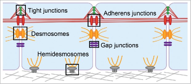
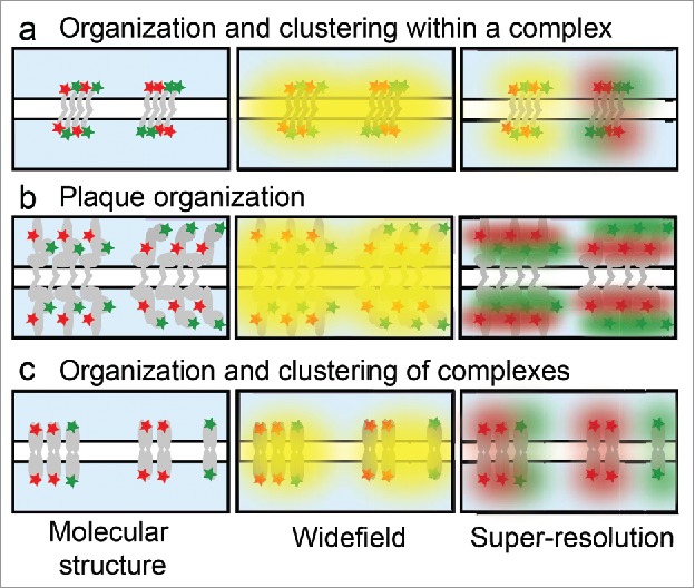
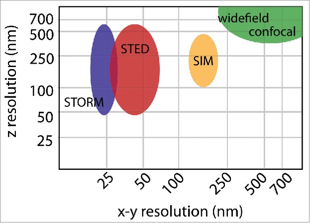
Cell junctions are critical for cell adhesion and communication in epithelial tissues. It is evident that the cellular distribution, size, and architecture of cell junctions play a vital role in regulating function. These details of junction architecture have been challenging to elucidate in part due to the complexity and size of cell junctions. A major challenge in understanding these features is attaining high resolution spatial information with molecular specificity. Fluorescence microscopy allows localization of specific proteins to junctions, but with a resolution on the same scale as junction size, rendering internal protein organization unobtainable. Super-resolution microscopy provides a bridge between fluorescence microscopy and nanoscale approaches, utilizing fluorescent tags to reveal protein organization below the resolution limit. Here we provide a brief introduction to super-resolution microscopy and discuss novel findings into the organization, structure and function of epithelial cell junctions.

Keywords: PALM; SIM; STED; STORM; adherens junction; desmosome; gap junction; hemidesmosome; tight junction.


Figures

Figure 1.
Figure 1.
Epithelial cell junctions. Tight junctions are localized apically on the lateral membrane of epithelial cells. They interact with the neighboring adherens junctions, and both junctions integrate with the actin cytoskeleton, shown in red. Desmosomes are located throughout the lateral membrane, below the adherens junctions, and associate with the intermediate filament cytoskeleton, shown in yellow. Gap junctions are also distributed throughout the lateral cell membrane where they form channels that allows passage of ions and small molecules between the two cells. In stratified epithelia, hemidesmosomes are localized to the basal membrane where they promote adhesion to the underlying substrate and integrate with intermediate filaments.
Figure 2.
Figure 2.
Protein organization in epithelial junctions. Illustration of different junctional molecular structures within the diffraction limit that cannot be distinguished by widefield microscopy, but are distinct by super-resolution. (a) Junctions can contain a homogenous mix of proteins (left) or distinct clusters (right). (b) Junctional plaque proteins are organized in layers parallel to the plasma membrane, with the red protein more distal (left) or proximal (right) to the membrane. (c) Junctions can have higher-order organization within the cell membrane with clusters of mixed junctions (left) or segregation of like junctions into distinct structures (right).
Figure 3.
Figure 3.
Spatial resolution. The resolution of widefield or confocal microscopy is determined by the numerical aperture of the objective and wavelength of the excitation light and is ∼250 nm in x-y and 600 nm in z. SIM doubles the resolution in all three dimensions achieving ∼125 nm lateral resolution and ∼300 nm axial resolution. STED and STORM can achieve a range of axial resolutions, depending on the use of TIRF or 3D imaging modalities. In general STORM achieves a slightly better x-y resolution of ∼20 nm, as opposed to ∼40 nm by STED. These resolutions are approximations based on commercially available systems. Variations in the resolution of each method depend on the properties and quality of the sample, as well as the particular optical setup.

References

    1. Rübsam M, Broussard JA, Wickström SA, Nekrasova O, Green KJ, Niessen CM. Adherens junctions and desmosomes coordinate mechanics and signaling to orchestrate tissue morphogenesis and function: An evolutionary perspective. Cold Spring Harb Perspect Biol. 2017. doi: 10.1101/cshperspect.a029207. PMID:28893859. - DOI - PMC - PubMed
    1. Xia S, Kanchanawong P. Nanoscale mechanobiology of cell adhesions. Semin Cell Dev Biol. 2017;71:53–67. doi: 10.1016/j.semcdb.2017.07.029. PMID:28754443. - DOI - PubMed
    1. Yano T, Kanoh H, Tamura A, Tsukita S. Apical cytoskeletons and junctional complexes as a combined system in epithelial cell sheets. Ann N Y Acad Sci. 2017;1405(1):32–43. doi: 10.1111/nyas.13432. PMID:28763830. - DOI - PubMed
    1. Maeda S, Nakagawa S, Suga M, Yamashita E, Oshima A, Fujiyoshi Y, Tsukihara T. Structure of the connexin 26 gap junction channel at 3.5 Å resolution. Nature. 2009;458:597–602. doi: 10.1038/nature07869. PMID:19340074. - DOI - PubMed
    1. Tamarin A, Sreebny LM. An analysis of desmosome shape, size, and orientation by the use of histometric and densitometric methods with electron microscopy. J Cell Biol. 1963;18:125–34. doi: 10.1083/jcb.18.1.125. PMID:19866631. - DOI - PMC - PubMed

Publication types

MeSH terms

LinkOut - more resources