Review
. 2018 May 1;124(5):1117-1139.
doi: 10.1152/japplphysiol.00070.2017. Epub 2018 Feb 8.

Exercise intolerance in Type 2 diabetes: is there a cardiovascular contribution?

Affiliations
Review

Exercise intolerance in Type 2 diabetes: is there a cardiovascular contribution?

Veronica J Poitras et al. J Appl Physiol (1985). .

Abstract

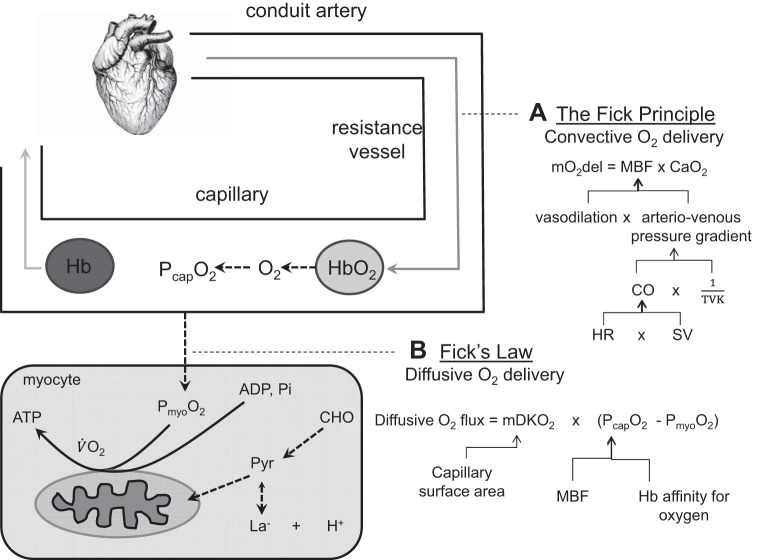
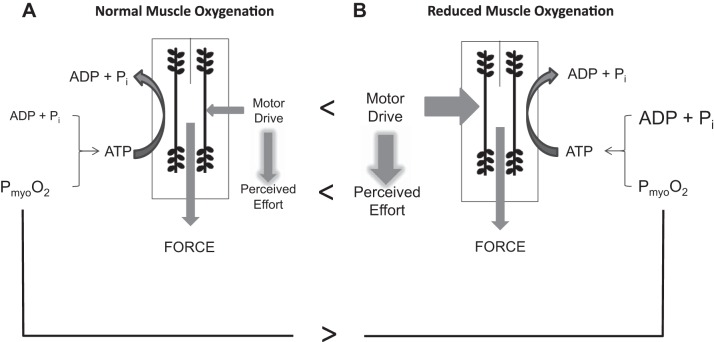
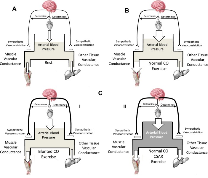
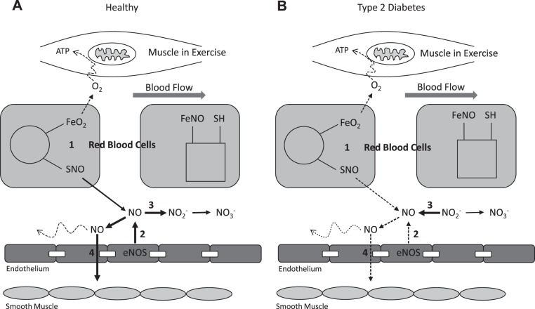
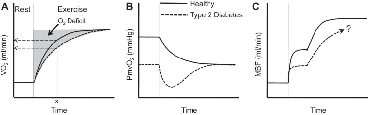
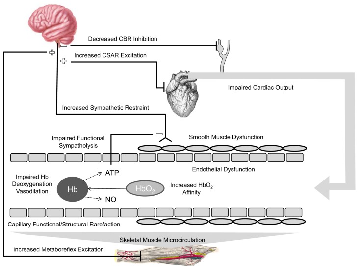
Physical activity is critically important for Type 2 diabetes management, yet adherence levels are poor. This might be partly due to disproportionate exercise intolerance. Submaximal exercise tolerance is highly sensitive to muscle oxygenation; impairments in exercising muscle oxygen delivery may contribute to exercise intolerance in Type 2 diabetes since there is considerable evidence for the existence of both cardiac and peripheral vascular dysfunction. While uncompromised cardiac output during submaximal exercise is consistently observed in Type 2 diabetes, it remains to be determined whether an elevated cardiac sympathetic afferent reflex could sympathetically restrain exercising muscle blood flow. Furthermore, while deficits in endothelial function are common in Type 2 diabetes and are often cited as impairing exercising muscle oxygen delivery, no direct evidence in exercise exists, and there are several other vasoregulatory mechanisms whose dysfunction could contribute. Finally, while there are findings of impaired oxygen delivery, conflicting evidence also exists. A definitive conclusion that Type 2 diabetes compromises exercising muscle oxygen delivery remains premature. We review these potentially dysfunctional mechanisms in terms of how they could impair oxygen delivery in exercise, evaluate the current literature on whether an oxygen delivery deficit is actually manifest, and correspondingly identify key directions for future research.

Keywords: cardiac dysfunction; exercise habits; microvascular flow; muscle blood flow; vasodilation.


Figures

Fig. 1.
Fig. 1.
Cardiovascular pathway for oxygen delivery to the mitochondria. The “flow” of oxygen has a convective component delivering oxygen to the capillaries, where a diffusive component determines the delivery from the capillaries into the myocytes. A: the Fick principle identifies the role of vasodilation and the arteriovenous pressure gradient in increasing convective oxygen delivery (mO2del) via increasing muscle blood flow (MBF). Arterial pressure is ultimately determined by the cardiac supply of blood to the arteries (cardiac output, CO) in balance with total systemic vascular tone (total vascular conductance, TVK). Heart rate (HR) and stroke volume (SV) responses determine CO. B: Fick’s law identifies the role of diffusive conductance (mDKO2) and the capillary plasma oxygen level (PcapO2) in determining the diffusion of oxygen into the myocyte. A combination of increased MBF supplying oxygen to the capillaries and reduced hemoglobin (Hb) affinity for oxygen determine PcapO2 during exercise. If Type 2 diabetes impairs oxygen delivery to the mitochondria, it would be as a result of problems with one or a number of these determinants along the cardiovascular pathway. Note that myocellular oxygenation (PmyoO2), which plays a key role in determining muscle metabolic and contractile function, can be different at the same V̇o2; changes in ADP and inorganic phosphate (Pi) can compensate for reductions in PmyoO2 (due to impaired convective and/or diffusive oxygen flow) to maintain V̇o2. CaO2, arterial oxygen content; CHO, carbohydrate, H+, hydrogen ion associated with lactate formation; La, lactate; Pyr, pyruvate.
Fig. 2.
Fig. 2.
Effect of PmyoO2 on muscle metabolism and contractile function. A: normal PmyoO2: schematic representation of the factors influencing muscle force production (i.e., ADP + Pi, PmyoO2, motor drive). Perceived effort is a function of the degree of motor drive for a given force production. B: a reduced PmyoO2 (comparison with normal indicated by >) necessitates both an increase in ADP + Pi levels and an increase in motor drive (comparison with normal indicated by <) to maintain the given aerobic supply of ATP and exercise intensity. This would be experienced as increased perceived effort (comparison with normal indicated by <) due to increased motor drive. [Borrowed with permission from Tschakovsky and Pyke (113).]
Fig. 3.
Fig. 3.
Schematic representation of the systemic circulation with arterial blood pressure as a reservoir determined by the balance between inflow (cardiac output) and outflow (muscle and other tissue blood flow). The two outflows are determined by their respective vascular conductance and the arterial driving pressure. Arrow size represents flow magnitude. A: rest represents rest. B: normal cardiac output (CO) response in submaximal exercise in which vasodilation in the exercising muscle is matched by an increase in cardiac output CO and potentially a decrease in other tissue blood flow due to sympathetic vasoconstriction, preserving the normal increases in arterial blood pressure and increasing muscle blood flow. Sympathetic vasoconstriction (magnitude indicated by thickness of neural pathway) also normally restrains exercising muscle vasodilation to some extent. The degree to which this restraint of peripheral vasodilation is required may be dictated by the magnitude of the cardiac output CO increase. C I illustrates the way in which a blunted CO response cardiac contribution to impaired exercising muscle blood flow would manifest. C I: blunted cardiac output; blunted CO increase requires an increase in sympathetic restraint of exercising muscle vasodilation and therefore blood flow to maintain arterial blood pressure. C II: normal CO response, but cardiac sympathetic afferent reflex (CSAR) increases sympathetic vasoconstriction and mediates increased arterial blood pressure and blunted muscle blood flow.
Fig. 4.
Fig. 4.
Schematic of proposed dysfunction of endocrine-like nitric oxide (NO) bioavailability. A: healthy: muscle oxygen consumption in exercise leads to red blood cell (RBC) deoxygenation and S-nitrosothiol (SNO) release (1) to supplement endothelial NO synthase (eNOS) NO production (2). Excess NO converts to nitrite (NO2), which is a biomarker of NO bioavailability (3), and eventually nitrate (NO3). NO initiates direct and conducted vasodilation to help match oxygen delivery to demand (4). B: Type 2 diabetes: muscle oxygen consumption in exercise leads to RBC deoxygenation, but SNO release is impaired (1). eNOS NO production is also impaired (2), such that plasma nitrite is now converted to NO (3), but this is not able to maintain required NO bioavailability. Therefore direct and conducted NO-mediated vasodilation (4) is inadequate to allow matching of oxygen delivery to muscle oxygen demand in exercise. FeNO, partially nitrosylated blood; FEO2,oxygenated blood; SH, thiol functional group.
Fig. 5.
Fig. 5.
Schematic of proposed exercise responses in persons with Type 2 diabetes (dashed lines) vs. healthy controls (solid lines). A: slowed V̇o2 kinetics in Type 2 diabetes. The magnitude of O2 deficit is determined by V̇o2 kinetics. Dashed arrows indicate a lower V̇o2 at time “x” for the dashed vs. solid curve. This is significant because it indicates that Type 2 diabetes results in the need for a greater reliance on substrate-level phosphorylation to meet the identical ATP demand. [Borrowed with permission from Lukin and Ralston (54).] B: overshoot of microvascular deoxygenation (PcapO2, partial pressure of oxygen in the capillaries) in Type 2 diabetes is suggestive of a greater MBF/V̇o2 mismatch at the onset of exercise. Responses in A and B are consistent with observations in the literature. C: hypothesized skeletal muscle blood flow (MBF) dynamics accounting for these responses; “?” indicates that in Type 2 diabetes a slower second phase response may depend on the exercise intensity, and there is inadequate consensus on whether steady state is impaired.
Fig. 6.
Fig. 6.
Schematic of potential central and peripheral cardiovascular impairments that could compromise steady-state convective and diffusive oxygen delivery to the contracting skeletal muscle myocyte, thereby contributing to exercise intolerance. The vessel shows capillary (left side) and upstream arteriolar (right side) levels of the vascular tree. Sympathetic restraint of exercising skeletal muscle vasodilation could be enhanced due to 1) reduced CBR inhibition of sympathetic neural outflow to compensate for inadequate CO and maintain arterial blood pressure, 2) CSAR stimulation in a diabetic heart, and/or 3) MMR activation. Impaired functional sympatholysis would reduce blunting of sympathetic restraint. The vasodilatory response to increased muscle metabolic demand for oxygen in contracting skeletal muscle could be compromised due to 1) endothelial and/or 2) smooth muscle dysfunction, and/or 3) impaired RBC release of ATP and/or nitric oxide (NO) with hemoglobin (Hb) deoxygenation. Diffusive flux of oxygen could be compromised by increased Hb affinity for O2 and/or structural and functional capillary rarefaction. CBR, carotid baroreflex; CO, cardiac output; CSAR, cardiac sympathetic afferent reflex; MMR, muscle metaboreflex.

Similar articles

Cited by

References

    1. American Diabetes Association Diagnosis and classification of diabetes mellitus. Diabetes Care 31, Suppl 1: S55–S60, 2008. doi:10.2337/dc08-S055. - DOI - PubMed
    1. Allen JD, Miller EM, Schwark E, Robbins JL, Duscha BD, Annex BH. Plasma nitrite response and arterial reactivity differentiate vascular health and performance. Nitric Oxide 20: 231–237, 2009. doi:10.1016/j.niox.2009.01.002. - DOI - PMC - PubMed
    1. Allen JD, Stabler T, Kenjale A, Ham KL, Robbins JL, Duscha BD, Dobrosielski DA, Annex BH. Plasma nitrite flux predicts exercise performance in peripheral arterial disease after 3 months of exercise training. Free Radic Biol Med 49: 1138–1144, 2010. doi:10.1016/j.freeradbiomed.2010.06.033. - DOI - PMC - PubMed
    1. Amann M, Romer LM, Pegelow DF, Jacques AJ, Hess CJ, Dempsey JA. Effects of arterial oxygen content on peripheral locomotor muscle fatigue. J Appl Physiol (1985) 101: 119–127, 2006. doi:10.1152/japplphysiol.01596.2005. - DOI - PubMed
    1. Anderson RJ, Freedland KE, Clouse RE, Lustman PJ. The prevalence of comorbid depression in adults with diabetes: a meta-analysis. Diabetes Care 24: 1069–1078, 2001. doi:10.2337/diacare.24.6.1069. - DOI - PubMed

Publication types

MeSH terms