. 2018 Mar 16;359(6381):1269-1273.
doi: 10.1126/science.aal3589. Epub 2018 Feb 1.

Astrocyte-derived interleukin-33 promotes microglial synapse engulfment and neural circuit development

Affiliations

Astrocyte-derived interleukin-33 promotes microglial synapse engulfment and neural circuit development

Ilia D Vainchtein et al. Science. .

Abstract

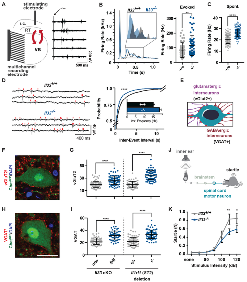
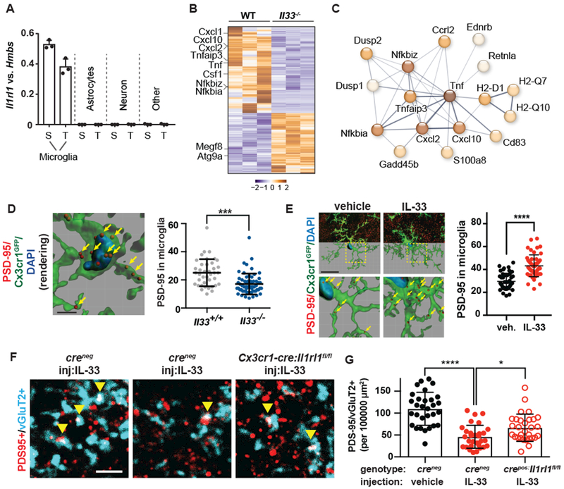
Neuronal synapse formation and remodeling are essential to central nervous system (CNS) development and are dysfunctional in neurodevelopmental diseases. Innate immune signals regulate tissue remodeling in the periphery, but how this affects CNS synapses is largely unknown. Here, we show that the interleukin-1 family cytokine interleukin-33 (IL-33) is produced by developing astrocytes and is developmentally required for normal synapse numbers and neural circuit function in the spinal cord and thalamus. We find that IL-33 signals primarily to microglia under physiologic conditions, that it promotes microglial synapse engulfment, and that it can drive microglial-dependent synapse depletion in vivo. These data reveal a cytokine-mediated mechanism required to maintain synapse homeostasis during CNS development.


Conflict of interest statement

Competing interests: None declared.

Figures

Figure 1:
Figure 1:. IL-33 is developmentally induced in synapse-associated astrocytes.
(A) Representative image of Il33mCherry with Aldh1l1-eGFP+ astrocytes and oligodendrocyte marker CC1 in spinal cord ventral horn (scale = 50 µm).(B) Gray matter restricted expression of Il33lacZ in the spinal cord at P30 (scale = 0.5 mm). (C, D) Il33lacZ increases in the visual thalamus (dLGN) during eye opening, normalized to sensorimotor thalamus (VB) (scale = 0.5 mm). (E) Representative images of Il33lacZ in P21 thalamus in littermate controls and after perinatal enucleation (scale = 0.5 mm). (F) Il33lacZ mean pixel intensity in dLGN. (G) Representative flow plot of spinal cord from Il33mCherry/Aldh1l1-eGFP mice at P15 with sorting gates indicated. (H) Heatmap of the top 444 differentially expressed genes in Il33-mCherry+ vs. mCherry astrocytes in spinal cord and thalamus (FC>2, pAdj<0.05), select candidates highlighted. Statistics: One-way ANOVA with Tukey’s post hoc comparison or student’s t-test. All points represent independent biological replicates. * p<0.05, **** p<0.0001.
Figure 2:
Figure 2:. IL-33 deficiency leads to excess synapses and abnormal thalamic and sensorimotor circuit function.
(A) Schematic of extracellular recording setup to measure circuit activity between ventrobasal (VB) and reticular thalamic nuclei (RT) with representative recording showing activity in five channels after stimulation of the internal capsule (i.c.) that contains cortical afferents. Red arrows indicate reciprocal VB-RT connections. (B) Average traces and quantification of mean firing rates reveal higher evoked firing in Il33−/−. (C) Quantification of mean firing rates in the absence of stimulation reveals increased spontaneous firing in Il33−/−. (D) Representative traces and quantification of intracellular patch-clamp recordings from neurons in the VB show increased miniature excitatory postsynaptic currents (mEPSCs) in Il33−/−. (E) Schematic of motor neuron synaptic afferents. (F, G) Representative image and quantification of excitatory inputs per motor neuron after conditional deletion of Il33 (hGFAPcre) or global deletion of Il1rl1. (H, I) Inhibitory (VGAT+) inputs in the same mice (scale = 25 µm). (J) Schematic of startle pathway. (K) Impaired sensorimotor startle in Il33−/− animals. Statistics: Data in B-C from WT: n=6-8 slices, 2 mice. KO: n=14-15 slices, 3 mice, points are individual recordings. Data in B analyzed by Mann-Whitney and C with student’s t test. Data in D from n=23-25 cells and 3-4 mice/group analyzed by Kolmogorov-Smirnov test. Data in F - I from n=3 animals, >75 neurons per genotype, student’s t-test; points are individual neurons. K is n=12/group, two-way ANOVA with Sidak’s multiple comparisons. *p<0.05, **p<0.01, ****p<.0001. B-I are mean±SD, K is mean±SEM.
Figure 3:
Figure 3:. IL-33 drives microglial synapse engulfment during development.
(A) Expression of Il1rl1 by qPCR of flow-sorted populations (S=spinal cord, T=thalamus.) (B) 484 differentially expressed genes in spinal cord microglia at pAdj<0.05. (C) Functionally associated gene clustering (STRING) identifies immune genes enriched in wild-type vs Il33−/− microglia. (D) PSD-95 puncta within microglia (yellow arrows) after IL-33 deletion (scale = 4 μM). (E) Representative image and quantification of engulfed PSD-95 in vehicle or IL-33 injected spinal cord (Scale= 20 µm). (F-G) Colocalized pre-and postsynaptic puncta in spinal cord ventral horn at P14 (yellow arrows) after IL-33 injection into control mice or in littermates with conditional deletion of Il1rl1 (Cx3cr1cre ; scale = 3 µm). Statistics: Points in A represent mice, in D-G individual microglia from N=3-5 animals/group, in G images from N=5 mice/group. In D, E student’s t-test and in G a one-way ANOVA with Tukey’s post hoc comparison, *p<0.05, ***p<0.001, ****p<0.0001, all data are mean±SD.

Comment in

References

    1. Molofsky AV et al., Astrocytes and disease: a neurodevelopmental perspective. Genes Dev. 26, 891–907 (2012). - PMC - PubMed
    1. Clarke LE, Barres BA, Emerging roles of astrocytes in neural circuit development. Nat Rev Neurosci. 14, 311–321 (2013). - PMC - PubMed
    1. Ransohoff RM, Cardona AE, The myeloid cells of the central nervous system parenchyma. Nature. 468, 253–262 (2010). - PubMed
    1. Salter MW, Beggs S, Sublime microglia: expanding roles for the guardians of the CNS. Cell. 158, 15–24 (2014). - PubMed
    1. Miyamoto A et al., Microglia contact induces synapse formation in developing somatosensory cortex. Nat Commun. 7, 12540 (2016). - PMC - PubMed

Publication types