Review
. 2018 Feb 7;97(3):494-510.
doi: 10.1016/j.neuron.2018.01.006.

Dopamine's Effects on Corticostriatal Synapses during Reward-Based Behaviors

Affiliations
Review

Dopamine's Effects on Corticostriatal Synapses during Reward-Based Behaviors

Nigel S Bamford et al. Neuron. .

Abstract

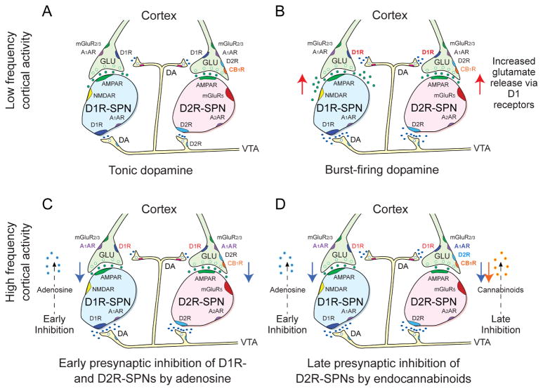
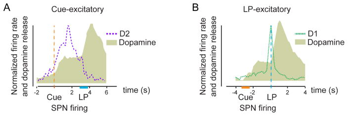
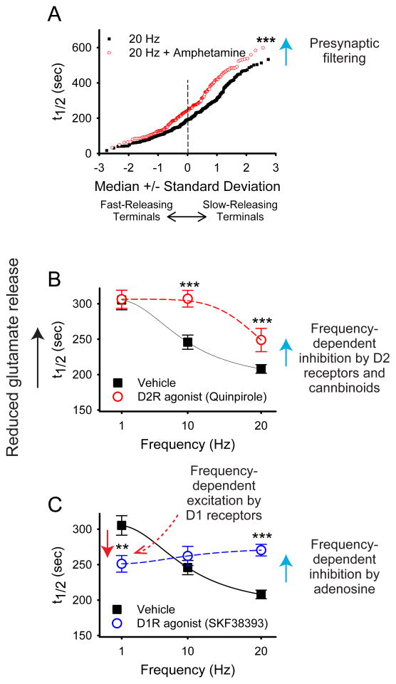
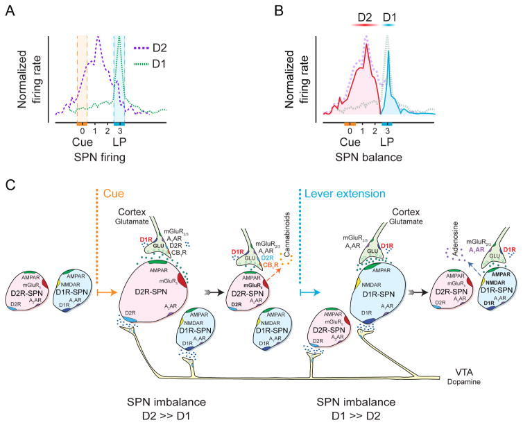
Many learned responses depend on the coordinated activation and inhibition of synaptic pathways in the striatum. Local dopamine neurotransmission acts in concert with a variety of neurotransmitters to regulate cortical, thalamic, and limbic excitatory inputs to drive the direct and indirect striatal spiny projection neuron outputs that determine the activity, sequence, and timing of learned behaviors. We review recent advances in the characterization of stereotyped neuronal and operant responses that predict and then obtain rewards. These depend on the local release of dopamine at discrete times during behavioral sequences, which, acting with glutamate, provides a presynaptic filter to select which excitatory synapses are inhibited and which signals pass to indirect pathway circuits. This is followed by dopamine-dependent activation of specific direct pathway circuits to procure a reward. These steps may provide a means by which higher organisms learn behaviors in response to feedback from the environment.

Keywords: direct pathway; indirect pathway; learning; motor; nucleus accumbens; reward; striatum; substantia nigra; synapse; ventral tegmental area.


Figures

Figure 1
Figure 1
Proposed model for how coincident cortical and dopaminergic inputs control rapid signal processing during operant behaviors in the NAc. Each panel shows D1-containing and D2-containing SPNs. D1-SPNs express AMPA, NMDA and A1A receptors, while D2-SPNs express AMPA, mGluR5 and A2A receptors. Cortical terminals to both types of SPNs present D1, A1A and mGluR2/3 receptors. Only cortical terminals to D2-SPNs present D2 and CB1 receptors. Dopamine projections from the VTA form synapses at both D1- and D2-containing SPNs. A. Low activity cortical and dopamine inputs. When dopamine neurons are tonically active, they produce low levels of striatal dopamine that inhibits the indirect corticoaccumbal pathway by binding to a fraction of pre-and postsynaptic high affinity D2 receptors. B. Low activity cortical and highly active dopamine inputs. High levels of dopamine reached during burst firing (due in part to saturation of dopamine reuptake) enhance corticoaccumbal activity through D1Rs that strengthen glutamate release from presynaptic terminals to both direct and indirect pathway SPNs. C. Highly activity cortical and dopamine input effects on the direct pathway. When corticoaccumbal stimulation of NMDA and AMPA receptors on direct SPNs are concurrent with high levels of dopamine that activate D1Rs, adenylate cyclase is activated and adenosine is released from direct pathway SPNs. The adenosine retrogradely inhibits presynaptic activity to both D1R and D2R-presenting SPNs via A1Rs. D. Highly activity cortical and dopamine input effects on the indirect pathway. High corticoaccumbal stimulation produces extrasynaptic overflow of glutamate, which activates group 1 mGLU receptors on indirect SPNs. When this is concurrent with high levels of dopamine that activate additional D2 receptors on indirect SPNs, the SPNs release endocannabinoids, which retrogradely inhibits presynaptic inputs to indirect SPNs via presynaptic CB1Rs. The most active corticoaccumbens release sites appear to escape presynaptic inhibition, although the means by which that occurs are unclear. Reproduced with permission from Wang et.al., J Physiol. 590:3743–69 (2012).
Figure 2
Figure 2
Dopamine release and SPN firing rate measured in the rat NAc during ICSS. Both responses are measured at the same carbon fiber electrode. Receptor subtype identified with controlled iontophoresis (see text). A. Responses at Cue-excitatory SPNs. All were determined to be D2R-containing cells. Dopamine release occurs immediately after cue initiation and rises further after the lever press (LP) due to electrical stimulus of the VTA/SN region. D2R-SPNs adjacent to the region of DA release fire following the cue. Their activity diminishes around lever extension (occurs at 2 s) and returns to baseline after the LP. B. Responses at LP-excitatory SPNs. Dopamine release follows an identical pattern as in A. LP-excitatory cells, that were predominantly D1R-containing SPNs, increase in firing around the time of the LP. D2R-responsive cells that responded around the LP predominantly displayed an inhibition (data not shown). Note that the large spike of dopamine release that reaches maximum values ~ 1s after lever press during ICSS is due to direct activation of dopamine axons by the stimulating electrode and is not an intrinsic CNS step in operant conditioning. Adapted from Owesson-White et.al., J Neurosci. 36:6011–21 (2016).
Figure 3
Figure 3
Modulation of stimulated release from glutamatergic terminals in mouse brain slices containing the NAc. Corticoaccumbal terminals in the NAc were loaded with FM1-43 during a pre-measurement interval, and then the destaining rate during a subsequent stimulation period at individual nerve terminals was monitored as t1/2 values (the time required for florescence to decay to half of its original value). A. Normal probability plot comparison of individual half-times of release in slices shows that the dopamine releaser amphetamine filters cortical activity by reducing exocytosis of FM1-43 from terminals with lowest probability of release (i.e. those with the highest t1/2). At 20 Hz, t1/2 = 207 s; n = 330. At 20 Hz with amphetamine, t1/2 = 264 s; n = 193; ***P < 0.001, Mann-Whitney. B. Distribution of mean t1/2 of release for slices containing the NAc as a function of frequency. The release of FM1-43 was reduced at higher frequencies (20 Hz) when the slice was exposed the D2R agonist quinpirole. This plot demonstrates that activation of D2 receptors inhibits glutamate release at higher frequencies (n = 80 – 96 terminals; ***P < 0.001 Mann-Whitney). C. A D1R agonist increased release at low frequencies (1 Hz). At higher frequencies of cortical stimulation (20 Hz), the D1R agonist reduced release by promoting presynaptic inhibition by adenosine (not shown). (n = 97 – 260 terminals; ***P < 0.001 Mann-Whitney). Reproduced with permission from Wang et.al., J Physiol. 590:3743–69 (2012).
Figure 4
Figure 4
Time course of D1 and D2 responses in the rat NAc during an ICSS trial consisting of cue presentation (0) followed two seconds later by lever extension and then a subsequent lever press (LP) to deliver and electrical stimulation to dopamine cell bodies. A. Time course of D2R-containing SPNs that are cue excitatory and D1R-containing SPNs that are LP-excitatory. The hatched areas indicate time variation of either cue or LP, as indicated in Figure 2. B. The differential responses of both sets of SPNs (D2 inhibitory cells were ignored in this representation). Firing rates from D1- and D2-SPNs (dashed) were subtracted to illustrate the imbalance in SPN activity following cue and LP. C. Temporal sequence of chemical activity at SPN synapses. The cue temporarily heightens D2R-SPN activity to create an imbalance in striatal output. The imbalance may alert and orient the animal, perhaps by promoting an urge to move and defining the motor sequence to be activated. The lever extension increases D1R-SPN activity, culminating in the lever press (LP).

References

    1. Adams RN. Probing brain chemistry with electroanalytical techniques. Anal Chem. 1976;48:1126A–1138A. - PubMed
    1. Albin RL, Young AB, Penney JB. The functional anatomy of basal ganglia disorders. Trends Neurosci. 1989;12:366–375. - PubMed
    1. Alexander GE, Crutcher MD. Functional architecture of basal ganglia circuits: neural substrates of parallel processing. Trends Neurosci. 1990;13:266–271. - PubMed
    1. Anzalone A, Lizardi-Ortiz JE, Ramos M, De Mei C, Hopf FW, Iaccarino C, Halbout B, Jacobsen J, Kinoshita C, Welter M, Caron MG, Bonci A, Sulzer D, Borrelli E. Dual control of dopamine synthesis and release by presynaptic and postsynaptic dopamine D2 receptors. J Neurosci. 2012;32:9023–9034. - PMC - PubMed
    1. Apicella P, Scarnati E, Ljungberg T, Schultz W. Neuronal activity in monkey striatum related to the expectation of predictable environmental events. J Neurophysiol. 1992;68:945–960. - PubMed

Publication types

LinkOut - more resources