Review
. 2018 Mar;34(1):1-18.
doi: 10.1016/j.cvfa.2017.10.006.

Enteric Immunity: Happy Gut, Healthy Animal

Affiliations
Review

Enteric Immunity: Happy Gut, Healthy Animal

Christopher C L Chase. Vet Clin North Am Food Anim Pract. 2018 Mar.

Abstract

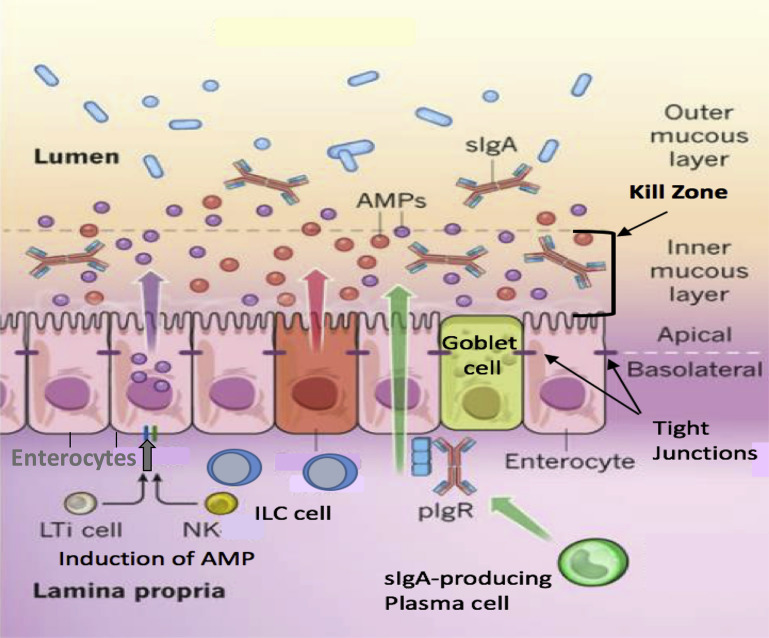
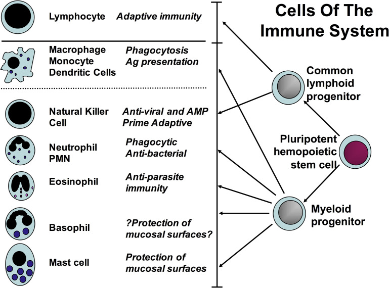
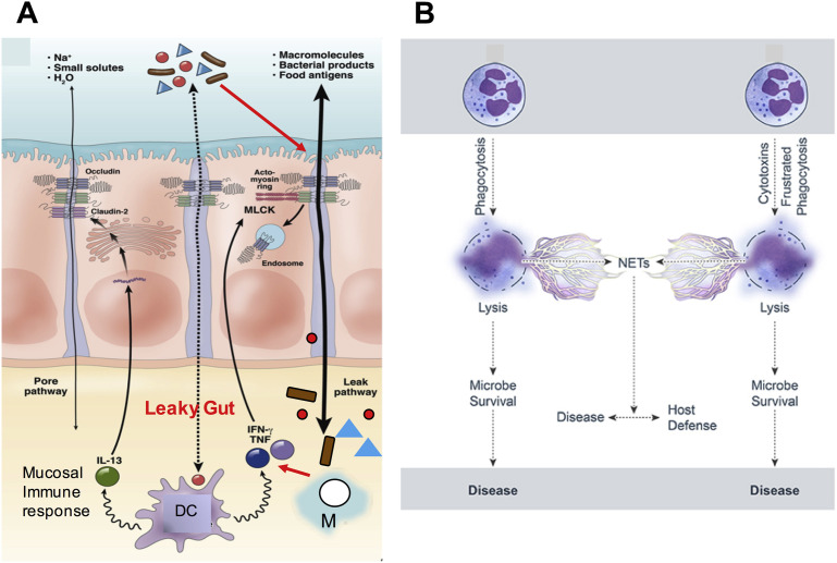
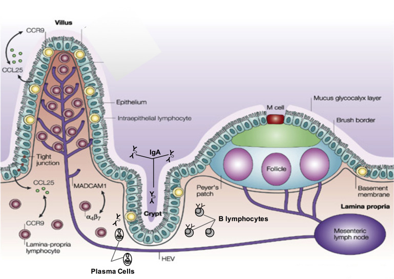
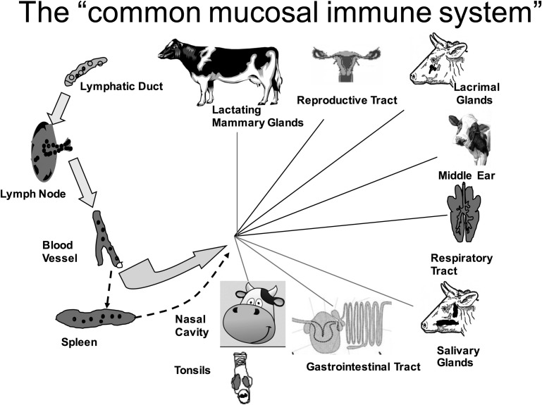
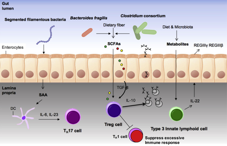
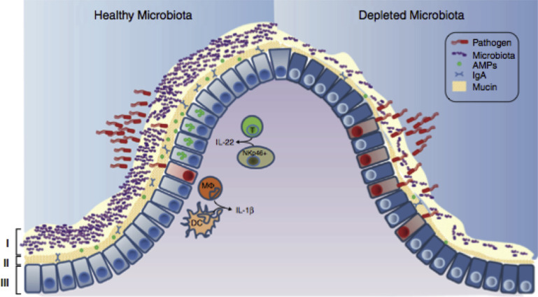
In this article, key concepts important for enteric immunity are discussed. The gastrointestinal tract is the largest immune organ of the body. The mucosal barrier, the tight junctions and the "kill zone," along with the gut mucosa and maintaining an "anti-inflammatory" state are essential for "good gut health." The microbiome, the microorganisms in the gastrointestinal tract, which has more cells then the entire animal's body, is essential for immune development, immune response, and maximizing ruminant productivity. Direct-fed microbials aid in both microbiome stability "homeostasis" and immune function.

Keywords: Bovine; Enteric; Immunology; Microbiome; Mucosal.


Figures

Fig. 1
Fig. 1
Development of the immune response in the bovine: from conception to puberty. The calf’s passive maternal immunity is only transferred after birth due to its unique placentation.
Fig. 2
Fig. 2
Organization of the gut lymphoid tissue. Lymphocytes can leave the surface epithelium (intraepithelial lymphocytes [IEL]) or Lamina propria (LP) via draining afferent lymphatics to mesenteric lymph nodes (MLNs), or via portal blood reaching the liver where induction of tolerance occurs. The M cells in the follicle-associated epithelium of PPs transport antigen to prime B cells in the isolated lymphoid follicles (ILF) of the PPs of the jejunum, ileum, and the large intestine. The continuous IPPs are a primary lymphoid organ responsible for B-cell development. The IPP can be up to 2 m long and constitute 80% to 90% of the intestinal lymphoid tissue. LN, lymph node.
Fig. 3
Fig. 3
Gut immune responses: the barrier, innate, and adaptive immune components.
Fig. 4
Fig. 4
The mucosal defenses of the GI tract. Distinct subpopulations of intestinal epithelial cells are integrated into a continuous, single-cell layer that is divided into apical and basolateral regions by tight junctions. Enterocytes sense the microbiota and their metabolites to induce the production of AMPs. Goblet cells produce mucin and mucous that is organized into a dense, more highly cross-linked inner proteoglycan gel that forms an adherent inner mucous layer, and a less densely cross-linked outer mucous layer. The outer layer is highly colonized by constituents of the microbiota. The inner mucous layer is largely impervious to bacterial colonization or penetration due to its high concentration of bactericidal AMPs, as well as commensals sIgA, which is moved from their basolateral surface, where it is bound by the polymeric Immunoglobulin receptor (pIgR), to the inner mucous layer. Responding to the microbiotal components, innate lymphoid cells (ILC), lymphoid tissue inducer cells (LTi), and NK produce cytokines, which stimulate AMP production and maintain the epithelial barrier.
Fig. 5
Fig. 5
The cells of the immune system. The innate and acquired immune cell lines have overlap with the macrophages and NK cells having important innate and acquired responses. Ag, antigen; PMN, polymorphonuclear cells.
Fig. 6
Fig. 6
Innate immunity and the mucosa. (A) Pathogenesis of leaky gut. The epithelial barrier normally restricts passage of luminal contents, including microbes and their products, but a small fraction of these materials do cross the tight junction. This diagram shows how DCs, and macrophages (M) react to these materials. These innate immune cells release cytokines that exert proinflammatory (TNF and interferon-gamma [IFN-γ]) and anti-inflammatory (IL-13) effects. If proinflammatory signals dominate and signal to the epithelium, MLCK can be activated to cause barrier dysfunction through the “leak pathway,” allowing an increase in the amount of luminal material presented to immune cells. In the absence of appropriate immune regulation, immune activation may cause further proinflammatory immune activation, cytokine release, and barrier loss, resulting in a self-amplifying cycle that can result in disease. (B) Neutrophil collateral damage from NET formation. Neutrophil lysis after phagocytosis. Cytolysis can be programmed, for example, necroptosis, or caused by direct damage. Neutrophil lysis is caused by cytolytic toxins, pore-forming agents, physical injury, or frustrated phagocytosis. This can result in the formation of NETs during neutrophil lysis. Hydrolytic enzymes–DNA complexes are released in the NETs, enhancing the proinflammatory response and tissue destruction, contributing to collateral damage and disease.
Fig. 7
Fig. 7
Mucosal immune system of the gut epithelium. The LP contains scattered T cells and lies beneath the epithelium, which contains intraepithelial lymphocytes (IEL). B cells are scattered in the LP but are more frequent in the crypt regions along with plasma cells that produce IgA that is transported and secreted into the lumen. M cells facilitate antigen uptake and delivery to the organized lymphoid tissues. T cells activated in the PP and mesenteric lymph node express mucosa specific receptors, which interact with cell-adhesion molecules on the HEVs, assisting in homing these T cells to the mucosal LP. The chemokine CCL25 produced by epithelial cells recruits lymphocytes expressing CCR9 receptors to the LP.
Fig. 8
Fig. 8
Lymphocyte circulation and common mucosal immune system of the bovine. As illustrated on the left side of the figure, lymphocyte circulation with lymphocytes entering the lymph nodes by afferent lymphatics and exiting by efferent lymphatics. The common mucosal system involves the circulation of B and T cells between lymphoid tissues on mucosal surfaces.
Fig. 9
Fig. 9
Gut microbiota and their products shape the development of epithelial cells and immunity. Segmented filamentous bacteria (related to Clostridium) promote the production of serum amyloid A (SAA) protein from epithelial cells, which activates DCs to produce IL-6 and IL-23, resulting in the generation of Th17 cells that are important for T-cell development. Clostridium consortium and Bacteroides fragilis produce short chain fatty acids (SCFAs) from dietary carbohydrates that induce directly or indirectly by the production of TGF-β by the enterocytes the differentiation of Treg cells to enhance IgA production and to help minimize inflammatory response. Diet- or microbiota-derived metabolites upregulate the number of IL-22-secreting type 3 innate lymphoid cells (ILC3s) that induce the production of defensins (AMP/HDP-REGIIIβ and REGIIIγ) from epithelial cells.
Fig. 10
Fig. 10
Healthy mucosal defenses and mucosal dysbiosis. The intestinal microbiota promotes 3 levels of protection against enteric infection. (I) Saturation of colonization sites and competition for nutrients by the microbiota limit pathogen association with host tissue. (II) Kill zone: Commensal microbes prime barrier immunity by driving expression of mucin, IgA, and AMPs that further prevents pathogen contact with host mucosa. (III) Finally, the microbiota enhances immune responses to invading pathogens. Enhanced immune protection is achieved by promoting IL-22 expression by T cells and NK cells, which increases epithelial resistance against infection, as well as priming secretion of IL-1b by intestinal monocytes (MΦ) and DCs, which promotes recruitment of inflammatory cells into the site of infection. In conditions in which the microbiota is absent, there is reduced competition, barrier resistance, and immune defense against pathogen invasion.

Similar articles

Cited by

References

    1. Chase C., Hurley D.J., Reber A.J. Neonatal immune development in the calf and its impact on vaccine response. Vet Clin North Am Food Anim Pract. 2008;24(1):87–104. - PMC - PubMed
    1. Barrington G.M. Bovine neonatal immunology. Vet Clin North Am Food Anim Pract. 2001;17:463–476. - PMC - PubMed
    1. Charleston B., Fray M.D., Baigent S. Establishment of persistent infection with non-cytopathic bovine viral diarrhoea virus in cattle is associated with a failure to induce type I interferon. J Gen Virol. 2001;82:1893–1897. - PubMed
    1. Wilson R.A., Zonai A., Rudas P. T-cell subsets in blood and lymphoid tissues obtained from fetal calves, maturing calves, and adult bovine. Vet Immunol Immunopath. 1996;53:49–60. - PubMed
    1. Kampen A.H., Olsen I., Tollersrud T. Lymphocyte subpopulations and neutrophil function in calves during the first 6 months of life. Vet Immunol Immunopath. 2006;113:53–63. - PubMed

LinkOut - more resources