Review
. 2018 Mar;34(1):155-184.
doi: 10.1016/j.cvfa.2017.10.010.

Clostridial Abomasitis and Enteritis in Ruminants

Affiliations
Review

Clostridial Abomasitis and Enteritis in Ruminants

Katharine M Simpson et al. Vet Clin North Am Food Anim Pract. 2018 Mar.

Abstract

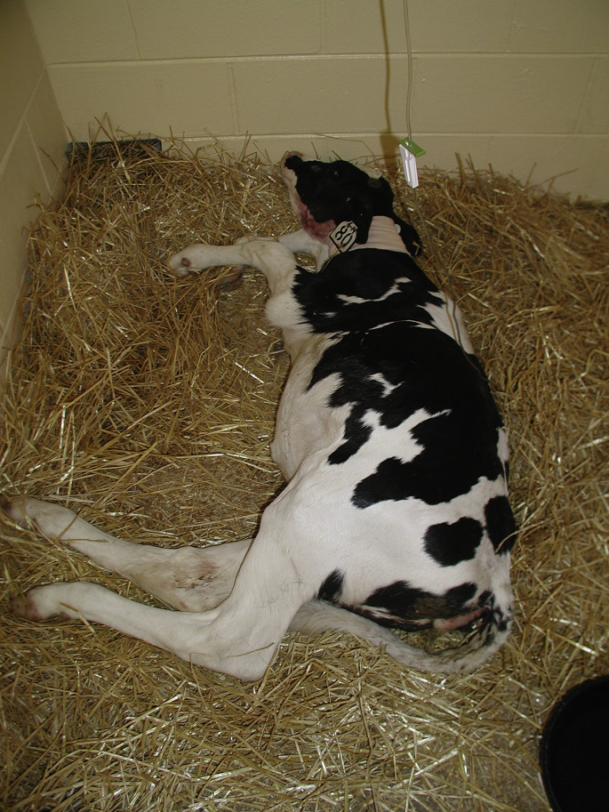
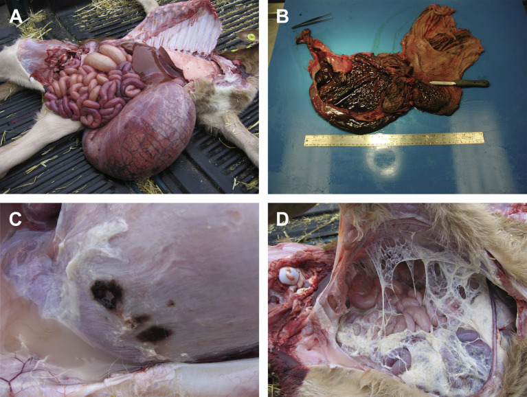
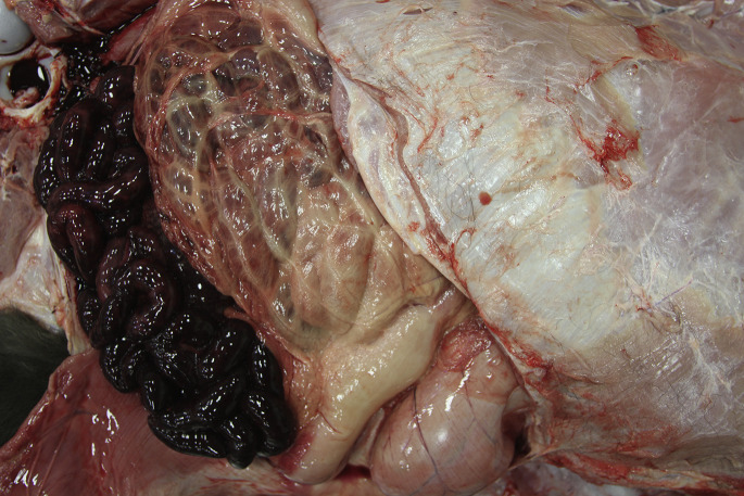
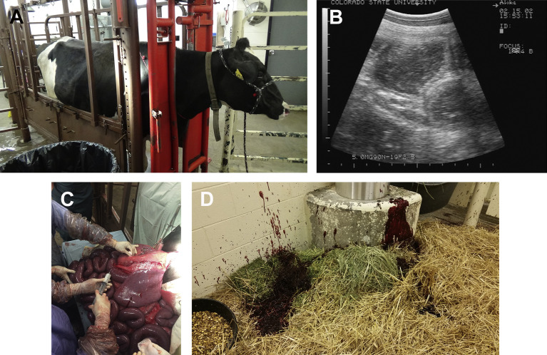
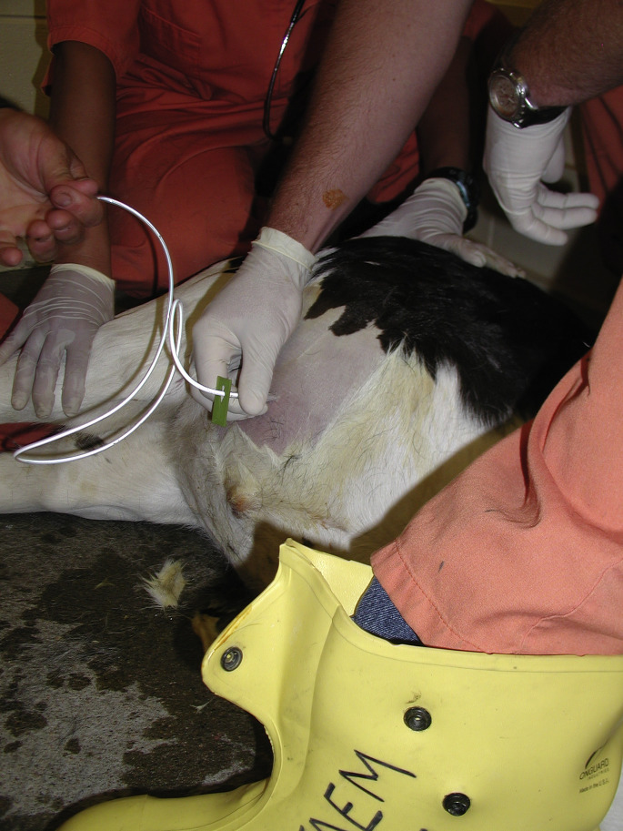
Clostridial abomasitis and enteritis are important alimentary diseases observed in all domestic ruminant species. These diseases most commonly result from overgrowth of Clostridium perfringens types A, B, C, D, and E with the associated release of bacterial exotoxins that result in necrosis of the abomasal or intestinal mucosa. Clostridium difficile may also be associated with enteritis in calves but is much less common than disease caused by C perfringens. This article reviews the causes, pathophysiology, clinical signs, diagnosis, treatment, and prevention of clostridial gastrointestinal diseases in ruminants. Particular emphasis is given to describing the various forms of disease and treatment of individual cases.

Keywords: Abomasitis; Clostridium difficile; Clostridium perfringens; Enteritis; Enterotoxemia; Ruminant.


Figures

Fig. 1
Fig. 1
A 3-week-old calf with abdominal distension caused by C perfringens type A abomasitis and enteritis.
Fig. 2
Fig. 2
Necropsy findings that may be observed in neonates with clostridial abomasitis. Common findings include grossly distended abomasum containing fluid and gas (A); diffuse, hemorrhagic to necrotizing inflammation of the abomasal mucosa (B); abomasal ulceration (C) with or without perforation resulting in fibrinous peritonitis (D).
Fig. 3
Fig. 3
Necropsy of a 3-day-old beef calf with C perfringens type A hemorrhagic enteritis. The cause was confirmed with anaerobic culture and mPCR genotyping.
Fig. 4
Fig. 4
Hemorrhagic C perfringens enteritis in an adult dairy cow. Note the right ventral abdominal distension caused by distension of the small intestine (A). Transabdominal ultrasonography shows dilated loops of small intestine up to 5 cm in diameter (B). A right flank exploratory laparotomy was performed and confirmed diffuse hemorrhagic enteritis (C). The cow received intraluminal procaine penicillin G in surgery. The following morning the cow had profuse hemorrhagic diarrhea (D), returned to eating, and recovered.
Fig. 5
Fig. 5
Gram stain of feces from a calf with clostridial enteritis (original magnification, ×1000). Note the group of large Gram positive rods within the background of other bacterial flora. Cytologic evidence of high numbers of large Gram-positive rods in ingesta or feces is suggestive of, but not definitive for, clostridial abomasitis and/or enteritis. The presence of white blood cells, red blood cells, and sloughed mucosal epithelium indicates inflammation with mucosal necrosis and hemorrhage. In early cases of abomasitis or enteritis, changes in fecal flora may not initially be observed.
Fig. 6
Fig. 6
Abomasocentesis performed in a 3-week-old Holstein calf in order to decompress a tightly distended abomasum. This procedure is best done with the calf in dorsal or left lateral recumbency. Following decompression, fluid samples can be obtained for culture and genotyping as well as cytology. Antibiotics and antitoxin can also be administered directly into the abomasum.
Fig. 7
Fig. 7
Beef calf with suspected C perfringens abomasitis. The calf showed marked abdominal distension and abomasal tympany at presentation (A). The calf was treated medically and the abdominal distension resolved within 24 hours (B).

References

    1. Lebrun M., Mainil J.G., Linden A. Cattle enterotoxaemia and Clostridium perfringens: description, diagnosis and prophylaxis. Vet Rec. 2010;167(1):13–22. - PubMed
    1. Uzal F.A., Songer J.G. Diagnosis of Clostridium perfringens intestinal infections in sheep and goats. J Vet Diagn Invest. 2008;20(3):253–265. - PubMed
    1. Songer J.G. Clostridial enteric diseases of domestic animals. Clin Microbiol Rev. 1996;9(2):216–234. - PMC - PubMed
    1. Lewis C.J. Control of important clostridial diseases of sheep. Vet Clin North Am Food Anim Pract. 2011;27(1):121–126. - PubMed
    1. Borriello S.P., Carman R.J., editors. Clostridia in gastrointestinal disease. CRC Press; Boca Raton (FL): 1992. pp. 195–221.

MeSH terms

LinkOut - more resources