

Jalili Syndrome: Cross-sectional and Longitudinal Features of Seven Patients With Cone-Rod Dystrophy and Amelogenesis Imperfecta
- PMID: 29421294
- PMCID: PMC5873517
- DOI: 10.1016/j.ajo.2018.01.029
Jalili Syndrome: Cross-sectional and Longitudinal Features of Seven Patients With Cone-Rod Dystrophy and Amelogenesis Imperfecta
Abstract
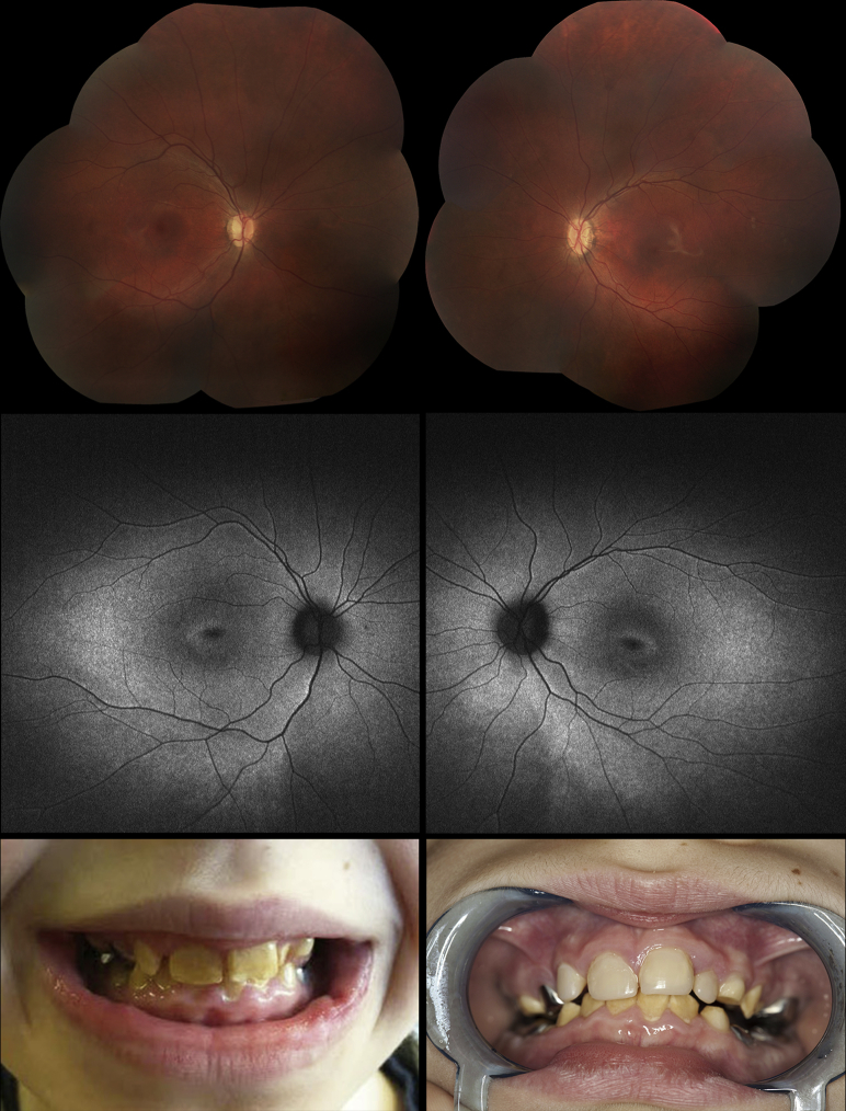
Purpose: To characterize a series of 7 patients with cone-rod dystrophy (CORD) and amelogenesis imperfecta (AI) owing to confirmed mutations in CNNM4, first described as "Jalili Syndrome."
Design: Retrospective observational case series.
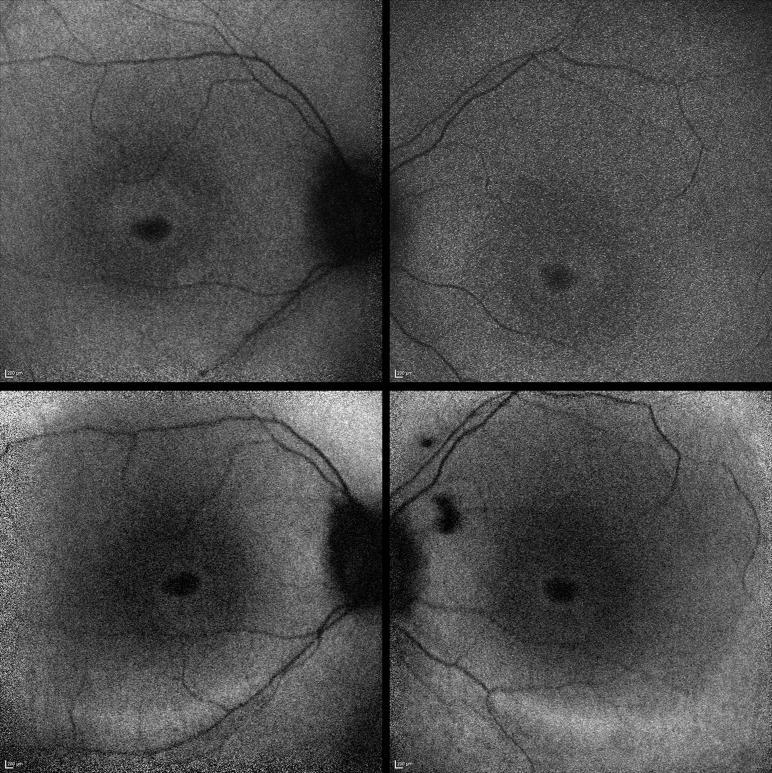
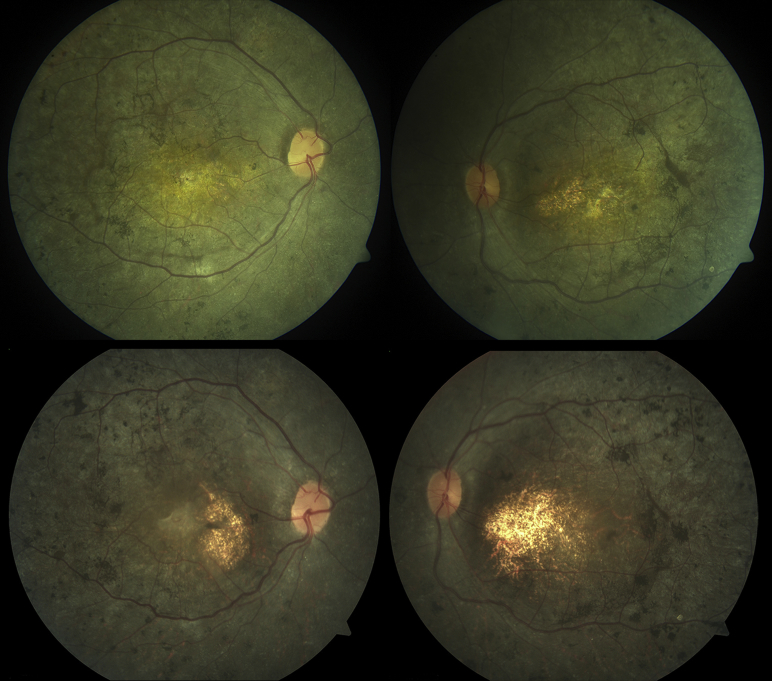
Methods: Seven patients from 6 families with Jalili Syndrome were identified at 3 tertiary referral centers. We systematically reviewed their available medical records, spectral-domain optical coherence tomography (SD-OCT), fundus autofluorescence imaging (FAF), color fundus photography, and electrophysiological assessments.
Results: The mean age at presentation was 6.7 years (range 3-16 years), with 6 male and 1 female patient. CNNM4 mutations were identified in all patients. The mean Snellen best-corrected visual acuity (BCVA) at presentation was 20/246 (range 20/98 to 20/399) in the right eye and 20/252 (range 20/98 to 20/480) in the left. Nystagmus was observed in all 7 patients, and photophobia was present in 6. Funduscopic findings at presentation were variable, ranging from only mild disc pallor to retinal vascular attenuation and macular atrophy. Multimodal imaging demonstrated disease progression in all 7 patients over time. Electroretinography uniformly revealed progressive cone-rod dysfunction.
Conclusions: Jalili Syndrome is a rare CORD associated with AI. We have further characterized its ocular phenotype, including describing SD-OCT, FAF, and electrophysiological features; and report several novel disease-causing sequence variants. Moreover, this study presents novel longitudinal data demonstrating structural and functional progression over time, allowing better informed advice on prognosis.
Copyright © 2018 The Authors. Published by Elsevier Inc. All rights reserved.
Figures
Similar articles
-
A novel mutation in CNNM4 is associated with a case of Jalili syndrome in Egypt.Doc Ophthalmol. 2025 Jun;150(3):189-196. doi: 10.1007/s10633-025-10018-1. Epub 2025 Apr 15. Doc Ophthalmol. 2025. PMID: 40232358 Free PMC article.
-
Expanding the genotypic spectrum of Jalili syndrome: Novel CNNM4 variants and uniparental isodisomy in a north American patient cohort.Am J Med Genet A. 2020 Mar;182(3):493-497. doi: 10.1002/ajmg.a.61484. Epub 2020 Feb 5. Am J Med Genet A. 2020. PMID: 32022389 Free PMC article.
-
Features, genetics and their correlation in Jalili syndrome: a systematic review.J Med Genet. 2019 Jun;56(6):358-369. doi: 10.1136/jmedgenet-2018-105716. Epub 2019 Jan 31. J Med Genet. 2019. PMID: 30705057
-
A new familial case of Jalili syndrome caused by a novel mutation in CNNM4.Ophthalmic Genet. 2017 Mar-Apr;38(2):161-166. doi: 10.3109/13816810.2016.1164192. Epub 2016 Apr 12. Ophthalmic Genet. 2017. PMID: 27070327
-
Cone pathway dysfunction in Jalili syndrome due to a novel familial variant of CNNM4 revealed by pupillometry and electrophysiologic investigations.Ophthalmic Genet. 2022 Apr;43(2):268-276. doi: 10.1080/13816810.2021.2002916. Epub 2021 Dec 7. Ophthalmic Genet. 2022. PMID: 34875963 Free PMC article.
Cited by
-
Novel homozygous nonsynonymous variant of CNNM4 gene in a Chinese family with Jalili syndrome.Mol Genet Genomic Med. 2022 Mar;10(3):e1860. doi: 10.1002/mgg3.1860. Epub 2022 Feb 12. Mol Genet Genomic Med. 2022. PMID: 35150469 Free PMC article.
-
Differential regulation of magnesium transporters Slc41, Cnnm and Trpm6-7 in the kidney of salmonids may represent evolutionary adaptations to high salinity environments.BMC Genomics. 2024 Nov 29;25(1):1156. doi: 10.1186/s12864-024-11055-x. BMC Genomics. 2024. PMID: 39614204 Free PMC article.
-
Functional and pathogenic insights into CNNM4 variants in Jalili syndrome.Sci Rep. 2024 Nov 23;14(1):29091. doi: 10.1038/s41598-024-80720-8. Sci Rep. 2024. PMID: 39580587 Free PMC article.
-
Acute Angle Closure Glaucoma in a Patient With Jalili-Smith Syndrome.Cureus. 2024 Sep 4;16(9):e68670. doi: 10.7759/cureus.68670. eCollection 2024 Sep. Cureus. 2024. PMID: 39371887 Free PMC article.
-
Structural Insights into the Intracellular Region of the Human Magnesium Transport Mediator CNNM4.Int J Mol Sci. 2019 Dec 12;20(24):6279. doi: 10.3390/ijms20246279. Int J Mol Sci. 2019. PMID: 31842432 Free PMC article.
References
-
- Michaelides M., Hardcastle A.J., Hunt D.M., Moore A.T. Progressive cone and cone-rod dystrophies: phenotypes and underlying molecular genetic basis. Surv Ophthalmol. 2006;51(3):232–258. - PubMed
-
- Aleman T.S., Cideciyan A.V., Volpe N.J., Stevanin G., Brice A., Jacobson S.G. Spinocerebellar ataxia type 7 (SCA7) shows a cone-rod dystrophy phenotype. Exp Eye Res. 2002;74(6):737–745. - PubMed
Publication types
MeSH terms
Substances
Grants and funding
LinkOut - more resources
Full Text Sources
Other Literature Sources
