

Genomic loci modulating retinal ganglion cell death following elevated IOP in the mouse
- PMID: 29421330
- PMCID: PMC5939594
- DOI: 10.1016/j.exer.2017.12.013
Genomic loci modulating retinal ganglion cell death following elevated IOP in the mouse
Abstract
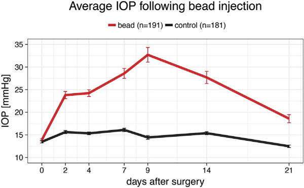
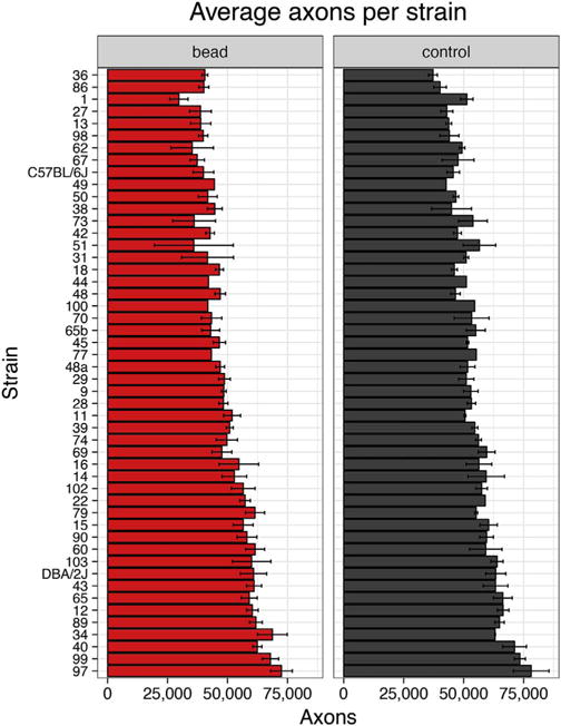
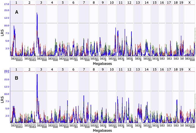
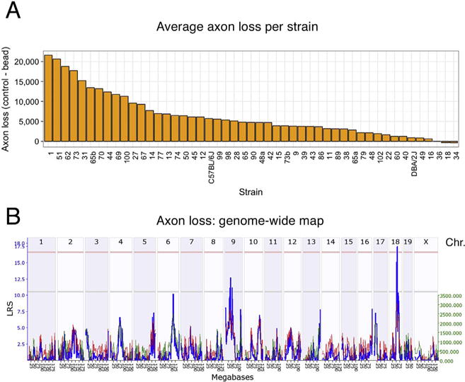
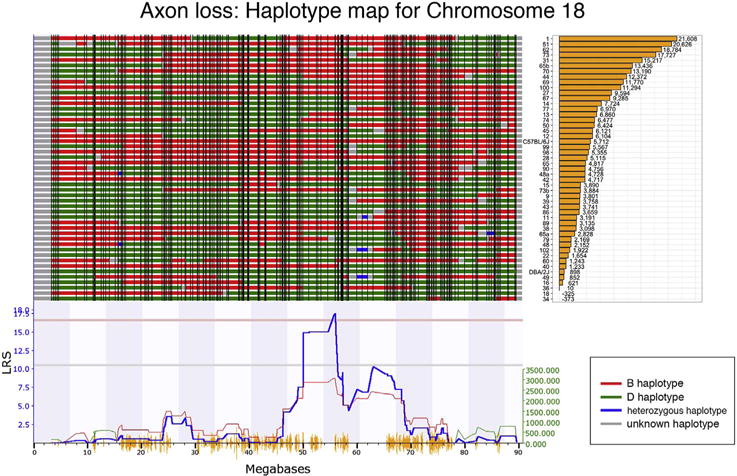
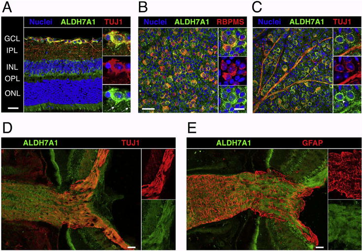
The present study was designed to identify genomic loci modulating the susceptibility of retinal ganglion cells (RGC) to elevated intraocular pressure (IOP) in the BXD recombinant inbred mouse strain set. IOP was elevated by injecting magnetic microspheres into the anterior chamber and blocking the trabecular meshwork using a handheld magnet to impede drainage. The IOP was then measured over the next 21 days. Only animals with IOP greater than 25 mmHg for two consecutive days or an IOP above 30 mmHg on a single day after microsphere-injection were used in this study. On day 21, mice were sacrificed and the optic nerve was processed for histology. Axons were counted for both the injected and the control eye in 49 BXD strains, totaling 181 normal counts and 191 counts associated with elevated IOP. The axon loss for each strain was calculated and the data were entered into genenetwork.org. The average number of normal axons in the optic nerve across all strains was 54,788 ± 16% (SD), which dropped to 49,545 ± 20% in animals with artificially elevated IOP. Interval mapping demonstrated a relatively similar genome-wide map for both conditions with a suggestive Quantitative Trait Locus (QTL) on proximal Chromosome 3. When the relative axon loss was used to generate a genome-wide interval map, we identified one significant QTL (p < 0.05) on Chromosome 18 between 53.6 and 57 Mb. Within this region, the best candidate gene for modulating axon loss was Aldh7a1. Immunohistochemistry demonstrated ALDH7A1 expression in mouse RGCs. ALDH7A1 variants were not significantly associated with glaucoma in the NEIGHBORHOOD GWAS dataset, but this enzyme was identified as part of the butanoate pathway previously associated with glaucoma risk. Our results suggest that genomic background influences susceptibility to RGC degeneration and death in an inducible glaucoma model.
Keywords: Butanoate pathway; GeneNetwork; Glaucoma model; QTL mapping; Systems genetics.
Copyright © 2018 The Authors. Published by Elsevier Ltd.. All rights reserved.
Figures
Similar articles
-
Genomic Locus Modulating IOP in the BXD RI Mouse Strains.G3 (Bethesda). 2018 May 4;8(5):1571-1578. doi: 10.1534/g3.118.200190. G3 (Bethesda). 2018. PMID: 29496776 Free PMC article.
-
Genomic locus modulating corneal thickness in the mouse identifies POU6F2 as a potential risk of developing glaucoma.PLoS Genet. 2018 Jan 25;14(1):e1007145. doi: 10.1371/journal.pgen.1007145. eCollection 2018 Jan. PLoS Genet. 2018. PMID: 29370175 Free PMC article.
-
The dark phase intraocular pressure elevation and retinal ganglion cell degeneration in a rat model of experimental glaucoma.Exp Eye Res. 2013 Jul;112:21-8. doi: 10.1016/j.exer.2013.04.008. Epub 2013 Apr 18. Exp Eye Res. 2013. PMID: 23603611 Free PMC article.
-
Using BXD mouse strains in vision research: A systems genetics approach.Mol Vis. 2020 Mar 6;26:173-187. eCollection 2020. Mol Vis. 2020. PMID: 32180682 Free PMC article. Review.
-
Gene therapies and gene product-based drug candidates for normalizing and preserving tissue functions in animal models of ocular hypertension and glaucoma.Mol Aspects Med. 2023 Dec;94:101218. doi: 10.1016/j.mam.2023.101218. Epub 2023 Nov 15. Mol Aspects Med. 2023. PMID: 37976898 Review.
Cited by
-
Protective Effect of Nicotinamide Riboside on Glucocorticoid-Induced Glaucoma: Mitigating Mitochondrial Damage and Extracellular Matrix Deposition.Invest Ophthalmol Vis Sci. 2024 Jul 1;65(8):1. doi: 10.1167/iovs.65.8.1. Invest Ophthalmol Vis Sci. 2024. PMID: 38949632 Free PMC article.
-
CircXPO5 Plays a Neuroprotective Function in the Lateral Geniculate Nucleus of Glaucoma by Regulating GRIN2A.Brain Sci. 2022 Jun 14;12(6):780. doi: 10.3390/brainsci12060780. Brain Sci. 2022. PMID: 35741665 Free PMC article.
-
Using retinal function to define ischemic exclusion criteria for animal models of glaucoma.Exp Eye Res. 2021 Jan;202:108354. doi: 10.1016/j.exer.2020.108354. Epub 2020 Nov 7. Exp Eye Res. 2021. PMID: 33171192 Free PMC article.
-
Commonalities of optic nerve injury and glaucoma-induced neurodegeneration: Insights from transcriptome-wide studies.Exp Eye Res. 2021 Jun;207:108571. doi: 10.1016/j.exer.2021.108571. Epub 2021 Apr 15. Exp Eye Res. 2021. PMID: 33844961 Free PMC article. Review.
-
Genomic Locus Modulating IOP in the BXD RI Mouse Strains.G3 (Bethesda). 2018 May 4;8(5):1571-1578. doi: 10.1534/g3.118.200190. G3 (Bethesda). 2018. PMID: 29496776 Free PMC article.
References
-
- Aboobakar IF, Johnson WM, Stamer WD, Hauser MA, Allingham RR. Major review: exfoliation syndrome; advances in disease genetics, molecular biology, and epidemiology. Exp Eye Res. 2016;154:88–103. - PubMed
-
- Bailey JN, Loomis SJ, Kang JH, Allingham RR, Gharahkhani P, Khor CC, Burdon KP, Aschard H, Chasman DI, Igo RP, Jr, Hysi PG, Glastonbury CA, Ashley-Koch A, Brilliant M, Brown AA, Budenz DL, Buil A, Cheng CY, Choi H, Christen WG, Curhan G, De Vivo I, Fingert JH, Foster PJ, Fuchs C, Gaasterland D, Gaasterland T, Hewitt AW, Hu F, Hunter DJ, Khawaja AP, Lee RK, Li Z, Lichter PR, Mackey DA, McGuffin P, Mitchell P, Moroi SE, Perera SA, Pepper KW, Qi Q, Realini T, Richards JE, Ridker PM, Rimm E, Ritch R, Ritchie M, Schuman JS, Scott WK, Singh K, Sit AJ, Song YE, Tamimi RM, Topouzis F, Viswanathan AC, Verma SS, Vollrath D, Wang JJ, Weisschuh N, Wissinger B, Wollstein G, Wong TY, Yaspan BL, Zack DJ, Zhang K, Study EN, Consortium A. Weinreb RN, Pericak-Vance MA, Small K, Hammond CJ, Aung T, Liu Y, Vithana EN, MacGregor S, Craig JE, Kraft P, Howell G, Hauser MA, Pasquale LR, Haines JL, Wiggs JL. Genome-wide association analysis identifies TXNRD2, ATXN2 and FOXC1 as susceptibility loci for primary open-angle glaucoma. Nat Genet. 2016;48:189–194. - PMC - PubMed
-
- Bailey JN, Yaspan BL, Pasquale LR, Hauser MA, Kang JH, Loomis SJ, Brilliant M, Budenz DL, Christen WG, Fingert J, Gaasterland D, Gaasterland T, Kraft P, Lee RK, Lichter PR, Liu Y, McCarty CA, Moroi SE, Richards JE, Realini T, Schuman JS, Scott WK, Singh K, Sit AJ, Vollrath D, Wollstein G, Zack DJ, Zhang K, Pericak-Vance MA, Allingham RR, Weinreb RN, Haines JL, Wiggs JL. Hypothesis-independent pathway analysis implicates GABA and acetyl-CoA metabolism in primary open-angle glaucoma and normal-pressure glaucoma. Hum Genet. 2014;133:1319–1330. - PMC - PubMed
-
- Bhave SV, Hoffman PL, Lassen N, Vasiliou V, Saba L, Deitrich RA, Tabakoff B. Gene array profiles of alcohol and aldehyde metabolizing enzymes in brains of C57BL/6 and DBA/2 mice. Alcohol Clin Exp Res. 2006;30:1659–1669. - PubMed
Publication types
MeSH terms
Substances
Grants and funding
LinkOut - more resources
Full Text Sources
Other Literature Sources
Molecular Biology Databases
Miscellaneous
