. 2018 Apr:161:216-227.
doi: 10.1016/j.biomaterials.2018.01.040. Epub 2018 Feb 3.

Harnessing macrophage-mediated degradation of gelatin microspheres for spatiotemporal control of BMP2 release

Affiliations

Harnessing macrophage-mediated degradation of gelatin microspheres for spatiotemporal control of BMP2 release

Ramkumar T Annamalai et al. Biomaterials. 2018 Apr.

Abstract

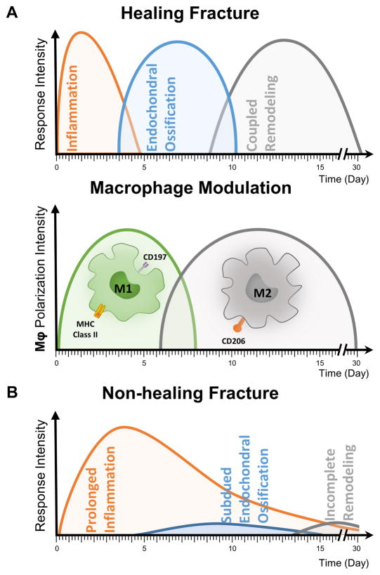
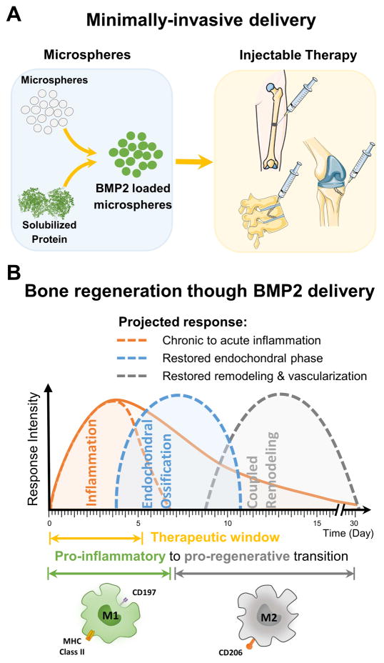
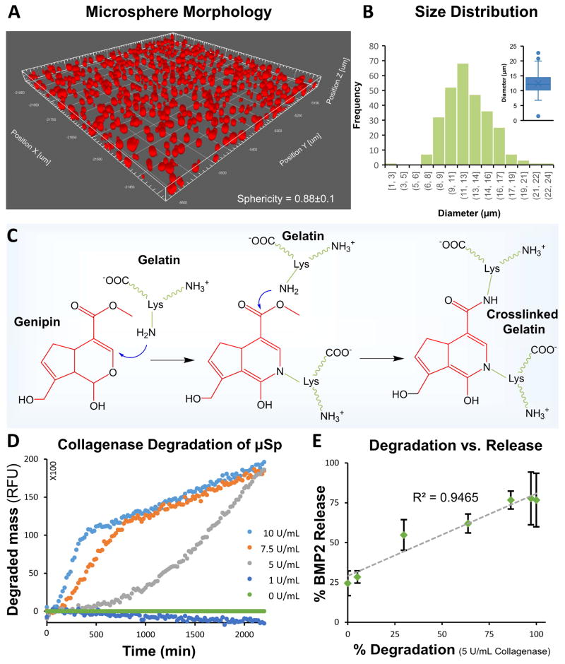
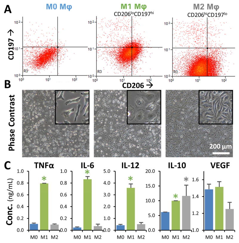
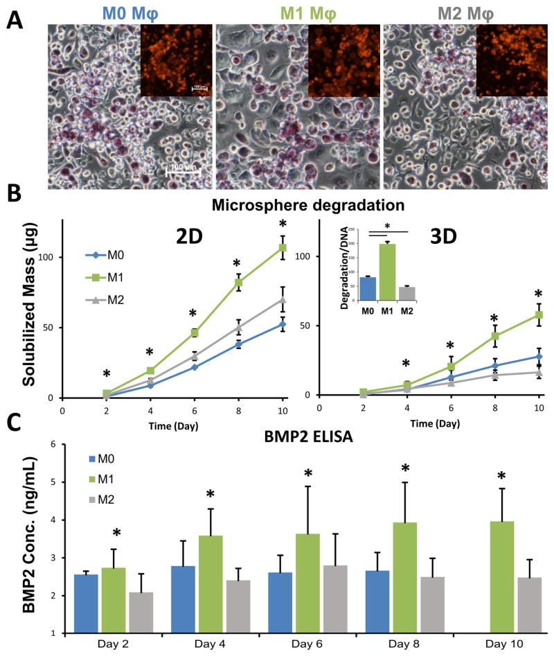
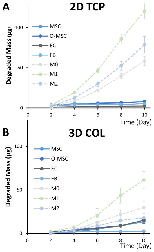
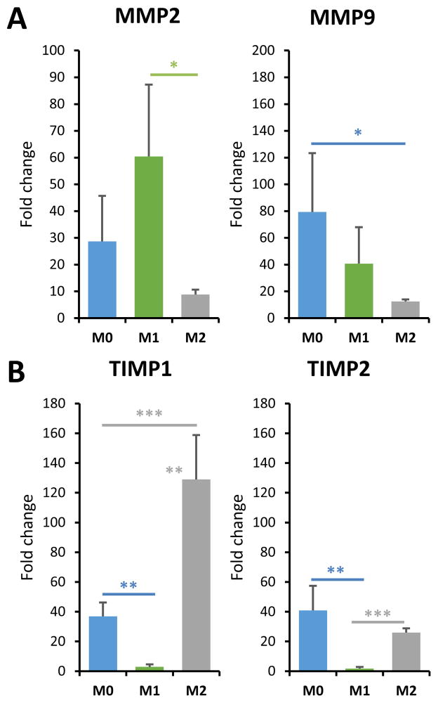
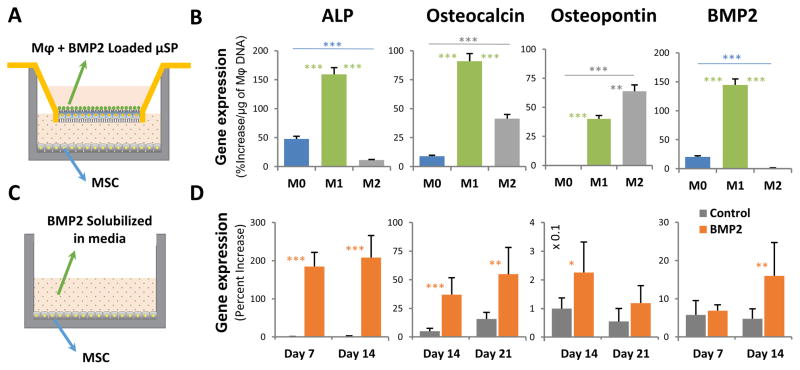
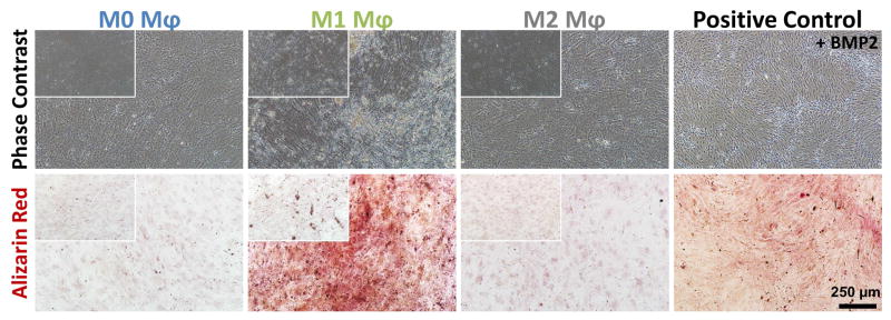
Biomaterials-based approaches to harnessing the immune and inflammatory responses to potentiate wound healing hold important promise. Bone fracture healing is characterized by an acute inflammatory phase, followed by a transition to a regenerative and repair phase. In this study, we developed genipin-crosslinked gelatin microspheres designed to be preferentially degraded by inflammatory (M1) macrophages. Highly crosslinked (>90%) microspheres allowed efficient incorporation of bioactive bone morphogenetic protein 2 (BMP2), a potent stimulator of osteogenesis in progenitor cells, via electrostatic interactions. Release of BMP2 was directly correlated with degradation of the gelatin matrix. Exposure of microspheres to polarized murine macrophages showed that degradation was significantly higher in the presence of M1 macrophages, relative to alternatively activated (M2) macrophages and unpolarized controls. Microsphere degradation in the presence of non-inflammatory cells resulted in very low degradation rates. The expression of matrix metalloproteinases (MMPs) and tissue inhibitors of MMP (TIMPs) by macrophages were consistent with the observed phenotype-dependent degradation rates. Indirect co-culture of BMP2-loaded microspheres and macrophages with isolated adipose-derived mesenchymal stem cells (MSC) showed that M1 macrophages produced the strongest osteogenic response, comparable to direct supplementation of the culture medium with BMP2. Controlled release systems that are synchronized with the inflammatory response have the potential to provide better spatiotemporal control of growth factor delivery and therefore may improve the outcomes of recalcitrant wounds.

Keywords: BMP; Bone tissue engineering; Controlled drug release; Immunomodulation; Inflammation; Macrophages.


Figures

Fig. 1
Fig. 1. Role of inflammatory cells in normal and pathological fracture healing
A) Normal fracture healing involves phases characterized by inflammation, endochondral ossification, and coupled tissue remodeling. The phenotype of local macrophages changes from pro-inflammatory (M1) to pro-reparative (M2) as bone repair proceeds. B) In non-healing fractures, the progression to the endochondral ossification is delayed or inhibited, such that new bone is not regenerated.
Fig. 2
Fig. 2. Delivery of therapeutic growth factors to potentiate bone regeneration
A) Gelatin microspheres can be designed to sequester BMP2 and can be used as a minimally-invasive method of delivering growth factor to bone wounds. B) Design of microspheres to be preferentially degraded by inflammatory macrophages allows delivery of BMP2 during the late inflammation phase of healing, and promotes progression to endochondral ossification, thereby preventing delayed healing.
Fig. 3
Fig. 3. Morphology, genipin crosslinking chemistry and enzymatic degradation of gelatin microspheres
A) Confocal image of microspheres show spherical morphology. B) Histogram showing the size distribution of crosslinked gelatin microspheres (inset shows the box plot of the size distribution). C) Genipin is used to crosslink lysine residues in gelatin via a two-step process. D) The degradation rate of crosslinked microspheres increased with an increase in collagenase concentration. E) Enzymatic degradation of the microspheres caused an initial burst release followed by sustained release of the remainder of the payload.
Fig. 4
Fig. 4. Characterization of macrophage phenotype
A) Flow cytometric analysis showed that M1-treated macrophages had increased CD197 expression and reduced CD206 expression (CD197hiCD206lo), while the M2-treated population showed increased CD206 expression and reduced CD197 expression (CD206hiCD197lo). B) M1 macrophages had a ‘broken-egg’ morphology, while M2 macrophages exhibited a more spindle-shaped morphology (insets show magnified cell morphology). C) Cytokine secretion by M1 macrophages showed significantly higher levels of TNFα, IL-6, Il-10, relative to M2 macrophages, which had increased secretion of IL10. *p<0.05
Fig. 5
Fig. 5. Cell-mediated degradation of microspheres leads to BMP2 release
A) Macrophages degraded microspheres through enzymatic degradation (insets show fluorescent microspheres). The genipin crosslinking changed the color of the gelatin microspheres to a blue/purple color under visible light. B) At the same cell density and microsphere mass, the rate of degradation was higher when cultured in 2D wells, compared to 3D collagen matrices. The normalized degradation rate of macrophages was higher under M1 conditions compared to that of M0 and M2 (inset). C) As a result of preferential degradation, the release of BMP2 from loaded microspheres was higher from M1 macrophages compared to M0 and M2. *p<0.05
Fig. 6
Fig. 6. Degradation rates of gelatin microspheres varied by cell type
A) In 2D cultures, macrophages of all phenotypes degraded microspheres at a faster rate than non-inflammatory cells. B) In 3D collagen gels, a similar trend was observed, though degradation by macrophages was slower in 3D than in 2D.
Fig. 7
Fig. 7. Macrophage polarization affects MMP and TIMP expression
A) The skewing of macrophages to the M1 phenotype enhanced MMP2 expression, while M2 macrophages showed suppressed MMP9 expression. B) M1 macrophages showed depressed TIMP expression while skewing to the M2 phenotype enhanced TIMP1 expression. *p<0.05, **p<0.005, ***p<0.0005.
Fig. 8
Fig. 8. BMP2 released through the degradation of microspheres stimulated MSC osteogenesis
A) A Transwell system was used to allow paracrine signaling between macrophage-degraded microspheres and cultured MSC. B) The BMP2 released by M1 macrophage-mediated degradation caused increased expression of osteogenic genes in MSC, compared to both M0 and M2 macrophages (p<0.01). C) Positive control cultures were exposed to a constant dose of 200 ng/ml of solubilized BMP2 in standard culture wells. D) Positive control cultures showed increased expression of osteogenic genes over time. *p<0.05, **p<0.01, ***p<0.005.
Fig. 9
Fig. 9. BMP2 released through macrophage-mediated degradation of microspheres stimulated MSC osteogenesis and calcification
Microspheres degraded by M1 macrophages had the largest osteogenic effect, similar to positive controls containing 200 ng/mL BMP2, and markedly higher than cultures exposed to M0 and M2 macrophages. The insets show the phase contrast and alizarin red staining of the corresponding negative control wells supplied with unloaded microspheres. There is no matrix deposition/staining on the unloaded microspheres on M0, M1 or M2 conditions.

References

    1. Claes L, Recknagel S, Ignatius A. Fracture healing under healthy and inflammatory conditions. Nature reviews Rheumatology. 2012;8(3):133–143. - PubMed
    1. Spiller KL, Freytes DO, Vunjak-Novakovic G. Macrophages modulate engineered human tissues for enhanced vascularization and healing. Annals of biomedical engineering. 2015;43(3):616–27. - PMC - PubMed
    1. Mountziaris PM, Mikos AG. Modulation of the Inflammatory Response for Enhanced Bone Tissue Regeneration, Tissue engineering. Part B. Reviews. 2008;14(2):179–186. - PMC - PubMed
    1. Brown BN, Ratner BD, Goodman SB, Amar S, Badylak SF. Macrophage polarization: An opportunity for improved outcomes in biomaterials and regenerative medicine. Biomaterials. 2012;33(15):3792–3802. - PMC - PubMed
    1. Spiller KL, Nassiri S, Witherel CE, Anfang RR, Ng J, Nakazawa KR, Yu T, Vunjak-Novakovic G. Sequential delivery of immunomodulatory cytokines to facilitate the M1-to-M2 transition of macrophages and enhance vascularization of bone scaffolds. Biomaterials. 2015;37:194–207. - PMC - PubMed

Publication types