. 2018 Jul 15;35(14):1604-1619.
doi: 10.1089/neu.2017.5457. Epub 2018 May 3.

Multimodal Characterization of the Late Effects of Traumatic Brain Injury: A Methodological Overview of the Late Effects of Traumatic Brain Injury Project

Affiliations

Multimodal Characterization of the Late Effects of Traumatic Brain Injury: A Methodological Overview of the Late Effects of Traumatic Brain Injury Project

Brian L Edlow et al. J Neurotrauma. .

Abstract

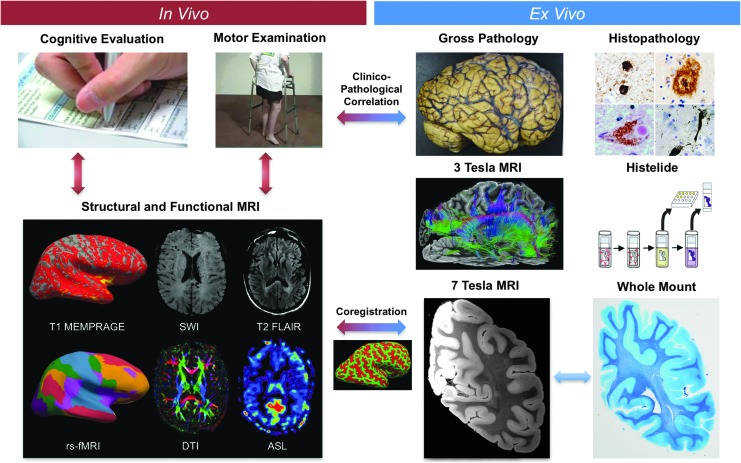
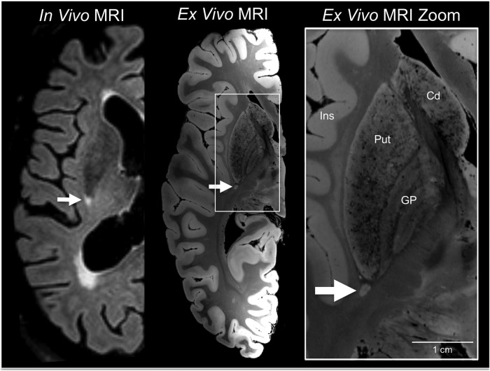
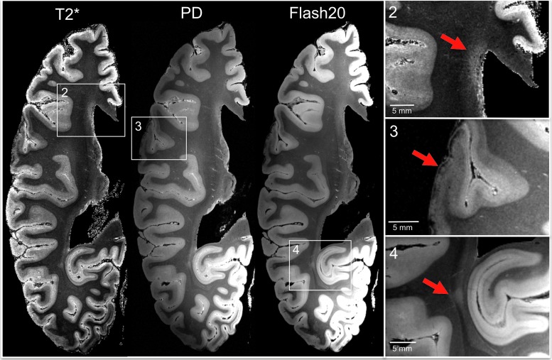
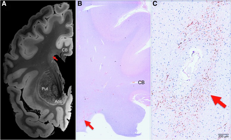
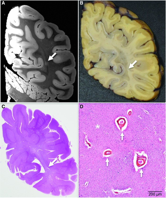
Epidemiological studies suggest that a single moderate-to-severe traumatic brain injury (TBI) is associated with an increased risk of neurodegenerative disease, including Alzheimer's disease (AD) and Parkinson's disease (PD). Histopathological studies describe complex neurodegenerative pathologies in individuals exposed to single moderate-to-severe TBI or repetitive mild TBI, including chronic traumatic encephalopathy (CTE). However, the clinicopathological links between TBI and post-traumatic neurodegenerative diseases such as AD, PD, and CTE remain poorly understood. Here, we describe the methodology of the Late Effects of TBI (LETBI) study, whose goals are to characterize chronic post-traumatic neuropathology and to identify in vivo biomarkers of post-traumatic neurodegeneration. LETBI participants undergo extensive clinical evaluation using National Institutes of Health TBI Common Data Elements, proteomic and genomic analysis, structural and functional magnetic resonance imaging (MRI), and prospective consent for brain donation. Selected brain specimens undergo ultra-high resolution ex vivo MRI and histopathological evaluation including whole-mount analysis. Co-registration of ex vivo and in vivo MRI data enables identification of ex vivo lesions that were present during life. In vivo signatures of postmortem pathology are then correlated with cognitive and behavioral data to characterize the clinical phenotype(s) associated with pathological brain lesions. We illustrate the study methods and demonstrate proof of concept for this approach by reporting results from the first LETBI participant, who despite the presence of multiple in vivo and ex vivo pathoanatomic lesions had normal cognition and was functionally independent until her mid-80s. The LETBI project represents a multidisciplinary effort to characterize post-traumatic neuropathology and identify in vivo signatures of postmortem pathology in a prospective study.

Keywords: MRI; dementia; neurodegeneration; neuropathology; traumatic brain injury.


Conflict of interest statement

No competing financial interests exist.

Dr. Fischl and Mr. Tirrell have financial interest in CorticoMetrics, a company whose medical pursuits focus on brain imaging and measurement technologies. Their interests were reviewed and are managed by Massachusetts General Hospital and Partners HealthCare in accordance with their conflict of interest policies.

Figures

<b>FIG. 1.</b>
FIG. 1.
Overview of study design. The Late Effects of Traumatic Brain Injury (LETBI) study integrates in vivo data (left) and ex vivo data (right) to comprehensively characterize post-traumatic neurodegeneration. LETBI participants undergo extensive cognitive, behavioral, and neurological examinations. In addition, structural and functional magnetic resonance imaging (MRI) are performed and blood samples are drawn for proteomic and genomic analysis to identify in vivo biomarkers of post-traumatic neuropathology. Autopsied brain specimens undergo gross pathological analysis, histopathological analysis including Histelide and whole–mount analysis, and ultra-high resolution ex vivo MRI on 3 Tesla and 7 Tesla MRI scanners. Co-registration of ex vivo and in vivo MRI data enables determination of whether pathologically relevant ex vivo lesions were present during life. In vivo signatures of postmortem pathology are then correlated with cognitive and behavioral data to characterize the clinical phenotype(s) associated with pathological lesions. Color image is available online at www.liebertpub.com/neu
<b>FIG. 2.</b>
FIG. 2.
White matter lesion identification on in vivo 3 Tesla magnetic resonance imaging (MRI) and ex vivo 7 Tesla MRI. An in vivo 3 Tesla MRI performed at age 88, after the patient's fifth TBI, revealed bihemispheric corona radiata and internal capsule white matter hyperintensities, including a punctate lesion at the posterior aspect of the putamen (arrow). The same lesion was identified by ex vivo 7 Tesla MRI (arrow), which was performed on the formalin-fixed right hemisphere. A zoomed view of the lesion is shown in the right panel (arrow) to demonstrate the increased anatomic precision provided by ex vivo MRI (200 μm resolution) for delineating the borders of the lesion with respect to nearby anatomic structures. Cd, caudate; GP, globus pallidus; Ins, insula; Put, putamen.
<b>FIG. 3.</b>
FIG. 3.
Ex vivo 7 Tesla magnetic resonance imaging (MRI) reveals focal lesions not detected by in vivo 3 Tesla MRI. Axial T2*, proton density (PD), and flash20 images synthesized from the multi-echo flash sequence are shown at the level of the superior border of the thalamus. These three datasets reveal two lesions that were not visualized on the in vivo MRI dataset (lesions 3 and 4) and confirm the presence of a lesion that was seen in vivo (lesion 2). Lesion 2 is a periventricular hyperintensity in the frontal lobe, suggesting leukoaraiosis. Lesion 3 is a curvilinear hypointensity that traverses the superficial layers of cerebral cortex in the frontal operculum, suggesting hemosiderin deposition. Lesion 4 is a punctate hyperintensity in the periventricular white matter of the occipital lobe, consistent with leukoaraiosis or possibly chronic traumatic axonal injury. Zoomed views of each lesion are shown on the inset panels, with red arrows indicating the lesions. Color image is available online at www.liebertpub.com/neu
<b>FIG. 4.</b>
FIG. 4.
Correlative analysis of 7 Tesla ex vivo magnetic resonance imaging (MRI) and histopathology for the periventricular lesion. (A) A 7 Tesla ex vivo MRI coronal image of periventricular signal abnormality (red arrow) is shown at the level of the nucleus accumbens (Acc). The image shown here is a flash20 synthesized image from a multi-echo flash sequence. (B) A coronal histopathological section at the same neuroanatomic location is shown (red arrow), stained with amyloid-precursor protein and counterstained with hematoxylin. (C) Microscopic analysis of the periventricular region revealed disrupted axonal transport, as evidenced by positive amyloid precursor protein staining (red arrow). The degree to which chronic microvascular disease and chronic traumatic axonal injury contributed to this axonal pathology cannot be definitively determined, but the perivascular location suggests a vascular etiology. Neuroanatomic landmarks: CB, cingulum bundle; Put, putamen. Color image is available online at www.liebertpub.com/neu
<b>FIG. 5.</b>
FIG. 5.
Correlative analysis of 7 Tesla ex vivo magnetic resonance imaging (MRI) and histopathology for the curvilinear cortical lesion. Zoomed views of axial, sagittal and coronal flash20 images synthesized from the 7 Tesla multi-echo flash sequence reveal a curvilinear hypointense signal abnormality (red arrows) in the subpial region of the frontal operculum. Hematoxylin and eosin (H + E) staining of this region revealed atherosclerosis of a vessel within the subarachnoid space. Staining with glial fibrillary acidic protein (GFAP) and Perls' iron stain revealed gliosis and hemosiderin deposition, respectively, in the underlying cerebral cortex. The inset in the bottom right panel shows a high-powered view of hemosiderin-containing microglia. These findings suggest the possibility that a healed contusion accounts for the signal abnormality seen on ex vivo MRI. Color image is available online at www.liebertpub.com/neu
<b>FIG. 6.</b>
FIG. 6.
Ex vivo 3 Tesla diffusion tractography reveals regional white matter injury. Transcallosal fiber tracts generated using a corpus callosum seed region are shown from a left lateral perspective for the index patient (left) and a representative control subject who died of non-neurological causes (right). Tractography analysis revealed a decrease in the relative density of transcallosal fiber tracts in the parietal region, compared with the corresponding region of fiber tracts in the control dataset (white arrows). Fiber tracts are color-coded according to their direction (top right inset). Color image is available online at www.liebertpub.com/neu
<b>FIG. 7.</b>
FIG. 7.
Co-registration of ex vivo and in vivo magnetic resonance imaging (MRI) data. Precise anatomic alignment of in vivo and ex vivo MRI datasets from an individual patient is challenging because postmortem fixation causes nonlinear deformations. A deformation is seen by comparing a sagittal image from the in vivo T1 multi-echo Magnetization Prepared Rapid Acquisition Gradient Echo dataset (top left) with a sagittal image from the ex vivo multi-echo flash dataset (top right). We developed a combined volume- and surface-based co-registration technique to address this challenge and obtain precise voxel-to-voxel match between the in vivo and ex vivo datasets. The inflated cortical surfaces generated from the in vivo and ex vivo datasets are shown in the bottom left and bottom right panels, respectively. Given that the topology of the cortical folds is invariant, this topology can be used to initialize a biomechanical nonlinear deformation. Color image is available online at www.liebertpub.com/neu
<b>FIG. 8.</b>
FIG. 8.
Co-registration-based lesion localization on in vivo magnetic resonance imaging (MRI). Using combined volume- and surface-based co-registration (as shown in Fig. 7), the punctate hyperintense lesion seen in the occipital white matter on the ex vivo MRI dataset (red circle, right panel) is colocalized to its precise anatomic coordinates in the in vivo MRI dataset (red circle, left panel). The ex vivo MRI dataset used in the co-registration was the flash20 parameter map synthesized from the multi-echo flash 7 Tesla acquisition (200 μm spatial resolution), and the in vivo dataset was the T1 multi-echo Magnetization Prepared Rapid Acquisition Gradient Echo dataset (1 mm spatial resolution). Although no lesion was detected in the occipital white matter during the initial review of the in vivo MRI dataset, there appears to be a punctate hypointensity on the in vivo MRI dataset (red circle, left panel) that was only appreciated after the co-registration procedure. This observation suggests that ex vivo MRI may reveal lesions that were initially undetected on in vivo MRI. Color image is available online at www.liebertpub.com/neu
<b>FIG. 9.</b>
FIG. 9.
Targeted whole–mount histopathological analysis based on ex vivo magnetic resonance imaging (MRI) lesion localization. The punctate hyperintense lesion in the occipital white matter identified on ex vivo 7 Tesla (7T) MRI is first localized in the coronal plane (A, white arrow), because this is the plane in which the gross pathological specimen is cut into ∼1.5 cm thick slabs. During an audiovisual consensus teleconference, the precise anatomic coordinates of the lesion seen on ex vivo 7T MRI are compared with surface landmarks seen on the serial coronal slabs using high resolution photographs of the tissue slabs. The investigators of the Late Effects of Traumatic Brain Injury study select a slab for whole–mount analysis (B) based upon its correspondence with the coronal MRI image that contained the lesion. Whole–mount analysis is then performed on this coronal slab (C), enabling targeted histopathological analysis (D) of the lesion identified on ex vivo MRI. This histopathological analysis revealed enlarged perivascular spaces (arrows), corpora amylacea, and white matter rarefaction (asterisk). A single pathological process, ischemia, is the likely cause of these pathological findings. Color image is available online at www.liebertpub.com/neu

Similar articles

Cited by

  • Intimate Partner Violence and Other Trauma Exposures in Females With Traumatic Brain Injury.
    de Souza NL, Kumar RG, Pruyser A, Blunt EE, Sanders W, Meydan A, Lawrence P, Venkatesan UM, Mac Donald CL, Hoffman JM, Bodien YG, Edlow BL, Dams-O'Connor K. de Souza NL, et al. J Neurotrauma. 2024 Feb;41(3-4):529-536. doi: 10.1089/neu.2023.0225. J Neurotrauma. 2024. PMID: 37974411 Free PMC article.
  • Long-Term Effects of Repeated Blast Exposure in United States Special Operations Forces Personnel: A Pilot Study Protocol.
    Edlow BL, Bodien YG, Baxter T, Belanger HG, Cali RJ, Deary KB, Fischl B, Foulkes AS, Gilmore N, Greve DN, Hooker JM, Huang SY, Kelemen JN, Kimberly WT, Maffei C, Masood M, Perl DP, Polimeni JR, Rosen BR, Tromly SL, Tseng CJ, Yao EF, Zürcher NR, Mac Donald CL, Dams-O'Connor K. Edlow BL, et al. J Neurotrauma. 2022 Oct;39(19-20):1391-1407. doi: 10.1089/neu.2022.0030. Epub 2022 Jun 29. J Neurotrauma. 2022. PMID: 35620901 Free PMC article.
  • Traumatic brain injury: progress and challenges in prevention, clinical care, and research.
    Maas AIR, Menon DK, Manley GT, Abrams M, Åkerlund C, Andelic N, Aries M, Bashford T, Bell MJ, Bodien YG, Brett BL, Büki A, Chesnut RM, Citerio G, Clark D, Clasby B, Cooper DJ, Czeiter E, Czosnyka M, Dams-O'Connor K, De Keyser V, Diaz-Arrastia R, Ercole A, van Essen TA, Falvey É, Ferguson AR, Figaji A, Fitzgerald M, Foreman B, Gantner D, Gao G, Giacino J, Gravesteijn B, Guiza F, Gupta D, Gurnell M, Haagsma JA, Hammond FM, Hawryluk G, Hutchinson P, van der Jagt M, Jain S, Jain S, Jiang JY, Kent H, Kolias A, Kompanje EJO, Lecky F, Lingsma HF, Maegele M, Majdan M, Markowitz A, McCrea M, Meyfroidt G, Mikolić A, Mondello S, Mukherjee P, Nelson D, Nelson LD, Newcombe V, Okonkwo D, Orešič M, Peul W, Pisică D, Polinder S, Ponsford J, Puybasset L, Raj R, Robba C, Røe C, Rosand J, Schueler P, Sharp DJ, Smielewski P, Stein MB, von Steinbüchel N, Stewart W, Steyerberg EW, Stocchetti N, Temkin N, Tenovuo O, Theadom A, Thomas I, Espin AT, Turgeon AF, Unterberg A, Van Praag D, van Veen E, Verheyden J, Vyvere TV, Wang KKW, Wiegers EJA, Williams WH, Wilson L, Wisniewski SR, Younsi A, Yue JK, Yuh EL, Zeiler FA, Zeldovich M, Zemek R; InTBIR Participants and Investigators. Maas AIR, et al. Lancet Neurol. 2022 Nov;21(11):1004-1060. doi: 10.1016/S1474-4422(22)00309-X. Epub 2022 Sep 29. Lancet Neurol. 2022. PMID: 36183712 Free PMC article. Review.
  • Alzheimer's Disease-Related Dementias Summit 2019: National Research Priorities for the Investigation of Traumatic Brain Injury as a Risk Factor for Alzheimer's Disease and Related Dementias.
    Dams-O'Connor K, Bellgowan PSF, Corriveau R, Pugh MJ, Smith DH, Schneider JA, Whitaker K, Zetterberg H. Dams-O'Connor K, et al. J Neurotrauma. 2021 Dec;38(23):3186-3194. doi: 10.1089/neu.2021.0216. J Neurotrauma. 2021. PMID: 34714152 Free PMC article.
  • Tractography-Pathology Correlations in Traumatic Brain Injury: A TRACK-TBI Study.
    Nolan AL, Petersen C, Iacono D, Mac Donald CL, Mukherjee P, van der Kouwe A, Jain S, Stevens A, Diamond BR, Wang R, Markowitz AJ, Fischl B, Perl DP, Manley GT, Keene CD, Diaz-Arrastia R, Edlow BL; TRACK-TBI Investigators. Nolan AL, et al. J Neurotrauma. 2021 Jun 15;38(12):1620-1631. doi: 10.1089/neu.2020.7373. Epub 2021 Feb 18. J Neurotrauma. 2021. PMID: 33412995 Free PMC article.

References

    1. Smith D.H., Johnson V.E., and Stewart W. (2013). Chronic neuropathologies of single and repetitive TBI: substrates of dementia? Nature Rev. Neurol. 9, 211–221 - PMC - PubMed
    1. Johnson V.E., Stewart W., and Smith D.H. (2010). Traumatic brain injury and amyloid-beta pathology: a link to Alzheimer's disease? Nat. Rev. Neurol. 11, 361–370 - PMC - PubMed
    1. McKee A.C., Stein T.D., Nowinski C.J., Stern R.A., Daneshvar D.H., Alvarez V.E., Lee H.S., Hall G., Wojtowicz S.M., Baugh C.M., Riley D.O., Kubilus C.A., Cormier K.A., Jacobs M.A., Martin B.R., Abraham C.R., Ikezu T., Reichard R.R., Wolozin B.L., Budson A.E., Goldstein L.E., Kowall N.W., and Cantu R.C. (2013). The spectrum of disease in chronic traumatic encephalopathy. Brain 136, 43–64 - PMC - PubMed
    1. Crane P.K., Gibbons L.E., Dams-O'Connor K., Trittschuh E., Leverenz J.B., Keene C.D., Sonnen J., Montine T.J., Bennett D.A., Leurgans S., Schneider J.A., and Larson E.B. (2016). Association of traumatic brain injury with late-life neurodegenerative conditions and neuropathologic findings. JAMA Neurol. 73, 1062–1069 - PMC - PubMed
    1. Health IOM COGWA (2008). Long-term consequences of traumatic brain injury. In: Gulf War and Health. National Academies Press: Washington, DC - PubMed

Publication types

MeSH terms