

Protection of nigral dopaminergic neurons by AAV1 transduction with Rheb(S16H) against neurotoxic inflammation in vivo
- PMID: 29422542
- PMCID: PMC5903818
- DOI: 10.1038/emm.2017.261
Protection of nigral dopaminergic neurons by AAV1 transduction with Rheb(S16H) against neurotoxic inflammation in vivo
Abstract
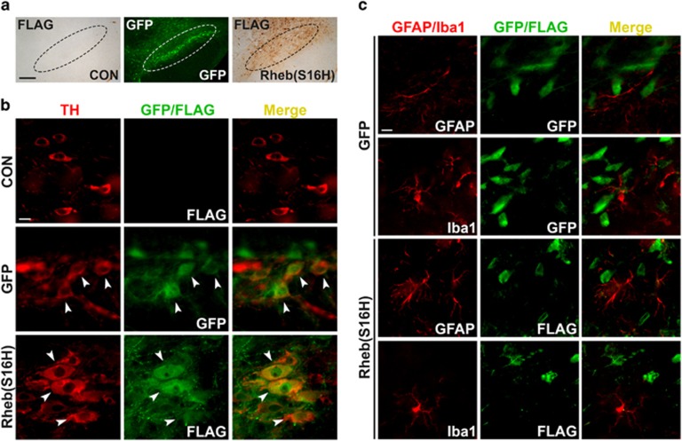
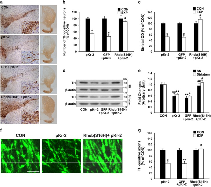
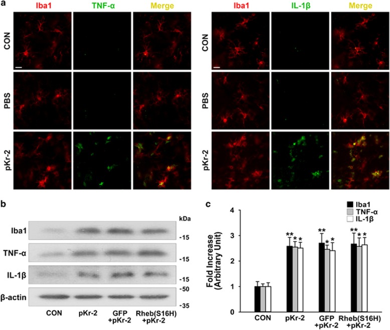
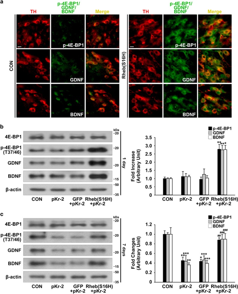
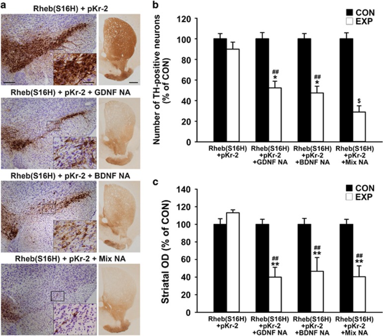
We recently reported that adeno-associated virus serotype 1 (AAV1) transduction of murine nigral dopaminergic (DA) neurons with constitutively active ras homolog enriched in brain with a mutation of serine to histidine at position 16 [Rheb(S16H)] induced the production of neurotrophic factors, resulting in neuroprotective effects on the nigrostriatal DA system in animal models of Parkinson's disease (PD). To further investigate whether AAV1-Rheb(S16H) transduction has neuroprotective potential against neurotoxic inflammation, which is known to be a potential event related to PD pathogenesis, we examined the effects of Rheb(S16H) expression in nigral DA neurons under a neurotoxic inflammatory environment induced by the endogenous microglial activator prothrombin kringle-2 (pKr-2). Our observations showed that Rheb(S16H) transduction played a role in the neuroprotection of the nigrostriatal DA system against pKr-2-induced neurotoxic inflammation, even though there were similar levels of pro-inflammatory cytokines, such as tumor necrosis factor-alpha (TNF-α) and interleukin-1-beta (IL-1β), in the AAV1-Rheb(S16H)-treated substantia nigra (SN) compared to the SN treated with pKr-2 alone; the neuroprotective effects may be mediated by the activation of neurotrophic signaling pathways following Rheb(S16H) transduction of nigral DA neurons. We conclude that AAV1-Rheb(S16H) transduction of neuronal populations to activate the production of neurotrophic factors and intracellular neurotrophic signaling pathways may offer promise for protecting adult neurons from extracellular neurotoxic inflammation.
Conflict of interest statement
The authors declare no conflict of interest.
Figures

Similar articles
-
Therapeutic Potential of AAV1-Rheb(S16H) Transduction against Neurodegenerative Diseases.Int J Mol Sci. 2021 Mar 17;22(6):3064. doi: 10.3390/ijms22063064. Int J Mol Sci. 2021. PMID: 33802760 Free PMC article. Review.
-
Neurotrophic interactions between neurons and astrocytes following AAV1-Rheb(S16H) transduction in the hippocampus in vivo.Br J Pharmacol. 2020 Feb;177(3):668-686. doi: 10.1111/bph.14882. Epub 2019 Dec 27. Br J Pharmacol. 2020. PMID: 31658360 Free PMC article.
-
Treatment with AAV1-Rheb(S16H) provides neuroprotection in a mouse model of photothrombosis-induced ischemic stroke.Neuroreport. 2020 Sep 9;31(13):971-978. doi: 10.1097/WNR.0000000000001506. Neuroreport. 2020. PMID: 32694311
-
Roles of Rheb(S16H) in substantia nigra pars compacta dopaminergic neurons in vivo.Biomed Rep. 2015 Mar;3(2):137-140. doi: 10.3892/br.2014.397. Epub 2014 Dec 9. Biomed Rep. 2015. PMID: 25798236 Free PMC article.
-
Upregulation of Neuronal Rheb(S16H) for Hippocampal Protection in the Adult Brain.Int J Mol Sci. 2020 Mar 16;21(6):2023. doi: 10.3390/ijms21062023. Int J Mol Sci. 2020. PMID: 32188096 Free PMC article. Review.
Cited by
-
Therapeutic Potential of AAV1-Rheb(S16H) Transduction against Neurodegenerative Diseases.Int J Mol Sci. 2021 Mar 17;22(6):3064. doi: 10.3390/ijms22063064. Int J Mol Sci. 2021. PMID: 33802760 Free PMC article. Review.
-
Neurotrophic interactions between neurons and astrocytes following AAV1-Rheb(S16H) transduction in the hippocampus in vivo.Br J Pharmacol. 2020 Feb;177(3):668-686. doi: 10.1111/bph.14882. Epub 2019 Dec 27. Br J Pharmacol. 2020. PMID: 31658360 Free PMC article.
-
Intracerebellar upregulation of Rheb(S16H) ameliorates motor dysfunction in mice with SCA2.Acta Pharmacol Sin. 2025 Jul;46(7):1852-1863. doi: 10.1038/s41401-025-01504-y. Epub 2025 Mar 3. Acta Pharmacol Sin. 2025. PMID: 40033054
-
Control of Reactive Oxygen Species for the Prevention of Parkinson's Disease: The Possible Application of Flavonoids.Antioxidants (Basel). 2020 Jul 3;9(7):583. doi: 10.3390/antiox9070583. Antioxidants (Basel). 2020. PMID: 32635299 Free PMC article. Review.
-
pKr-2 induces neurodegeneration via upregulation of microglial TLR4 in the hippocampus of AD brain.Brain Behav Immun Health. 2023 Jan 18;28:100593. doi: 10.1016/j.bbih.2023.100593. eCollection 2023 Mar. Brain Behav Immun Health. 2023. PMID: 36798617 Free PMC article.
References
-
- Dauer W, Przedborski S. Parkinson's disease: mechanisms and models. Neuron 2003; 39: 889–909. - PubMed
-
- Rascol O, Payoux P, Ory F, Ferreira JJ, Brefel-Courbon C, Montastruc JL. Limitations of current Parkinson's disease therapy. Ann Neurol 2003; 53(Suppl 3): S3–12 discussion S12-15. - PubMed
-
- Pezzoli G, Zini M. Levodopa in Parkinson's disease: from the past to the future. Expert Opin Pharmacother 2010; 11: 627–635. - PubMed
-
- Block ML, Hong JS. Chronic microglial activation and progressive dopaminergic neurotoxicity. Biochem Soc Trans 2007; 35: 1127–1132. - PubMed
Publication types
MeSH terms
Substances
Supplementary concepts
LinkOut - more resources
Full Text Sources
Other Literature Sources
Molecular Biology Databases
