. 2018 Feb 9;50(2):e441.
doi: 10.1038/emm.2017.262.

Poly(rC) binding protein 2 acts as a negative regulator of IRES-mediated translation of Hr mRNA

Affiliations

Poly(rC) binding protein 2 acts as a negative regulator of IRES-mediated translation of Hr mRNA

Jeong-Ki Kim et al. Exp Mol Med. .

Abstract

During the hair follicle (HF) cycle, HR protein expression is not concordant with the presence of the Hr mRNA transcript, suggesting an elaborate regulation of Hr gene expression. Here we present evidence that the 5' untranslated region (UTR) of the Hr gene has internal ribosome entry site (IRES) activity and this activity is regulated by the binding of poly (rC) binding protein 2 (PCBP2) to Hr mRNA. Overexpression and knockdown of PCBP2 resulted in a decrease in Hr 5' UTR IRES activity and an increase in HR protein expression without changing mRNA levels. We also found that this regulation was disrupted in a mutant Hr 5' UTR that has a mutation responsible for Marie Unna hereditary hypotrichosis (MUHH) in both mice and humans. These findings suggest that Hr mRNA expression is regulated at the post-transcriptional level via IRES-mediated translation control through interaction with PCPB2, but not in MUHH.


Conflict of interest statement

The authors declare no conflict of interest.

Figures

Figure 1
Figure 1
The 5′ UTR of Hr has IRES activity. (a) Schematic diagrams of the Hr-5′ UTR-X constructs which includes the 5′ UTR and CDS of Hr mRNA. Western blotting was performed using lysates of HEK293T cells transfected with the Hr-5′ UTR-X construct. (b) Rapamycin (Rapa)- or cycloheximide (CHX)-treated HEK293T cells were harvested at indicated time points, followed by the determination of HR protein expression levels by western blotting. Dimethyl sulfoxidewas used as a vehicle control treatment. (c) Schematic diagrams of the pRF bicistronic vectors containing the 5′ UTRs from mouse Hr or human HR with forward (pRF_m695 and pRF_h690) or reverse (pRF_m695rev and pRF_h690rev) orientations, and EMCV IRES elements (pRF_EMCV). The reporter constructs were transfected into HEK293T cells, and the IRES activities were measured and normalized by β-galactosidase activity 48 h after transfection. (d) PAM212 mouse keratinocytes were transfected with pRF_m695, and IRES activities were measured after 48 h. (e) Schematic diagrams of the pRF reporter vectors that contain several deletion fragments of the Hr 5′ UTR. The plasmid pRF_m695 includes the full-length Hr 5′ UTR, and the numbers associated with each of the constructs represent the nucleotide positions of the Hr 5′ UTR sequences. IRES activities were measured 48 h after transfection. All luciferase experiments were performed three times in triplicate, and transfection efficiencies were normalized against β-galactosidase activity. Activities are expressed as the mean±s.e.m. **P<0.01, ***P<0.001, ****P<0.0001.
Figure 2
Figure 2
Poly(rC) binding protein 2 interactions with the 5′ UTR of Hr mRNA and expression patterns during the HF cycle. (a, b) RNA pull-down experiments were performed using FLAG-PCBP2 overexpressing HEK293T cell lysates or purified GST-PCBP2 protein with or without a biotinylated Hr 5′ UTR probe (Biotin-m695). PCBP2 protein was detected by western blotting. (c) A competition analysis was performed in a UV cross-linking experiment. A radiolabeled Hr 5′ UTR RNA probe was incubated with different amounts of purified PCBP2 protein (+ 100 ng/rxn, ++ 200 ng/rxn, +++ 300 ng/rxn). The signal shows a concentration-dependent increase of the interaction, which was decreased specifically by the addition of unlabeled Hr 5′ UTR RNA. GST protein was used as a negative control, and signals were visualized by autoradiography. (d) Mouse skin sections from each time point were subjected to hematoxylin and eosin staining or immunostaining using the anti-PCBP2 antibody. Hoechst dye was used for counterstaining. Scale bar, 200 μm. (e, f) Western blotting was performed using mouse skin tissue from each time point. The quantitative densitometry of PCBP2 expression was presented as the mean±s.e.m. of three mice per time point.
Figure 3
Figure 3
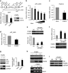
PCBP2 negatively regulates the IRES activity of the Hr 5′ UTR and HR protein expression. (a) The IRES activities were measured in HEK293T cells co-transfected with pRF_m695 and pFLAG-PCBP2 at 48 h after transfection. The expression of the FLAG-PCBP2 protein was confirmed by western blotting. (b) The IRES activities of pRF_m695 were measured at 48 h after transfection with various concentrations of FLAG-PCBP2. (c) PAM212 mouse keratinocytes were transfected with pRF_m695 and FLAG-PCBP2, and then IRES activities were measured. (d) The IRES activity of human HR 5′ UTR (pRF_h690) was measured in HEK293T cells co-transfected with the pFLAG-PCBP2 expression construct. (e) SiPCBP2 or a scrambled control were transfected into HEK293T cells. At 48 h post transfection, protein extracts were subjected to western blot analysis and expression level was determined using quantitative densitometry of the western blots from three independent experiments. (f) HEK293T cells were transfected with siPCBP2s or a scrambled control and incubated for 24 h. Then, the pRF_m695 reporter construct was transfected and further incubated. Twenty-four hours later, the IRES activities were measured, and cell lysates were subjected to western blotting. (g) HEK293T cells were co-transfected with the pCMV-FL-mHr and pFLAG-PCBP2 constructs. Forty-eight hours later, cells were harvested and HR expression determined by western blot analysis. Actin and GAPDH were used as loading controls, and GFP was used for normalization of the transfection efficiency. Additionally, Hr mRNA expression levels were determined by quantitative real time PCR, and the relative expression level was determined against Gapdh mRNA expression. (h) Increasing amounts of the pFLAG–PCBP2 construct were transfected into the HR stable cell line. Cell lysates were subjected to western blot analysis at 48 h post transfection. RT-PCR revealed no change in the Hr mRNA expression level. (i) HR-expressing stable cells were transfected with siPCBP2s, and cell lysates were subjected to western blotting after 48 h of incubation. All experiments for IRES activity were performed three times in triplicate, and transfection efficiencies were normalized by β-galactosidase activity. Activities are expressed as the mean±SEM. *P<0.05, **P<0.01, ***P<0.001, ****P<0.0001.
Figure 4
Figure 4
MUHH mutant forms of the 5′ UTR were not affected by PCBP2 negative regulation in both mice and humans. (a) PCBP2 mRNA expression levels were determined by quantitative real time PCR using wild-type and HrHp mutant P28 mouse skin tissues, and the relative expression level was determined against Gapdh mRNA expression. (b) The IRES activities of pRF_m695 (wild type) and pRF_mT403A (HrHp mutant type) were measured 48 h after transfection. (c) The pRF_mT403A and pFLAG-PCBP2 constructs were co-transfected into HEK293T cells. The IRES activities were measured 48 h after transfection. The expression of the FLAG-PCBP2 protein was confirmed by western blotting. (d) HEK293T cells were co-transfected with the pCMV-FL-mHr_T403A and the pFLAG-PCBP2 constructs. Forty-eight hours later, cells were harvested and HR expression determined by western blot analysis. GAPDH was used as a loading control, and GFP was used for normalization of the transfection efficiency. (e, f) RNA pull-down experiments were performed using FLAG-PCBP2 overexpressing HEK293T cell lysates with a wild-type (Biotin-m695) or mutant (Biotin-mT403A) biotinylated Hr 5′ UTR probe. The PCBP2 protein was detected by western blotting. (g) The pRF_hT-320C and pFLAG-PCBP2 constructs were co-transfected into HEK293T cells. The IRES activities were measured 48 h after transfection. The expression of the FLAG-PCBP2 protein was confirmed by western blotting. All experiments for IRES activity were performed three times in triplicate, and transfection efficiencies were normalized by β-galactosidase activity. Activities are expressed as the mean±s.e.m. *P<0.05.
Figure 5
Figure 5
Model for the regulation of HR expression via IRES-mediated translational control by PCBP2 in wild type and MUHH during HF cycling. In wild type, PCBP2 inhibits the IRES activity of Hr 5′ UTR in the anagen phase, then this inhibition is lifted at the catagen phase as PCBP2 expression decreases. However, in the MUHH mutant, the IRES activity of the Hr 5′ UTR is not inhibited by PCBP2 during the anagen phase, even though the PCBP2 expression is high because of the weak interaction between PCBP2 and Hr mRNA. Consequently, it results in abnormal overexpression of the HR protein during the whole HF cycle.

Similar articles

Cited by

References

    1. Hsieh JC, Sisk JM, Jurutka PW, Haussler CA, Slater SA, Haussler MR et al. Physical and functional interaction between the vitamin D receptor and hairless corepressor, two proteins required for hair cycling. J Biol Chem 2003; 278: 38665–38674. - PubMed
    1. Moraitis AN, Giguere V, Thompson CC. Novel mechanism of nuclear receptor corepressor interaction dictated by activation function 2 helix determinants. Mol Cell Biol 2002; 22: 6831–6841. - PMC - PubMed
    1. Potter GB, Beaudoin GM 3rd, DeRenzo CL, Zarach JM, Chen SH, Thompson CC. The hairless gene mutated in congenital hair loss disorders encodes a novel nuclear receptor corepressor. Genes Dev 2001; 15: 2687–2701. - PMC - PubMed
    1. Thompson CC. Thyroid hormone-responsive genes in developing cerebellum include a novel synaptotagmin and a hairless homolog. J Neurosci 1996; 16: 7832–7840. - PMC - PubMed
    1. Beaudoin GM 3rd, Sisk JM, Coulombe PA, Thompson CC. Hairless triggers reactivation of hair growth by promoting Wnt signaling. Proc Natl Acad Sci USA 2005; 102: 14653–14658. - PMC - PubMed

Publication types