. 2017 Dec 14;9(3):3188-3197.
doi: 10.18632/oncotarget.23225. eCollection 2018 Jan 9.

Hypoxia-inducible factor-1α activates transforming growth factor-β1/Smad signaling and increases collagen deposition in dermal fibroblasts

Affiliations

Hypoxia-inducible factor-1α activates transforming growth factor-β1/Smad signaling and increases collagen deposition in dermal fibroblasts

Xu Mingyuan et al. Oncotarget. .

Abstract

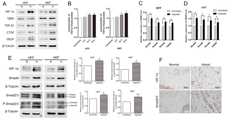
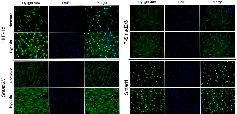
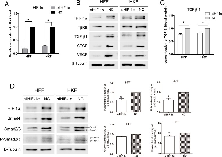
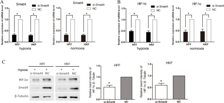
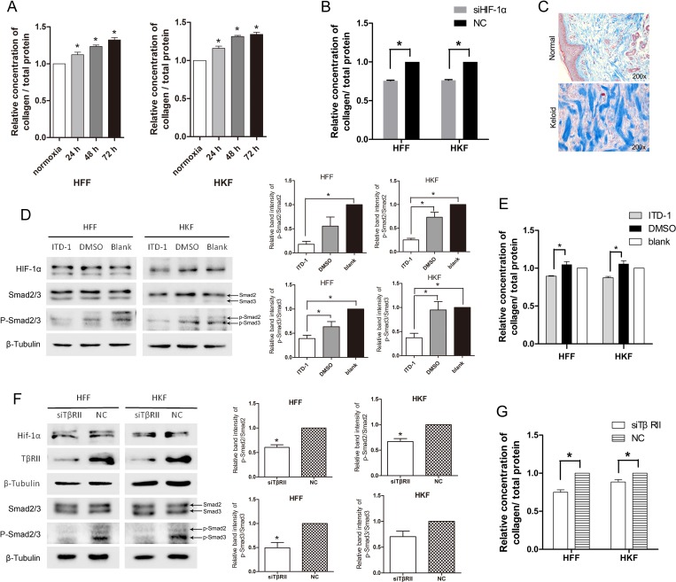
Hypoxia of local tissue occurs during the scar formation; however, the degree of ischemia and hypoxia in the central areas of keloids is more serious than those in normal scars. Hypoxia-induced factor (HIF), is one of the main cellular responses to hypoxia, allowing cells to adapt to low-oxygen conditions. We investigated the correlation among hypoxia, transforming growth factor-β1/Smad signaling and collagen deposition. Hypoxia up-regulated TGF-β1, Smad2/3, p-Smad2/3, Smad4, and total collagen in both normal and keloid fibroblasts via HIF-1α, which was attenuated by HIF-1α inhibition, but TβRII levels were not significantly altered. Silencing Smad4 under hypoxia decreased the mRNA and protein levels of HIF-1α, suggesting up-regulated Smad4 may also plays a role in promoting HIF-1α. Finally, we examined the role of the TGF-β1/Smad pathway in collagen deposition. When TβRII was inhibited by ITD-1 under hypoxic conditions, p-Smad2/3 levels and collagen deposition decreased. When inhibited TβRII by siRNA under normoxia, the levels of p-Smad2/3, Smad4 and collagen deposition also decreased. This result demonstrated that hypoxia promoted TGF-β1/Smad signaling via HIF-1α and that both HIF-1α and the TGF-β1/Smad signaling promotes collagen deposition in hypoxia, which is an important mechanism of keloid formation.

Keywords: collagen; hypoxia; hypoxia-Inducible factor-1α; keloid; transforming growth factor-β1/Smad.


Conflict of interest statement

CONFLICTS OF INTEREST None of the authors has a financial interest in any of the products, devices, or drugs mentioned in this manuscript.

Figures

Figure 1
Figure 1. Hypoxia promoted TGF-β/Smad signaling in HFFs and HKFs
(A) HIF-1α, TGF-β1, VEGF and CTGF protein levels were up-regulated by 24 h of hypoxia (1% O2) exposure, but TβRII expression did not obviously differ between the normoxia and hypoxia groups. (B) ELISA was used to detect secreted TGF-β1 in serum-free medium after 6 h, 12 h and 18 h of exposure to normoxia (21% O2) or hypoxia (1% O2). (C, D) Quantitative reverse transcriptase-PCR (qRT-PCR) analyses of Smad2, Smad3, Smad4 and TGF-β1 mRNA levels in HFFs and HKFs after 24 h of hypoxia or normoxia exposure. Bars show the means±SE of three independent experiments (n = 3); *represents P < 0.05. (E) Western blotting shows the protein levels of HIF-1α, Smad2/3, p-Smad2/3 and Smad4 after 24 h of hypoxia or normoxia exposure. The histogram shows the protein band intensity ratio of p-Smad2 to Smad2 and p-Smad3 to Smad3. (F) The expression of HIF-1α and Smad2/3 was tested using immunohistochemistry analysis.
Figure 2
Figure 2. HIF-1α, Smad2/3, p-Smad2/3 and Smad4 was enhanced following treatment with 1% hypoxia
The protein expression and intracellular localization of HIF-1α, Smad2/3, p-Smad2/3 and Smad4 were detected by immunofluorescence staining in HFFs under normoxia or 24 h of hypoxia (1% O2). HIF-1α and Smad4 localized mainly in the nucleus and Smad2/3 mainly in cytoplasm.
Figure 3
Figure 3. siHIF-1α inhibited TGF-β/Smad signaling in HFFs and HKFs
(A) qRT-PCR showing clear knockdown of HIF-1α by siHIF-1α transfection for 48 h. (B, D) 48 h after transfection, HFFs and HKFs were transferred to a 1% O2 hypoxia incubator for 24 h, and siHIF-1α inhibited HIF-1α, TGF-β1, Smad4, Smad2/3, p-Smad2/3, CTGF and VEGF levels. The histogram shows the protein band intensity ratio of p-Smad2 to Smad2 and p-Smad3 to Smad3. (C) Before transferring cells to the hypoxia incubator, we replaced the culture media with serum-free media, and both the siHIF-1α and NC groups were treated with 1% O2 for 12 h. The level of secreted TGF-β1 in the serum-free media was then measured.
Figure 4
Figure 4. siSmad4 inhibits HIF-1α in hypoxia
(A) qRT-PCR showing clear knockdown of Smad4 by siSmad4 transfection for 48 h. (B) 24 h after transfection, HFFs and HKFs were treated with 1% hypoxia or normoxia for 24 h. 48 h after transfection, the expression of mRNA level of HIF-1α was down-regulated in the group silencing Smad4. (C) After 72 h transfected by si-Smad4 and 24 h hypoxia treatment, the protein level of HIF-1α was down-regulated in the si-Smad4 group compared with the negative control group. The histogram shows the protein band intensity ratio of HIF-1α to β-Tubulin.
Figure 5
Figure 5. Total collagen deposition was promoted by acute hypoxia via HIF-1α and the TGF-β signaling pathway
(A) The ratio of deposited collagen to the total protein concentration was elevated after 24 h, 48 h, 72 h of hypoxia in both HFFs and HKFs. (B) At 48 h after transfection, HFFs and HKFs were transferred to a 1% O2 hypoxia incubator for 24 h. Collagen deposition was clearly inhibited in the siHIF-1α group compared with the NC group. (C) Masson staining for collagen fiber (blue) of normal and keloid tissues. (D) Treatment with 5 μM ITD-1(dissolved in DMSO), DMSO, and blank, respectively, for 24 h under 1% O2 condition. ITD-1 inhibited the phosphorylation of Smad2/3 by inhibiting TβRII. Meanwhile, HIF-1α and Smad2/3 levels remained stable in all three groups. The histogram shows the protein band intensity ratio of p-Smad2 to Smad2 and p-Smad3 to Smad3. (E) Collagen deposition was reduced in the ITD-1-treated HFFs and HKFs. (F) siTβRII reduced the protein levels of TβRII, p-Smad2/3 at 72 h after transfection in normoxia. HIF-1α protein levels remained stable in the two groups. The histogram shows the protein band intensity ratio of p-Smad2 to Smad2 and p-Smad3 to Smad3. (G) Collagen deposition was clearly inhibited in the siTβRII group compared with the NC group.

References

    1. Ikeda M, Naitoh M, Kubota H, Ishiko T, Yoshikawa K, Yamawaki S, Kurokawa M, Utani A, Nakamura T, Nagata K, Suzuki S. Elastic fiber assembly is disrupted by excessive accumulation of chondroitin sulfate in the human dermal fibrotic disease, keloid. Biochem Biophys Res Commun. 2009;390:1221–8. https://doi.org/10.1016/j.bbrc.2009.10.125. - DOI - PubMed
    1. Touchi R, Ueda K, Kurokawa N, Tsuji M. Central regions of keloids are severely ischaemic. J Plast Reconstr Aesthet Surg. 2016;69:e35–41. https://doi.org/10.1016/j.bjps.2015.11.006. - DOI - PubMed
    1. Celeste CJ, Deschene K, Riley CB, Theoret CL. Regional differences in wound oxygenation during normal healing in an equine model of cutaneous fibroproliferative disorder. Wound Repair Regen. 2011;19:89–97. https://doi.org/10.1111/j.1524-475X.2010.00639.x. - DOI - PubMed
    1. Schreml S, Szeimies RM, Prantl L, Karrer S, Landthaler M, Babilas P. Oxygen in acute and chronic wound healing. Br J Dermatol. 2010;163:257–68. https://doi.org/10.1111/j.1365-2133.2010.09804.x. - DOI - PubMed
    1. Weir L, Robertson D, Leigh IM, Vass JK, Panteleyev AA. Hypoxia-mediated control of HIF/ARNT machinery in epidermal keratinocytes. Biochim Biophys Acta. 2011;1813:60–72. https://doi.org/10.1016/j.bbamcr.2010.11.014. - DOI - PubMed

LinkOut - more resources