. 2017 Dec 14;9(3):3641-3652.
doi: 10.18632/oncotarget.23248. eCollection 2018 Jan 9.

Epithelial-mesenchymal crosstalk induces radioresistance in HNSCC cells

Affiliations

Epithelial-mesenchymal crosstalk induces radioresistance in HNSCC cells

Teresa Bernadette Steinbichler et al. Oncotarget. .

Erratum in

Abstract

Objective: Epithelial-mesenchymal crosstalk (EMC) contributes to tumor progression, chemoresistance and acquisition of a mesenchymal phenotype (EMT) of cancer cells. This study aims to investigate the effects of EMC on radioresistance in head and neck squamous cell carcinoma (HNSCC) cells.

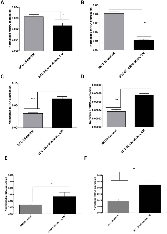
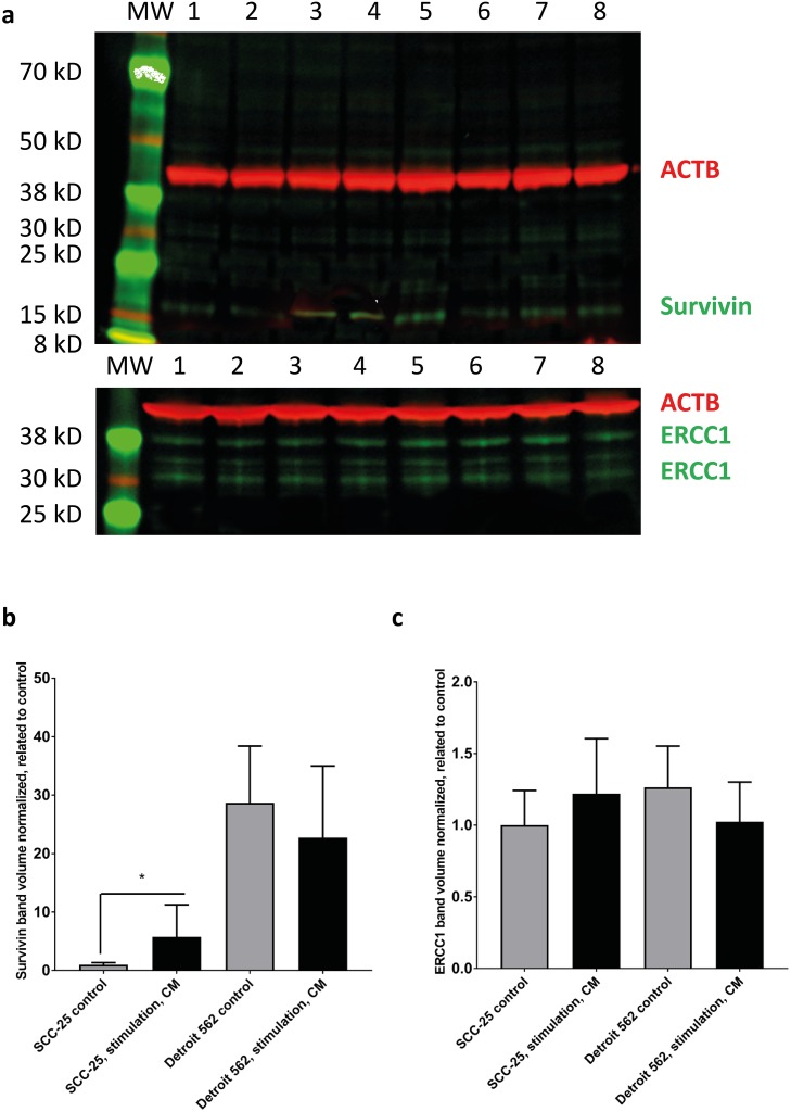
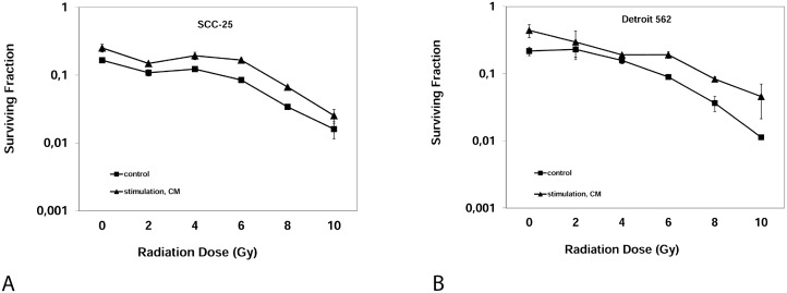
Methods: In tumor cell lines, the response of HNSCC cells, stimulated with EMC conditioned medium (CM), to irradiation was evaluated with viability and clonogenic assays. Dose modifying factors (DMF) were calculated from the results of clonogenic assays. Potential pathways involved in radioresistance were analyzed with quantitative Real-Time PCR and western blot.

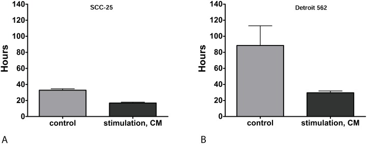
Results: CM significantly reduced the doubling time of SCC-25 cells (from 32.8 hours to 16.8 hours, p=0.0001) and Detroit 562 cells (from 88.5 hours to 29.6 hours, p=0.014). Further it increased clonogenic survival after irradiation. The DMF of CM was 2.04 ± 0.43 (mean ± standard deviation) for SCC-25 cells (p=0.015) and 2.14 ± 0.34 for Detroit 562 cells (p=0.008). Treatment with CM more than tripled the ERCC1 and survivin gene expression in SCC-25 cells.

Conclusion: EMC induced pathways involved in cell survival and DNA repair and led to increased radioresistance in HNSCC cells.

Keywords: ERCC1; chemoresistance; clonogenic assays; epithelial to mesenchymal transition; survivin.


Conflict of interest statement

CONFLICTS OF INTEREST The authors declare that they have no competing interests.

Figures

Figure 1
Figure 1
(A) Doubling time of SCC-25 in hours: Doubling times were calculated in non-irradiated cells. Control: following treatment of SCC-25 cells with standard albumin medium. CM: after treatment of SCC-25 with co-culture conditioned medium. Stimulation with CM reduced the doubling time in SCC-25 cells from 32.8 +/- 2.4 hours to 16.8 +/- 1.6 hours compared to the control medium (p=0.0001). (B) Doubling time of Detroit 562 in hours: Control: after treatment of Detroit 562 cells with standard albumin medium. CM: after treatment of Detroit 562 with co-culture conditioned medium. In Detroit 562 cells, stimulation with CM reduced doubling time from 88.5 +/- 34.7 hours (mean +/- SD) to 29.6 +/- 3.3 hours compared to the control medium (p= 0.014).
Figure 2
Figure 2
Real-time PCR analysis of EMT-like (A-D) and radioresistance - related (E-F) gene expression changes in SCC-25 cells in control and CM-treated conditions. SCC-25 cells were treated for 72 hours with CM, followed by total RNA isolation, reverse transcription, and real-time PCR. Relative gene expression was calculated with the −δδCt method for E-cadherin (A), desmoplakin (B), vimentin (C), MMP-9 (D), ERCC1 (E), and survivin (F) in control and CM-treated conditions, where β-actin was used as housekeeping gene.
Figure 3
Figure 3. Western blot analysis of survivin and ERCC1 in SCC-25 and Detroit 562 cells in control and CM - treated conditions
SCC-25 and Detroit 562 cells were treated with albumin - containing (control) or with CM medium (stimulation, CM) and subjected to western blot analysis of β-actin (red), survivin (green) or ERCC1 (green) in a two channel near infrared fluorescence detection system. β-actin loading control (42 kD), a single band was detected at 700 nm and is presented in red, survivin or ERCC1 were detected at 800 nm and are presented in green. (a) Survivin was detected as a single band in both SCC-25 (lanes 1-4) and Detroit 562 cells (lanes 5-8). ERCC1 was detected as multiple bands with two more intensive ones at 38 and 30 kD in both cell lines. More bands might be present in certain cell lines, which was also described by the data sheet of the antibody provider. Lanes 1-2: SCC-25 control, lanes 3-4: SCC-25 stimulation with CM, lanes 5-6: Detroit 562 control, lanes 7-8: Detroit 562 stimulation with CM; MW: molecular weight standard. (b-c) Column diagrams representing mean and standard error of measurement of normalized (to loading control, β-actin) survivin (b) and ERCC1 (c) band volumes from 8 gel lanes for each SCC-25 and Detroit 562 control and CM stimulated protein samples. The mean band volume of SCC-25 control was set as “1”. *: p < 0.05.
Figure 4
Figure 4
Clonogenic Assay: Surviving fraction of SCC-25 cells (A) or Detroit 562 cells (B) after irradiation with 0, 2, 4, 6, 8, 10 Gray after three weeks. Control: Albumin medium treated, stimulation, CM: EMC-conditioned medium.

References

    1. Warner L, Chudasama J, Kelly CG, Loughran S, McKenzie K, Wight R, Dey P. Radiotherapy versus open surgery versus endolaryngeal surgery (with or without laser) for early laryngeal squamous cell cancer. Cochrane Database Syst Rev. 2014 - PMC - PubMed
    1. Skvortsova I, Debbage P, Kumar V, Skvortsov S. Radiation resistance: cancer stem cells (CSCs) and their enigmatic pro-survival signaling. Semin Cancer Biol. 2015;35:39–44. - PubMed
    1. Steinbichler TB, Metzler V, Pritz C, Riechelmann H, Dudas J. Tumor-associated fibroblast-conditioned medium induces CDDP resistance in HNSCC cells. Oncotarget. 2015;7:2508–18. https://doi.org/10.18632/oncotarget.6210. - DOI - PMC - PubMed
    1. Smith A, Teknos TN, Pan Q. Epithelial to mesenchymal transition in head and neck squamous cell carcinoma. Oral Oncol. 2013;49:287–92. - PMC - PubMed
    1. Orimo A, Weinberg RA. Stromal fibroblasts in cancer: a novel tumor-promoting cell type. Cell Cycle. 2006;5:1597–601. - PubMed