. 2018 Jan 16:4:3.
doi: 10.1038/s41421-017-0002-1. eCollection 2018.

Dual roles of endothelial FGF-2-FGFR1-PDGF-BB and perivascular FGF-2-FGFR2-PDGFRβ signaling pathways in tumor vascular remodeling

Affiliations

Dual roles of endothelial FGF-2-FGFR1-PDGF-BB and perivascular FGF-2-FGFR2-PDGFRβ signaling pathways in tumor vascular remodeling

Kayoko Hosaka et al. Cell Discov. .

Abstract

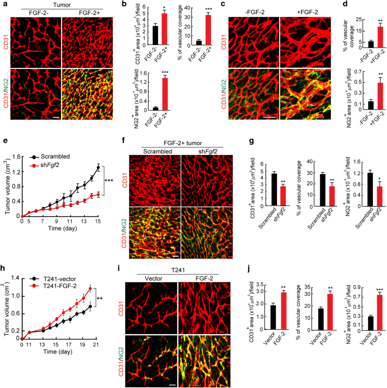
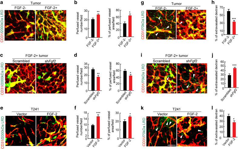
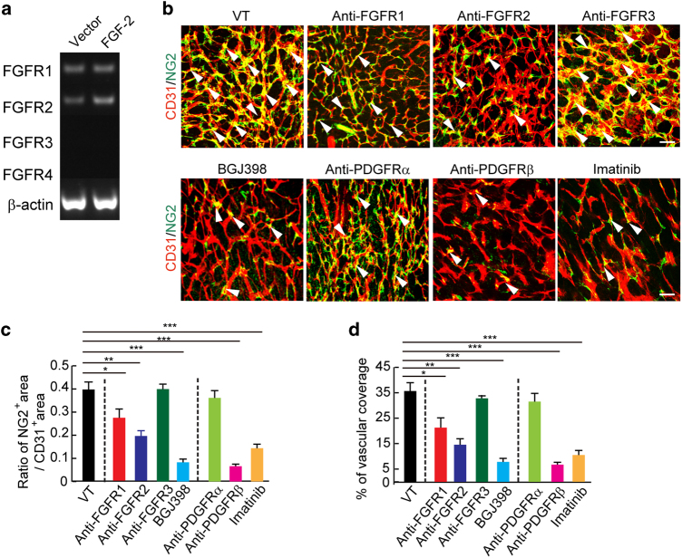
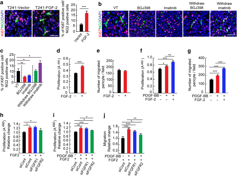
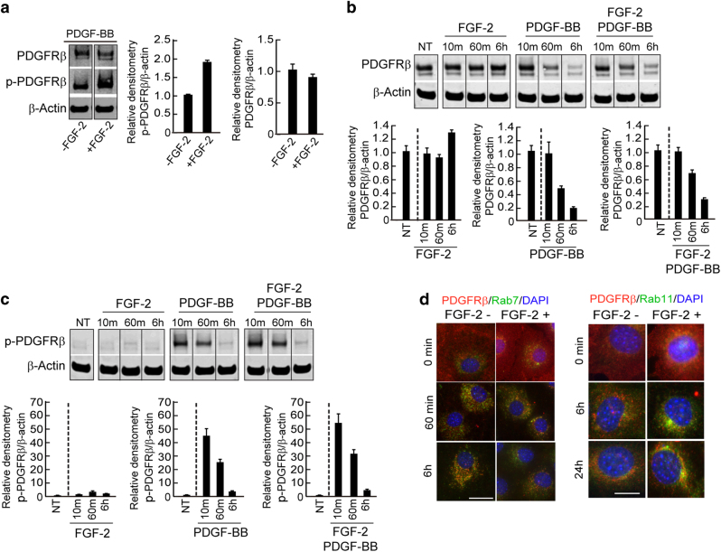
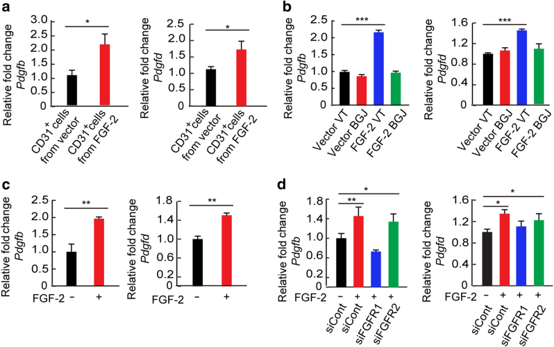
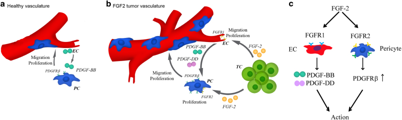
Perivascular cells are important cellular components in the tumor microenvironment (TME) and they modulate vascular integrity, remodeling, stability, and functions. Here we show using mice models that FGF-2 is a potent pericyte-stimulating factor in tumors. Mechanistically, FGF-2 binds to FGFR2 to stimulate pericyte proliferation and orchestrates the PDGFRβ signaling for vascular recruitment. FGF-2 sensitizes the PDGFRβ signaling through increasing PDGFRβ levels in pericytes. To ensure activation of PDGFRβ, the FGF-2-FGFR1-siganling induces PDGF-BB and PDGF-DD, two ligands for PDGFRβ, in angiogenic endothelial cells. Thus, FGF-2 directly and indirectly stimulates pericyte proliferation and recruitment by modulating the PDGF-PDGFRβ signaling. Our study identifies a novel mechanism by which the FGF-2 and PDGF-BB collaboratively modulate perivascular cell coverage in tumor vessels, thus providing mechanistic insights of pericyte-endothelial cell interactions in TME and conceptual implications for treatment of cancers and other diseases by targeting the FGF-2-FGFR-pericyte axis.


Conflict of interest statement

The authors declare that they have no competing interests.

Figures

Fig. 1
Fig. 1. FGF-2-induced angiogenesis, pericyte recruitment, and tumor growth in vivo.
a Tumor microvessel and pericyte contents. CD31+ endothelial cell (red) and NG2+ pericyte (green) signals in FGF-2+ and FGF-2 tumors. Bar = 50 μm. b Quantification of microvessel density, vascular coverage by pericytes, and NG2+ pericyte area (n = 7 random fields; n = 4 mice for each group). c CD31+ endothelial (red) and NG2+ pericyte (green) signals in matrigels containing FGF-2- tumors with and without FGF-2 protein. Bar = 50 μm. d Quantification of vascular coverage by pericytes and NG2+ pericyte area in matrigels with and without FGF-2 protein (n = 7 random fields; n = 4 mice for each group). e Tumor growth rates of scrambled-shRNA and Fgf2-shRNA-transfected FGF-2+ tumors (n = 4–6 animals/group). f CD31+ endothelial (red) and NG2+ pericyte (green) contents in scrambled-shRNA and Fgf2-shRNA-transfected FGF-2+ tumors. Bar = 50 μm. g Quantification of microvascular density, vascular coverage by pericytes, and NG2+ pericyte area in scrambled-shRNA and Fgf2-shRNA-transfected FGF-2+ tumors (n = 7 random fields; n = 4 mice for each group). h Tumor growth rates of T241-vector and T241-FGF-2 fibrosarcomas (n = 6 animals/group). i CD31+ endothelial (red) and NG2+ pericyte (green) contents in T241-vector and -FGF-2 fibrosarcomas. Bar = 50 μm. j Quantification of microvessel density, vascular coverage by pericytes, and NG2+ pericyte area in T241-vector and T241–FGF-2 fibrosarcomas (n = 7 random fields; n = 4 mice for each group). Vessels and pericytes were visualized using whole mount staining. All data as means ± S.E.M. *P < 0.05, **P < 0.01 and ***P < 0.001
Fig. 2
Fig. 2. Vascular perfusion and permeability in FGF-2+ and FGF-2- tumors in vivo.
a, c, e CD31+ tumor vasculature (red) and perfusion of 2000-kDa dextran (green) in FGF-2+ and FGF, scrambled-shRNA and Fgf2-shRNA-transfected FGF-2+, and T241-vector and T241-FGF-2 tumors. Yellow color indicates double positive signals and perfused vessels. Bar = 50 μm. b, d, f Quantification of blood perfusion in in FGF-2+ and FGF, scrambled-shRNA and Fgf2-shRNA-transfected FGF-2+, and T241-vector and T241-FGF-2 tumors (n = 10 random fields; n = 3 mice for each group). g, i, k CD31+ tumor vasculature (red) and leakiness of 70-kDa dextran (green) in FGF-2+ and FGF, scrambled-shRNA and Fgf2-shRNA-transfected FGF-2+, and T241-vector and T241-FGF-2 tumors. Arrowheads indicate extravagated dextran (green). Intravascular dextran molecules are in yellow color. Bar = 50 μm. h, j, l Quantification of vascular permeability of 70-kDa dextran in FGF-2+ and FGF, scrambled-shRNA and Fgf2-shRNA-transfected FGF-2+, and T241-vector and T241-FGF-2 tumors (n = 10 random fields; n = 3 mice for each group). Images are shown using whole mount staining. All data as means ± S.E.M.; Student’s t test, *P < 0.05, **P < 0.01 and ***P < 0.001
Fig. 3
Fig. 3. FGFRs and PDGFRs in pericyte recruitment.
a RT-PCR analysis of mRNA expression levels of FGF receptors in pericytes freshly isolated from T241-vector and T241–FGF-2 tumors using magnetic bead separation. Beta-actin serves as a control. b CD31+ endothelial (red) and NG2+ pericyte (green) signals in vehicle (VT)-, anti-FGFR1 neutralizing antibody-, anti-FGFR2 neutralizing antibody treated-, anti-FGFR3 neutralizing antibody-, BGJ398-, anti-PDGFRα neutralizing antibody-, anti-PDGFRβ neutralizing antibody-, and imatinib-treated FGF-2+ tumors. Arrowheads indicate pericyte-associated vessels. Images are presented using whole mount staining. Bar = 50 μm. c, d Quantification of NG2+ pericyte area versus the total CD31+ microvessels and vascular coverage. (n = 7 random fields; n = 4 mice for each group). All data as means ± S.E.M.; Student’s t test, *P < 0.05, **P < 0.01 and ***P < 0.001
Fig. 4
Fig. 4. Interplay between the FGF-2–FGFR2 and PDGF-B–PDGFR signaling in perivascular functions.
a Ki67+ (red) and NG2+ pericyte (green) in T241-vector and T241–FGF-2 tumor tissue. Cell nuclei were counterstained with DAPI (blue). Allows point to Ki67+-NG2+ double positive pericytes. Bar = 50 μm. Images are obtained using immunohistochemistry. Quantification of percentages of Ki67+-NG2+ double positive pericytes tumors (n = 15 random fields; n = 4 mice for each group). b Ki67+ (red) and NG2+ pericyte (green) in vehicle (VT)-, BGJ398-, and imatinib-treated FGF-2+ tumor tissue. Allows point to Ki67+-NG2+ double positive pericytes. Withdrawal experimental settings were performed at day 6 after drug cessation. Images are acquired using immunohistochemistry. Bar = 50 μm. c Quantification of percentages of Ki67 +-NG2+ double positive pericytes in VT-, BGJ398-, or imatinib-treated FGF-2+ tumors (n = 15 random fields; n = 4 mice for each group). d Pericyte in vitro proliferation after FGF-2 stimulation (n = 6 samples/group). e Pericyte in vitro migration after FGF-2 stimulation (n = 6 samples/group) P = 0.43. f Pericyte in vitro proliferation after PDGF-BB stimulation with or without FGF-2 pretreatment (n = 6 samples/group). g Pericyte in vitro migration after PDGF-BB stimulation with or without FGF-2 pretreatment (n = 6 samples/group). h FGF-2-induced proliferation of scrambled-siRNA-, Fgfr1-siRNA-, or Fgfr2-siRNA-transfected pericytes in vitro (n = 6 samples/group). i PDGF-BB-induced proliferation of scrambled-siRNA-, Fgfr1-siRNA-, or Fgfr2-siRNA-transfected pericytes with or without FGF-2 pretreatment in vitro (n = 6 samples/group). j PDGF-BB-induced migration of scrambled-siRNA-, Fgfr1-siRNA-, or Fgfr2-siRNA-transfected pericytes with or without FGF-2 pretreatment in vitro (n = 6 samples/group). All data as means ± S.E.M.; Student’s t test, *P < 0.05, **P < 0.01 and ***P < 0.001. All in vitro experiments were repeated at least twice
Fig. 5
Fig. 5. FGF-2-induced sensitization of PDGF-BB–PDGFR signaling by enhancing PDGFRβ recycling.
a Total and phosphorylated PDGFRβ protein levels in PDGF-BB-stimulated pericytes with or without FGF-2 pretreatment. β-actin levels were used as standard loading controls. Data were presented as means ± S.E.M. (n = 3). b Time-course analysis of PDGFRβ protein levels in FGF-2-, PDGF-BB-, or FGF-2 plus PDGF-BB-stimulated pericytes. β-actin levels were used as standard loading controls; NT no treatment. c Time-course analysis of phosphorylated PDGFRβ protein levels in FGF-2, PDGF-BB-, or FGF-2 plus PDGF-BB-stimulated pericytes. β-actin levels were used as standard loading controls. NT no treatment. d PDGF-BB-stimulated and non-stimulated pericytes were stained with specific anti-PDGFRβ, Rab7, and Rab11 antibodies with or without FGF-2 pretreatment. Nuclei were counterstained with DAPI. Bar = 25 μm (left) and 15 μm (right). All data as standardized values. All experiments were repeated three times
Fig. 6
Fig. 6. FGF-2-stimulated endothelial Pdgfb and Pdgfd expression in vivo and in vitro.
a Pdgfb and Pdgfd mRNA levels in CD31+ ECs freshly isolated from T241-vector or T241–FGF-2 tumors (n = 3 samples; n = 3 mice for each group). b Pdgfb and Pdgfd mRNA levels in CD31+ ECs freshly isolated from VT- or BGJ398-treated T241-vector and -FGF-2 tumors (n = 3 samples; n = 3 mice for each group). c Pdgfb and Pdgfd mRNA levels in cultivated CD31+ ECs in response to FGF-2 stimulation (n = 3 samples/group). d Pdgfb and Pdgfd mRNA levels in FGF-2-stimulated cultivated CD31+ ECs transfected with scrambled-RNA, siFgfr1-RNA, or siFgfr2-RNA (n = 3 samples/group). All data as means ± S.E.M.; Student’s t test, *P < 0.05, **P < 0.01 and ***P < 0.001. All in vitro experiments were repeated at least twice
Fig. 7
Fig. 7. Mechanisms of FGF-2-induced dual effects on endothelial cells and pericytes on vascular remodeling.
a In healthy vasculatures, angiogenic endothelial cell-derived PDGF-BB promotes pericyte recruitment. b In FGF-2+ tumors, tumor-derived FGF-2 stimulates endothelial PDGF-BB and PDGF-DD production through the FGFR1 signaling pathway. In addition to endothelial function, FGF-2 stimulates pericyte proliferation and migration through the direct pericyte effect on the FGFR-2 signaling and indirect mechanism by sensitizing PDGFRβ signaling on pericytes. c The FGF-2-FGFR1 endothelial and FGF-2-FGFR2 pericyte signaling pathways in vascular remodeling. EC, endothelial cell; PC, pericyte; TC, tumor cell

References

    1. Alderton G. Tumour microenvironment: to me, to you. Nat. Rev. Cancer. 2013;13:756–757. doi: 10.1038/nrc3618. - DOI - PubMed
    1. Cao Y, Zhong W, Sun Y. Improvement of antiangiogenic cancer therapy by understanding the mechanisms of angiogenic factor interplay and drug resistance. Semin. Cancer Biol. 2009;19:338–343. doi: 10.1016/j.semcancer.2009.05.001. - DOI - PubMed
    1. Danovi S. Tumour microenvironment: as time goes by. Nat. Rev. Cancer. 2016;16:342–343. doi: 10.1038/nrc.2016.53. - DOI - PubMed
    1. von Tell D, Armulik A, Betsholtz C. Pericytes and vascular stability. Exp. Cell Res. 2006;312:623–629. doi: 10.1016/j.yexcr.2005.10.019. - DOI - PubMed
    1. Abramsson A, et al. Analysis of mural cell recruitment to tumor vessels. Circulation. 2002;105:112–117. doi: 10.1161/hc0102.101437. - DOI - PubMed