. 2018 Jan;7(1):56-68.
doi: 10.7762/cnr.2018.7.1.56. Epub 2018 Jan 30.

Dual Effects of High Protein Diet on Mouse Skin and Colonic Inflammation

Affiliations

Dual Effects of High Protein Diet on Mouse Skin and Colonic Inflammation

Xuelei Cui et al. Clin Nutr Res. 2018 Jan.

Abstract

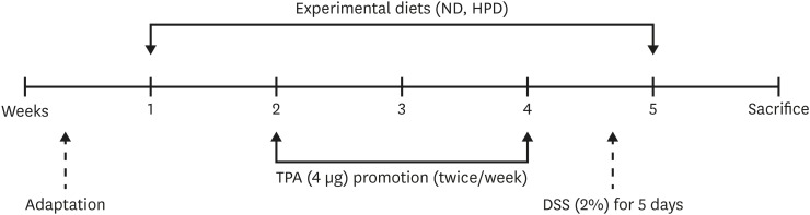
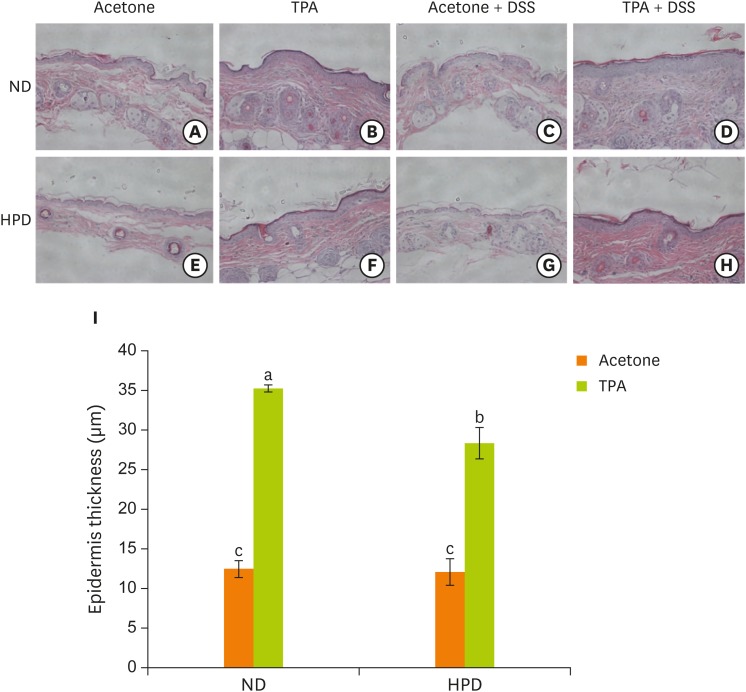
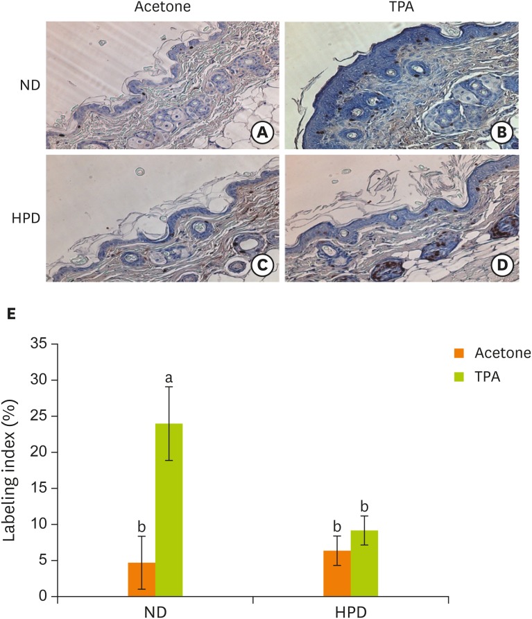
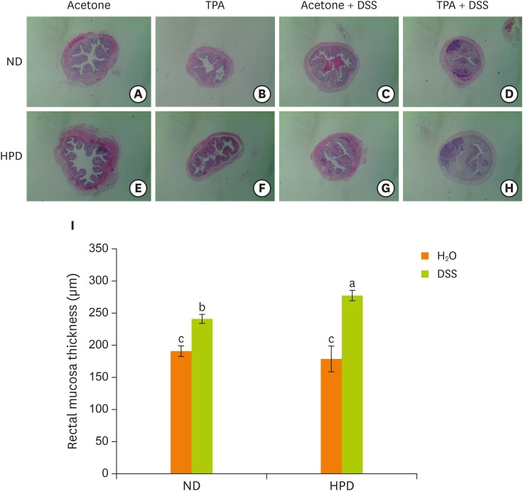
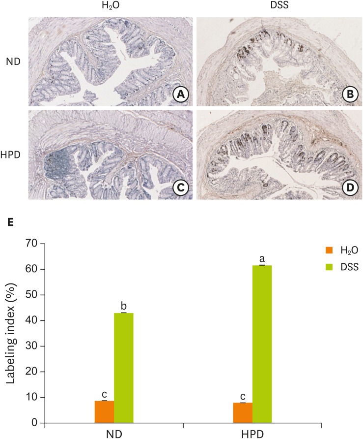
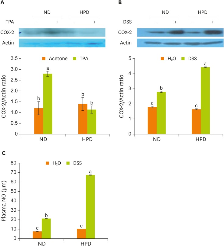
Chronic inflammation is a major etiology of cancer. Accumulating epidemiological and experimental evidences suggest that intake of high protein diet (HPD) is associated with colitis-associated colon cancer, however, most of the studies were confined in colon. Systemic influence of HPD on inflammation indices in different tissues of an organism has never been studied. We therefore investigated the effect of HPD on mouse skin and colonic inflammation using the well characterized inflammation induction protocol in both tissues (12-O-tetradecanoylphorbol-13-acetate [TPA] for skin and dextran sodium sulfate [DSS] for colon). ICR mice were grouped to normal diet (ND, 20% casein) or HPD (50% casein) groups. In each diet group, mice were treated with either vehicle (acetone or H2O), TPA, TPA and DSS, or DSS. Experimental diet was fed for total 4 weeks. After 1 week of diet feeding, 6.5 nmol of TPA was topically applied twice a week for 2 weeks on the shaved mouse dorsal skin. Drinking water containing 2% DSS was administered for 7 days at the final week of experiment. The results showed that TPA-induced skin hyperplasia, epidermal cell proliferation, and cyclooxygenase-2 (COX-2) expression were reduced in HPD group compared to ND group. In contrast, HPD increased DSS-induced colon mucosal hyperplasia, colonocyte proliferation, COX-2 expression, and plasma nitric oxide compared to ND group. This suggests that HPD exerts differential effect on different tissue inflammation which implies efficacy of protein intervention to human also should be monitored more thoroughly.

Keywords: Colon; Dietary Protein; Inflammation; Mouse; Skin.


Conflict of interest statement

Conflict of Interest: The authors declare no competing financial interests.

Figures

Figure 1
Figure 1
Schematic representation of study design. Five-week-old female ICR mouse were acclimated for 1 week and then randomly grouped to ND (20% casein) and HPD (50% casein) groups. In each diet group, mice were treated with either vehicle (acetone or H2O), TPA, TPA and DSS, or DSS. Experimental diet was fed for total 4 weeks. After 1 week of diet feeding, 6.5 nmol (4 μg) of TPA was topically applied twice a week for 2 weeks on the shaved mouse dorsal skin. DSS in drinking water (2%, wt/v) was administered for 5 days at the final week of the experiment. ND, normal diet; HPD, high protein diet; TPA, 12-O-tetradecanoylphorbol-13-acetate; DSS, dextran sodium sulfate.
Figure 2
Figure 2
Effect of HPD on mouse epidermal hyperplasia. (A-H) Histological analysis of the skin of ND (upper panel) and HPD fed mice (lower panel) with or without TPA and/or DSS treatment. Tissue sections were stained with H & E and photographed at 100×. (I) Epidermal thickness was measured microscopically. Each value represents the mean ± standard error of the epidermal thickness from 3 random tissue sections in each animal and 3 mice/group. ND, normal diet; HPD, high protein diet; TPA, 12-O-tetradecanoylphorbol-13-acetate; DSS, dextran sodium sulfate; H & E, hematoxylin and eosin. Means with different letters are significantly different at p < 0.05 by Duncan's multiple range test. In the graph, alphabets are assigned sequentially starting from a high number to a.
Figure 3
Figure 3
Effect of HPD on basal cell proliferation in mouse skin. Skin sections were immunostained with an antibody against BrdU and photographed at 200× magnification. Dorsal skins of mice fed (A) ND and treated with acetone, (B) ND and treated with TPA (4 μg of TPA, twice a week for 2 weeks), (C) HPD and treated with acetone, (D) HPD and treated with TPA. (E) The index represents the percentage of BrdU-positive cells relative to the total number of basal cells in the interfollicular epidermis in each experiment group. Each value represents the mean ± standard error of the labeling indices from 5 random tissue sections in each animal and 3 mice/group. HPD, high protein diet; BrdU, 5-bromo-2′-deoxyuridine; ND, normal diet; TPA, 12-O-tetradecanoylphorbol-13-acetate. Means with different letters are significantly different at p < 0.05 by Duncan's multiple range test. In the graph, alphabets are assigned sequentially starting from a high number to a.
Figure 4
Figure 4
Effect of HPD on mouse mucosal hyperplasia. The distal part of large intestine was removed and fixed in 10% formalin. (A-H) Histological analysis of the skin of ND (upper panel) and HPD fed mice (lower panel) with or without DSS and/or TPA treatment. Tissue sections were stained with H & E and photographed at 100×. (I) Mucosal thickness was measured microscopically. Each value represents the mean ± standard error of the mucosal thickness from 3 random tissue sections in each animal and 3 mice/group. HPD, high protein diet; ND, normal diet; DSS, dextran sodium sulfate; TPA, 12-O-tetradecanoylphorbol-13-acetate; H & E, hematoxylin and eosin. Means with different letters are significantly different at p < 0.05 by Duncan's multiple range test. In the graph, alphabets are assigned sequentially starting from a high number to a.
Figure 5
Figure 5
Effect of HPD on mucosal cell proliferation in mouse colon. The distal part of large intestine was removed and fixed in 10% formalin. Rectal sections of mice fed (A) ND and administered with H2O, (B) ND and administered with DSS (2%), (C) HPD and administered with H2O, (D) HPD and administered with DSS (2%) were immunostained with an antibody against BrdU and photographed at 200× magnification. (E) The index represents the percentage of BrdU-positive cells relative to the total number of mucosal cells in each experiment group. Each value represents the mean ± standard error of the labeling indices from 5 random tissue sections in each animal and 3 mice/group. HPD, high protein diet; ND, normal diet; DSS, dextran sodium sulfate; BrdU, 5-bromo-2′-deoxyuridine. Means with different letters are significantly different at p < 0.05 by Duncan's multiple range test. In the graph, alphabets are assigned sequentially starting from a high number to a.
Figure 6
Figure 6
Expression of inflammatory protein in tissues and the production of plasma NO. Mouse dorsal skin (A) and the distal part of large intestine (B) were removed and homogenized. The protein levels were determined by immunoblotting with the appropriate antibodies, as indicated. (C) Plasma NO levels were measured in all mice. Values are presented as the mean ± standard error. NO, nitric oxide; ND, normal diet; HPD, high protein diet; TPA, 12-O-tetradecanoylphorbol-13-acetate; COX-2, cyclooxygenase-2; DSS, dextran sodium sulfate. Means with different letters are significantly different at p < 0.05 by Duncan's multiple range test. In the graph, alphabets are assigned sequentially starting from a high number to a.

References

    1. Hanahan D, Weinberg RA. Hallmarks of cancer: the next generation. Cell. 2011;144:646–674. - PubMed
    1. Mantovani A, Allavena P, Sica A, Balkwill F. Cancer-related inflammation. Nature. 2008;454:436–444. - PubMed
    1. Balkwill F, Charles KA, Mantovani A. Smoldering and polarized inflammation in the initiation and promotion of malignant disease. Cancer Cell. 2005;7:211–217. - PubMed
    1. Ogilvie GK. Interventional nutrition for the cancer patient. Clin Tech Small Anim Pract. 1998;13:224–231. - PubMed
    1. Kurzer M, Meguid MM. Cancer and protein metabolism. Surg Clin North Am. 1986;66:969–1001. - PubMed