. 2018 Jul;59(4):432-443.
doi: 10.1111/vru.12605. Epub 2018 Feb 8.

Abrogation of fluid suppression in intracranial postcontrast fluid-attenuated inversion recovery magnetic resonance imaging: A clinical and phantom study

Affiliations

Abrogation of fluid suppression in intracranial postcontrast fluid-attenuated inversion recovery magnetic resonance imaging: A clinical and phantom study

Peter J Dickinson et al. Vet Radiol Ultrasound. 2018 Jul.

Abstract

Postcontrast, fluid-attenuated inversion recovery (FLAIR) sequences are reported to be of variable value in veterinary and human neuroimaging. The source of hyperintensity in postcontrast-T2 FLAIR images is inconsistently reported and has implications for the significance of imaging findings. We hypothesized that the main source of increased signal intensity in postcontrast-T2 FLAIR images would be due to gadolinium leakage into adjacent fluid, and that the resulting gadolinium-induced T1 shortening causes reappearance of fluid hyperintensity, previously nulled on precontrast FLAIR images. A retrospective, descriptive study was carried out comparing T2 weighted, pre- and postcontrast T1 weighted and pre- and postcontrast weighted T2 FLAIR images in a variety of intracranial diseases in dogs and cats. A prospective, experimental, phantom, in vitro study was also done to compare the relative effects of gadolinium concentration on T2 weighted, T1 weighted, and FLAIR images. A majority of hyperintensities on postcontrast-T2 FLAIR images that were not present on precontrast FLAIR images were also present on precontrast T2 weighted images, and were consistent with normal or pathological fluid filled structures. Phantom imaging demonstrated increased sensitivity of FLAIR sequences to low concentrations of gadolinium compared to T1 weighted sequences. Apparent contrast enhancement on postcontrast-T2 FLAIR images often reflects leakage of gadolinium across normal or pathology specific barriers into fluid-filled structures, and hyperintensity may therefore represent normal fluid structures as well as pathological tissues. Findings indicated that postcontrast-T2 FLAIR images may provide insight into integrity of biological structures such as the ependymal and subarachnoid barriers that may be relevant to progression of disease.

Keywords: MRI; brain; cat; dog; gadolinium.


Figures

Fig 1.
Fig 1.
Evolution of longitudinal magnetization (MZ) for different tissues without (dark gray arrows) and with added gadolinium (light gray arrows) between the inversion pulse and excitation pulse of a typical fluid attenuating inversion recovery pulse sequence. The magnitude of MZ at the time of excitation determines the maximum possible signal intensity of a tissue in the resulting image. A 180° inversion pulse flips the equilibrium MZ from positive to negative, and then MZ recovers for each tissue according to their T1 relaxation times. Short to intermediate T1 relaxation times result in negligible gain in MZ for fat (solid oval) and a small gain of MZ for brain gray matter (dotted oval). In the absence of gadolinium, MZ of normal cerebrospinal fluid (CSF) reaches the null point at the inversion time and contributes zero signal to the image. Addition of gadolinium to CSF causes MZ to cross the null point prior to the inversion time, resulting in positive MZ that contributes signal to the image (dashed circles). RF = radiofrequency.
Fig 2.
Fig 2.
Effect of gadopentetate dimeglumine (GPDM) concentration on the qualitative appearance of water (Water), water + 2.4 g/dL bovine serum albumin (Albumin), and 2% w/v agarose (Agarose) on T1-weighted (T1W), T2-weighted (T2W), and T2-weighted fluid attenuating inversion recovery (PC-T2FLAIR) images. Conspicuous contrast enhancement of water is observed at lower gadolinium concentrations on FLAIR images than on T1W images, but signal intensity decreases on T2W and FLAIR images at concentration of gadolinium (>0.28 mM). All images are displayed with window width = 2000 and window level = 1000.
Fig 3.
Fig 3.
Signal intensity of water (A) and 2% agarose (B) as a function of gadopentetate dimeglumine (GPDM) concentration for T1-weighted (T1W), T2-weighted (T2W), and T2-weighted fluid attenuating inversion recovery (FLAIR) images. Signal intensity of water peaks at lower concentrations of GPDM on FLAIR images than on T1W images. Agarose exhibits a modest (3.3x) peak increase in signal intensity on FLAIR images compared to the marked (7.4x) increase in signal intensity apparent on T1W images.
Fig 4.
Fig 4.
PC-T2FLAIR hyperintensity involving ventricular structures distant to primary lesions. A-E and J-N (T1W, T1W + contrast, T2W, FLAIR and FLAIR + contrast images) (F, H, O, Q pre-contrast FLAIR, G, I, P, R post-contrast FLAIR). PC-T2FLAIR hyperintensity (E, arrow) is present involving the lateral ventricle contralateral to an oligodendroglioma impinging on the opposite ventricle. Hyperintensity is also seen involving the 3rd ventricle (G), 4th ventricle and adjacent subarachnoid space (H) distal to the tumor (arrowheads). PC-T2FLAIR hyperintensity associated with an extraaxial caudal fossa mass (N, arrowhead) is also present involving the 3rd ventricle (P, arrowhead) and C1 spinal cord subarachnoid space (R, arrowhead), also distant from the primary lesion. PC-T2FLAIR hyperintensities localize spatially with T2 hyperintensities that are suppressed on pre-contrast FLAIR imaging.
Fig 5.
Fig 5.
Caudal fossa glioblastoma (A), meningioma (B) and lymphoma (C). Hyperintensity associated with intratumoral fluid accumulations present on T2W images (arrowheads, A, B) is suppressed on FLAIR images and reappears on PC-T2FLAIR images due to loss of fluid signal suppression. Leptomeningeal infiltration by lymphoma (C) with associated delineation of sulcal subarachnoid CSF on PC-T2FLAIR images (C, arrowheads) is more extensive than on T1W post-contrast images.
Fig 6.
Fig 6.
Bacterial meningoencephalitis (A, B), bacterial otitis media-interna (C) and meningoencephalitis of undetermined origin (presumed immune–mediated) (D). PC-T2FLAIR hyperintensities (arrowheads) not associated with T1W post-contrast signal can be seen associated with both ventricular (A) and subarachnoid (B) structures, and correlating with T2W hyperintensities that are suppressed on FLAIR images. Abrogation of labyrinthine fluid suppression in the cochlea on the right side (C, arrowhead) correlated with clinical signs of vestibular dysfunction in a cat with bilateral disease. Asymmetrical enhancement of the cochlea on T1C images is present though less conspicuous. Loss of fluid suppression on PC-T2FLAIR images with no associated post-contrast T1W signal (D, arrowhead) is present in a dog with inflammatory CSF, and correlates with T2W hyperintensity.
Fig 7.
Fig 7.
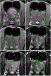
Post-operative, PC-T2FLAIR hyperintensity associated with the local subarachnoid spaces following removal of an olfactory/frontal lobe meningioma. Pre-surgery transverse T1W post-contrast image (A), post-surgery T1W (B), T1W post-contrast (C), T2W (D), FLAIR (E) and PC-T2FLAIR (F) images. While post-surgery T1W post-contrast and PC-T2FLAIR hyperintensity localizations on the side of the surgery essentially overlap, PC-T2FLAIR hyperintensities most consistently recapitulate T2W signal. In contrast, abrogation of subarachnoid fluid suppression is minimal in the contralateral frontal cortex.
Fig 8.
Fig 8.
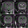
T1W (A), T2W (B), FLAIR (C, D) images all acquired pre-contrast administration. Regional loss of suppression of CSF signal from the temporal horn of the lateral ventricle (C) has occurred secondary to local field inhomogeneity due to the presence of a metallic endotracheal cuff spring (arrow heads). Suppression is reestablished following removal of the metallic spring (D).

Similar articles

Cited by

References

    1. Merhof K, Lang J, Durr S, Stahl C, Gorgas D. Use of contrast-enhanced fluid-attenuated inversion recovery sequence to detect brain lesions in dogs and cats. J Vet Intern Med. 2014;28: 1263–1267. - PMC - PubMed
    1. Falzone C, Rossi F, Calistri M, Tranquillo M, Baroni M. Contrast-enhanced fluid-attenuated inversion recovery vs. contrast-enhanced spin echo T1-weighted brain imaging. Vet Radiol Ultrasound. 2008;49: 333–338. - PubMed
    1. Bozzao A, Floris R, Fasoli F, Fantozzi LM, Colonnese C, Simonetti G. Cerebrospinal fluid changes after intravenous injection of gadolinium chelate: assessment by FLAIR MR imaging. Eur Radiol. 2003;13: 592–597. - PubMed
    1. Lee EK, Lee EJ, Kim S, Lee YS. Importance of Contrast-Enhanced Fluid-Attenuated Inversion Recovery Magnetic Resonance Imaging in Various Intracranial Pathologic Conditions. Korean J Radiol. 2016;17: 127–141. - PMC - PubMed
    1. Mathews VP, Caldemeyer KS, Lowe MJ, Greenspan SL, Weber DM, Ulmer JL. Brain: gadolinium-enhanced fast fluid-attenuated inversion-recovery MR imaging. Radiology. 1999;211: 257–263. - PubMed