. 2018 May;66(5):1118-1130.
doi: 10.1002/glia.23305. Epub 2018 Feb 9.

Differential local tissue permissiveness influences the final fate of GPR17-expressing oligodendrocyte precursors in two distinct models of demyelination

Affiliations

Differential local tissue permissiveness influences the final fate of GPR17-expressing oligodendrocyte precursors in two distinct models of demyelination

Giusy T Coppolino et al. Glia. 2018 May.

Abstract

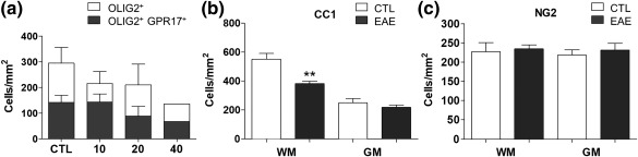
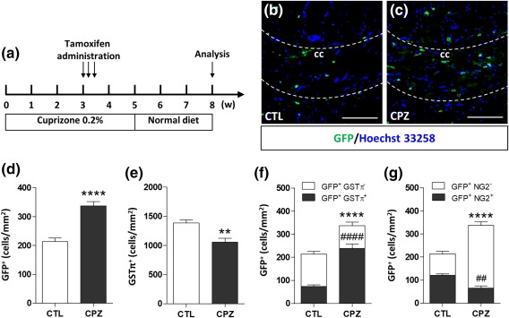
Promoting remyelination is recognized as a novel strategy to foster repair in neurodegenerative demyelinating diseases, such as multiple sclerosis. In this respect, the receptor GPR17, recently emerged as a new target for remyelination, is expressed by early oligodendrocyte precursors (OPCs) and after a certain differentiation stage it has to be downregulated to allow progression to mature myelinating oligodendrocytes. Here, we took advantage of the first inducible GPR17 reporter mouse line (GPR17-iCreERT2 xCAG-eGFP mice) allowing to follow the final fate of GPR17+ cells by tamoxifen-induced GFP-labeling to unveil the destiny of these cells in two demyelination models: experimental autoimmune encephalomyelitis (EAE), characterized by marked immune cell activation and inflammation, and cuprizone induced demyelination, where myelin dysfunction is achieved by a toxic insult. In both models, demyelination induced a strong increase of fluorescent GFP+ cells at damaged areas. However, only in the cuprizone model reacting GFP+ cells terminally differentiated to mature oligodendrocytes, thus contributing to remyelination. In EAE, GFP+ cells were blocked at immature stages and never became myelinating oligodendrocytes. We suggest these strikingly distinct fates be due to different permissiveness of the local CNS environment. Based on previously reported GPR17 activation by emergency signals (e.g., Stromal Derived Factor-1), we propose that a marked inflammatory milieu, such as that reproduced in EAE, induces GPR17 overactivation resulting in impaired downregulation, untimely and prolonged permanence in OPCs, leading, in turn, to differentiation blockade. Combined treatments with remyelinating agents and anti-inflammatory drugs may represent new potential adequate strategies to halt neurodegeneration and foster recovery.

Keywords: G protein-coupled receptor; animal models; differentiation; multiple sclerosis; oligodendrocyte precursor cells.


Figures

Figure 1
Figure 1
Confocal images of control mouse spinal cord immunolabeled for GPR17 and other markers. (a) Distribution of GPR17+‐cells (in red) in the spinal cord of an adult mouse. Cell nuclei were labeled with Hoechst 33258 (in blue). GPR17+‐cells expressed the oligodendroglial marker OLIG2 (b and inset b′). GPR17 staining showed co‐localization with both the early OPC marker NG2 (c and inset c′) and the more mature marker CC1 (d and inset d′). No GPR17 positivity was found in microglia (IBA1+ cells), astrocytes (GFAP+ cells) and neurons (MAP2+ cells) (e, f, and g). Micrographs were taken at the confocal microscope (Zeiss LSM 510 Meta). Scale bars: 200 µm (a), 50 µm (b–g), 20 µm (b′, c′, and d′) [Color figure can be viewed at http://wileyonlinelibrary.com]
Figure 2
Figure 2
Oligodendrocyte alterations during EAE. (a) Animals were analyzed on 10, 20, and 40‐day post immunization (DPI). We reported oligodendrocyte (OLIG2+‐cells) loss at each time‐point as expected in this disease; most of these cells were GPR17‐expressing OPCs and immature oligodendrocytes; in particular, upon acute EAE (20 DPI) there was a significant decrease of CC1+ mature oligodendrocytes (b). At this time, the NG2+ OPCs were almost the same in both white (WM) and gray (GM) matter (c). Unpaired two‐tailed Student's t test; **p < .01 compared with control from two independent experiments
Figure 3
Figure 3
Characterization of GPR17 subpopulations and changes of GPR17 expression in acute EAE in spinal cord. Animals were analyzed 21 days after immunization (acute EAE). Upon acute phase, there was a significant increase of GPR17+/NG2+ cells (OPCs), only in the white matter (WM; a and b); conversely, the number of GPR17+/CC1+ cells (immature oligodendrocytes) was apparently unchanged in both WM and GM (c, d). Data are the mean ± SEM of cervical, thoracic and lumbar sections; (CTL n = 3, EAE n = 5). Unpaired two‐tailed Student's t test; ****p < .0001 compared with control from two independent experiments. (e) By means of real‐time PCR, a significant up‐regulation of GPR17 was found in spinal cord of mice after acute EAE compared with controls and this correlated with the increased expression of inflammatory cytokines (f). Histograms show the fold change value ± SEM compared with control set to 1. Two‐tailed Mann‐Whitney student t test, *p ≤ .05, **p < .01 from three independent experiments. (g) GPR17 up‐regulation was also confirmed by means of in situ hybridization at the lesion site (black arrows) indicate cells with increased levels of Gpr17 mRNA. Scale bar 100 μm. (h) A local up‐regulation of GPR17+ cells (white arrows) was observed after EAE induction in the same area where inflammatory cells infiltrate the tissue (characterized by a high number of nuclei, in blue), bordered by IBA1+ activated microglial cells. Scale bar 50 μm [Color figure can be viewed at http://wileyonlinelibrary.com]
Figure 4
Figure 4
Identity of recombinant cells in the adult spinal cord. (a) Schematic representation of the transgenic alleles in GPR17‐iCreERT2xCAG‐eGFP mice showing tamoxifen‐responsive recombination of the CAG‐GFP allele to induce GFP in cells expressing GPR17. (b and c) Quantification of OLIG2+ cells among GFP+ cells in the spinal cord reveals that nearly all recombined cells belong to the oligodendroglial lineage. Many GFP+ cells were also found positive for GPR17 (d) and the NG2 marker (e). Although they were not abundant, we detect the presence of some GFP+GST+ cells (f). Vice versa, GFP+ cells are not positive for the markers of microglia, IBA1 (g), neurons, NEUN (h) and astrocytes, GFAP (i). Images were taken at the confocal microscope (Zeiss LSM 510 Meta). Scale bar: 10 µm (c and f), 5 µm (c′), 50 µm (d–i) [Color figure can be viewed at http://wileyonlinelibrary.com]
Figure 5
Figure 5
Reaction and fate of recombined cells in the spinal cord of GPR17‐iCreERT2xCAG‐eGFP mice after acute EAE. (a) Mice received tamoxifen by oral gavage three times (every other day), starting 14 days before EAE induction, and were analyzed in the EAE acute phase (day 21 after immunization). (b) Clinical scores of mice during acute EAE. Error bars represent mean of CS ± SEM. (c) Quantification of the number of GFP+ cells in whole spinal cord white (WM) and grey (GM) matter of EAE mice compared with controls. Quantification of GFP+/NG2+‐ and GFP+/GSTπ+‐ cells in both WM (d and e) and GM (f and g) in spinal cord of EAE mice. Data are the mean ± SEM; (CTL n = 2, EAE n = 5). Unpaired two‐tailed Student's t test; #p < .01, *< .05, ***p < .001 compared with control. (h and i). Representative micrographs showing myelination (fluoromyelin red staining), GFP+ cells (green fluorescence) and nuclei (in blue) in the area of the fasciculus gracilis (FG) in control and EAE mice. The accumulation of nuclei in the FG corresponds to infiltrating cells. A decrease in myelination level coupled to a strong increase in the number of GFP+ cells was found in the area. Scale bars: 100 µm. The distribution graphs show the percentage of myelination (j) and the number of GFP+ cell/mm2 (k) in the fasciculus gracilis of the spinal cord in EAE mice compared with controls. Data are the mean ± SEM of cervical, thoracic and lumbar sections; (CTL n = 15, EAE n = 41). Unpaired two‐tailed Student's t test; ****p < .0001. (l) The number of GFP+ cells was correlated to the extent of demyelination for different FG sections of EAE mice by performing a Pearson's correlation test (r = .585; p value = .0001). The analysis showed a strong association between the two variables [Color figure can be viewed at http://wileyonlinelibrary.com]
Figure 6
Figure 6
Reaction of GPR17+ OPCs in the corpus callosum of cuprizone fed mice. (a) Mice received a 2% cuprizone‐supplemented diet for up to 5 weeks (W) to induce demyelination and were then switched to normal diet to allow spontaneous remyelination. Expression profile of Mbp (b), Gpr17 (c), and IL‐1β (d) genes in the corpus callosum during demyelination (1, 3, and 5 weeks of cuprizone diet indicated as W0, W1, W3, and W5) and during the remyelination phase after cuprizone withdrawal (7 weeks, 7W). Mbp expression followed the typical already published pattern, with a marked decrease at W1–W5, followed by recovery at W7; in the case of Gpr17, an initial decrease was followed by increased expression at later phases of demyelination (3W and 5W). Gpr17 up‐regulation persisted also after cuprizone withdrawal (W7). As expected, IL‐1β expression increased overtime, peaking at W5. Data are the mean ± SEM of four animals for each time‐point; one‐way ANOVA with Tukey's multiple comparison post test; *p < .05, **p < .01, ***p < .001 compared with W0. (e–i) Qualitative immunostaining showing GPR17 expression (in red) during demyelination and remyelination phases in corpus callosum (highlighted by the white dotted lines). The red inset in the brain's drawing refers to the area where the IHC analysis reported in e–i was performed. Scale bar = 200 µm [Color figure can be viewed at http://wileyonlinelibrary.com]
Figure 7
Figure 7
The pool of expanded GPR17+ OPCs undergoes terminal maturation in response to cuprizone‐induced demyelination. (a) GPR17‐iCreERT2xCAG‐eGFP mice received a 0.2% cuprizone (CPZ)‐supplemented diet for up to 5 weeks (W) to induce demyelination and were then switched to normal diet to allow spontaneous remyelination. Tamoxifen induction was performed at 3W after beginning the cuprizone diet. Analyses were performed at the end of the experimental protocol (8W). (b and c) Qualitative immunostaining showing GFP expression (in green) in corpus callosum (cc) merged with Hoechst 33258 (in blue) to label cell nuclei. (d and e) Quantification of the number of GFP+‐cells and GSTπ+ cells in cc of CPZ fed mice compared with mice receiving standard diet (CTL). (f and g) Quantification of the number of GFP+/GSTπ+ cells and GFP+/NG2+ compared with GFP+ cells in cc of CPZ fed mice (n = 3). Data are the mean ± SEM. Unpaired t test; **p < .01, ****p < .0001, ####p < .0001, compared with mice receiving the standard diet (CTL). Scale bar = 25 µm [Color figure can be viewed at http://wileyonlinelibrary.com]

References

    1. Boda, E. , Viganò, F. , Rosa, P. , Fumagalli, M. , Labat‐Gest, V. , Tempia, F. , … Buffo, A. (2011). The GPR17 receptor in NG2 expressing cells: Focus on in vivo cell maturation and participation in acute trauma and chronic damage. Glia, 59, 1958–1973. - PubMed
    1. Calderon, T. M. , Eugenin, E. A. , Lopez, L. , Kumar, S. S. , Hesselgesser, J. , Raine, C. S. , & Berman, J. W. (2006). A role for CXCL12 (SDF‐1alpha) in the pathogenesis of multiple sclerosis: Regulation of CXCL12 expression in astrocytes by soluble myelin basic protein. Journal of Neuroimmunology, 177, 27–39. - PubMed
    1. Ceruti, S. , Viganò, F. , Boda, E. , Ferrario, S. , Magni, G. , Boccazzi, M. , … Abbracchio, M. P. (2011). Expression of the new P2Y‐like receptor GPR17 during oligodendrocyte precursor cell maturation regulates sensitivity to ATP‐induced death. Glia, 59, 363–378. - PubMed
    1. Ceruti, S. , Villa, G. , Genovese, T. , Mazzon, E. , Longhi, R. , Rosa, P. , … Abbracchio, M. P. (2009). The P2Y‐like receptor GPR17 as a sensor of damage and a new potential target in spinal cord injury. Brain: A Journal of Neurology, 132, 2206–2218. - PubMed
    1. Chen, Y. , Wu, H. , Wang, S. , Koito, H. , Li, J. , Ye, F. , … Lu, Q. R. (2009). The oligodendrocyte‐specific G protein‐coupled receptor GPR17 is a cell‐intrinsic timer of myelination. Nature Neuroscience, 12, 1398–1406. - PMC - PubMed

Publication types

MeSH terms

Substances