

Review
doi: 10.1038/eye.2017.298.
Epub 2018 Feb 9.
Elusive drusen and changing terminology of AMD
Affiliations
- PMID: 29424832
- PMCID: PMC5944644
- DOI: 10.1038/eye.2017.298
Item in Clipboard
Review
Elusive drusen and changing terminology of AMD
Eye (Lond).
2018 May.
Abstract
The first descriptions of ageing macula disorder (AMD), be it under other names, appeared in 1855 and 1868. The earliest accounts of AMD linked the presence of drusen with visual loss. It took a century before these connections between drusen and AMD were generally accepted by medical science and in clinical articles. The first signs of AMD appear in the region of the choriocapillaris, Bruch's membrane and the retinal pigment epithelium. The pathogenesis of drusen and of AMD is still uncertain. This is reflected in the wide variation in nomenclature of both, since the first publications.
Conflict of interest statement
The authors declare that they have no conflict of interest.
Figures
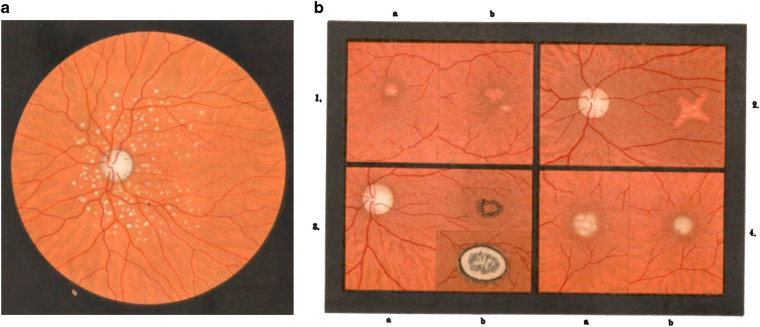
(a) Drusen of the vitreous membrane of the choroid. Haab wrote: These drusen, also a change in old age, have nothing to do with the macular disorder depicted here and should be distinctly separated from this. [20] (b) Images of senile macular disease. [20] 1. a b Macula of right and left eye from a 70-year-old person; 2. No legend given. 3. a Beginning signs of disease; 3. b 6 months later; 4. a b Right and left eye from a 74-year-old female
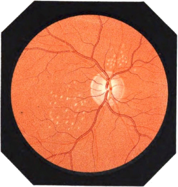
First color image of drusen. These were called: Nodular exudates in the choroid. Table 1 [23]
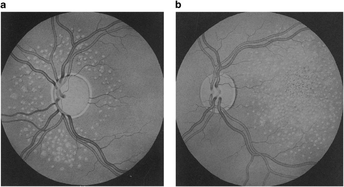
(a) Inferred association between 'posterieur' glaucoma with total disc cupping and circumpapillary drusen. [27] (b) Drusen and pigmentary changes in the fundus
First color images ever of the eye fundus [39] from thesis by Van Trigt [67]
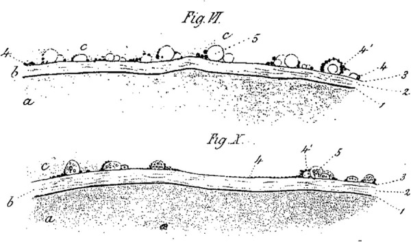
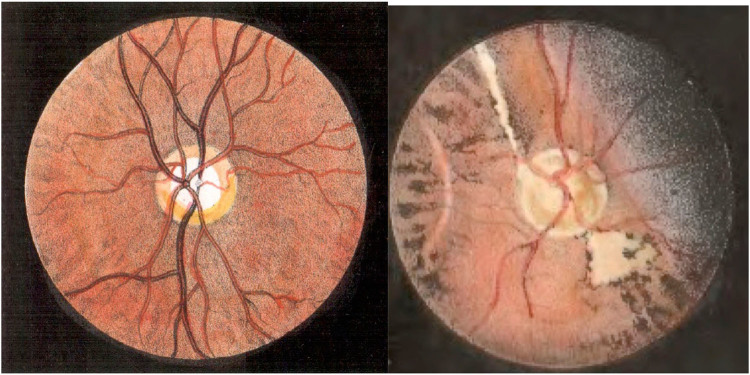
Large drusen (5) pressing against the retina (c) [14]. aa is choroid, b probably Bruch’s membrane, c retina in which drusen protrude
(a) Central guttate choroiditis [42] (b) Central senile areolar choroidal atrophy [43]
Disciform macular degeneration. Male, 79 years old. Round center, over 2.5 diopter prominence [60]
References
-
- Bird AC, Bressler NM, Bressler SB, Chisholm IH, Coscas G, Davis MD, et al. An international classification and grading system for age-related maculopathy and age-related macular degeneration. The International ARM Epidemiological Study Group. Surv Ophthalmol. 1995;39:367–374. doi: 10.1016/S0039-6257(05)80092-X. - DOI - PubMed
-
- Duke-Elder S, Wybar KC. The anatomy of the visual system II. The vascular tunic In:The anatomy of the visual system. Kimpton:London, 1961.
Publication types
MeSH terms
LinkOut - more resources
Full Text Sources
Other Literature Sources
Medical
