. 2018 Feb 9;11(1):8.
doi: 10.1186/s13041-018-0352-0.

Poly-arginine R18 and R18D (D-enantiomer) peptides reduce infarct volume and improves behavioural outcomes following perinatal hypoxic-ischaemic encephalopathy in the P7 rat

Affiliations

Poly-arginine R18 and R18D (D-enantiomer) peptides reduce infarct volume and improves behavioural outcomes following perinatal hypoxic-ischaemic encephalopathy in the P7 rat

Adam B Edwards et al. Mol Brain. .

Abstract

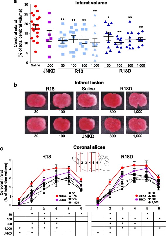
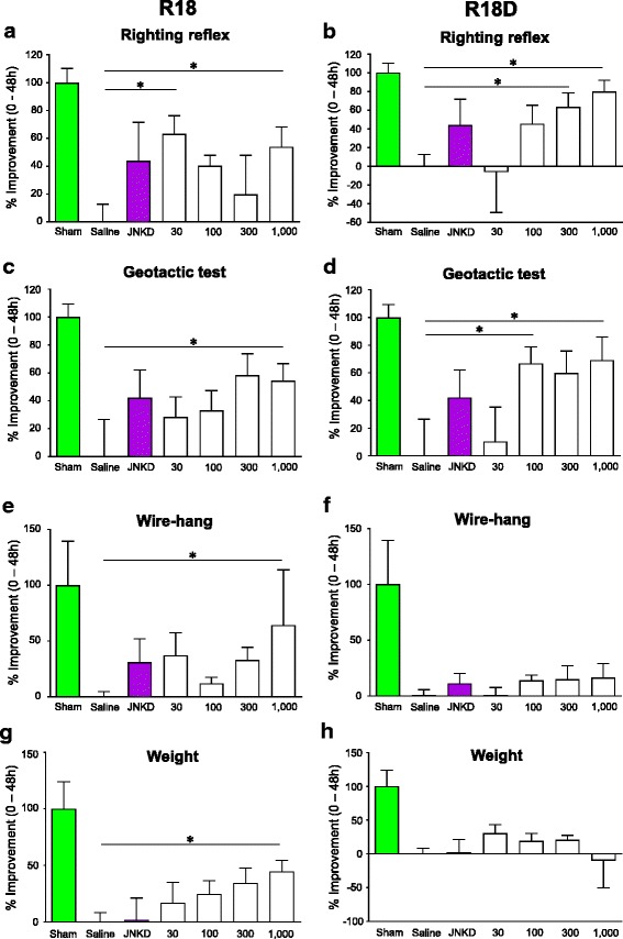
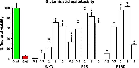
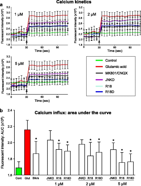
We examined the neuroprotective efficacy of the poly-arginine peptide R18 and its D-enantiomer R18D in a perinatal hypoxic-ischaemic (HI) model in P7 Sprague-Dawley rats. R18 and R18D peptides were administered intraperitoneally at doses of 30, 100, 300 or 1000 nmol/kg immediately after HI (8% O2/92%N2 for 2.5 h). The previously characterised neuroprotective JNKI-1-TATD peptide at a dose of 1000 nmol/kg was used as a control. Infarct volume and behavioural outcomes were measured 48 h after HI. For the R18 and R18D doses examined, total infarct volume was reduced by 25.93% to 43.80% (P = 0.038 to < 0.001). By comparison, the JNKI-1-TATD reduced lesion volume by 25.27% (P = 0.073). Moreover, R18 and R18D treatment resulted in significant improvements in behavioural outcomes, while with JNKI-1-TATD there was a trend towards improvement. As an insight into the likely mechanism underlying the effects of R18, R18D and JNKI-1-TATD, the peptides were added to cortical neuronal cultures exposed to glutamic acid excitotoxicity, resulting in up to 89, 100 and 71% neuroprotection, respectively, and a dose dependent inhibition of neuronal calcium influx. The study further confirms the neuroprotective properties of poly-arginine peptides, and suggests a potential therapeutic role for R18 and R18D in the treatment of HIE.

Keywords: Cationic arginine-rich peptides (CARPs); Hypoxia-ischaemia; Hypoxic-ischaemic encephalopathy; Neuroprotection; Poly-arginine peptides; R18.


Conflict of interest statement

Ethics approval and consent to participate

Not applicable

Consent for publication

Not applicable

Competing interests

B.P. Meloni and N.W. Knuckey are named inventors of several patent applications regarding the use of arginine-rich peptides as neuroprotective agents. The other authors declare no conflict of interest.

Publisher’s Note

Springer Nature remains neutral with regard to jurisdictional claims in published maps and institutional affiliations.

Figures

Fig. 1
Fig. 1
Percentage infarct volume; percentage infarct volume, representative images of coronal brain slices and percentage infarct volume in brain slices for the different treatment groups as determined 48 h after HI. Treatments were administered intraperitoneally (saline or R18, R18D and JNKI-1-TATD; doses in nmol/kg) immediately after hypoxia. a Percentage of infarct volume when compared to total brain volume. b Representative TTC coronal brain slice 2, from saline and peptide treated animals. Minor adjustments to brightness and contrast has been made to improve digital images. c Infarct volume analysis in 2 mm coronal brain slices (slices numbered 1–6 from rostral to caudal) from saline and peptide treated animals. Statistical significance is expressed in the table. Values are mean ± SE; *P < 0.05, **P < 0.01 when compared to saline. JNKD = JNKI-1-TATD
Fig. 2
Fig. 2
Behavioural measurements using righting reflex, negative geotactic response, wire-hang test and weight gain 48 h after HI. Treatments were administered intraperitoneally (saline, R18, R18D or JNKI-1-TATD; doses in nmol/kg). The sham procedure group was assessed 48 h following sham-surgery. a R18 righting reflex percentage improvement from 0 to 48 h. b R18D righting reflex percentage improvement from 0 to 48 h. c R18 negative geotactic response percentage improvement from 0 to 48 h. d R18D negative geotactic response percentage improvement from 0 to 48 h. e R18 wire-hang percentage improvement from 0 to 48 h. f R18D wire-hang percentage improvement from 0 to 48 h. g R18 weight percentage improvement from 0 to 48 h. h R18D weight percentage improvement from 0 to 48 h. Values are mean ± SE. *P < 0.05 when compared to saline. JNKD = JNKI-1-TATD
Fig. 3
Fig. 3
Glutamic acid excitotoxicity model; R18, R18D and JNKI-1-TATD dose response study. Peptides present in neuronal cultures 10 min before and during 5 min glutamic acid exposure. Neuronal viability measured 24 h after glutamic acid exposure. Concentration of peptide in μmol/L. MTS data were expressed as percentage neuronal viability with no insult (100% viability) and glutamic acid control (5% viability). Values are mean ± SE; n = 4; *P < 0.05 when compared to no glutamic acid control. Cont = no treatment control. Glut = glutamic acid control. JNKD = JNKI-1-TATD
Fig. 4
Fig. 4
Intracellular calcium assessment using Fura-2 AM after glutamic acid exposure in primary neuronal cultures. a Fluorescent Fura-2 AM tracers; fluorescent intensity (FI) of neuronal cultures 30 s before and after the addition (arrow) of glutamic acid (100 μM final concentration). Peptides (1, 2 and 5 μM) or glutamate receptor blockers (MK801/CNQX; 5 μM/5 μM) were added to neuronal cultures for 10 min and removed (time = 0) before glutamic acid addition (time = 30 s). Values are mean ± SE; n = 3. b Trapezoidal area under the curve (AUC) approximation of calcium kinetic tracers. AUC is determined at 35 s (point after glutamic acid addition. Values are mean ± SE; n = 3; *P < 0.05 when compared to glutamic acid control. Cont = no treatment control. Glut = glutamic acid control. Blkrs = glutamate receptor blockers (MK801/CNQX; 5 μM/5 μM). JNKD = JNKI-1-TATD

References

    1. Kurinczuk JJ, White-Koning M, Badawi N. Epidemiology of neonatal encephalopathy and hypoxic-ischaemic encephalopathy. Early Hum Dev. 2010;86(6):329–338. doi: 10.1016/j.earlhumdev.2010.05.010. - DOI - PubMed
    1. Graham EM, Ruis KA, Hartman AL, Northington FJ, Fox HE. A systematic review of the role of intrapartum hypoxia-ischemia in the causation of neonatal encephalopathy. Am J Obstet Gynecol. 2008;199(6):587–595. doi: 10.1016/j.ajog.2008.06.094. - DOI - PubMed
    1. Vannucci RC, Perlman JM. Interventions for perinatal hypoxic-ischemic encephalopathy. Pediatrics. 1997;100(6):1004–1014. doi: 10.1542/peds.100.6.1004. - DOI - PubMed
    1. Shankaran S. Therapeutic hypothermia for neonatal encephalopathy. Curr Treat Options Neurol. 2012;14(6):608–619. doi: 10.1007/s11940-012-0200-y. - DOI - PMC - PubMed
    1. Ferrer-Montiel AV, et al. Selected peptides targeted to the NMDA receptor channel protect neurons from excitotoxic death. Nat Biotechnol. 1998;16(3):286–291. doi: 10.1038/nbt0398-286. - DOI - PubMed

Publication types

MeSH terms