

Damage in a Distal Radius Fracture Model Treated With Locked Volar Plating After Simulated Postoperative Loading
- PMID: 29426604
- PMCID: PMC6035079
- DOI: 10.1016/j.jhsa.2017.12.019
Damage in a Distal Radius Fracture Model Treated With Locked Volar Plating After Simulated Postoperative Loading
Abstract
Purpose: "Damage" is an engineering term defining a period between a state of material perfection and the onset of crack initiation. Clinically, it is a loss of fixation due to microstructural breakdown, indirectly measured as a reduction of stiffness of the bone-implant construct, normalized by the cross-sectional area and length of the bone. The purpose of this study was to characterize damage in a cadaver model of extra-articular distal radius fracture with dorsal comminution treated using 2-column volar distal radius plates.
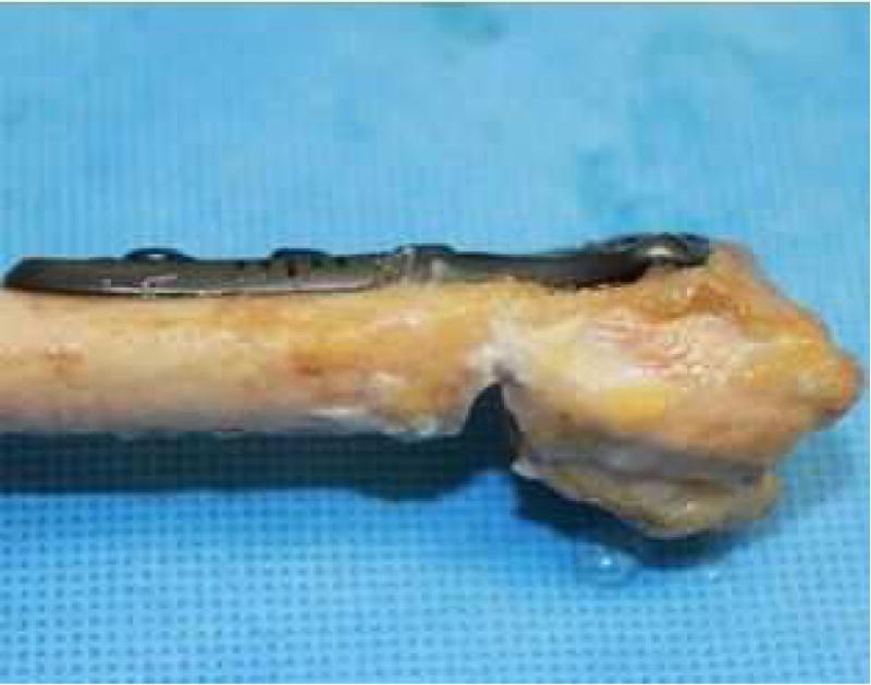
Methods: Ten matched distal radii were randomly divided into 2 groups: group I specimens were treated with a volar distal radius plate with an independent, 2-tiered scaffold design; group II specimens (contralateral limbs) were treated with a volar plate with a single-head design for enhanced ulnar buttressing. Specimens were cyclically loaded to simulate a 6-month postoperative load-bearing period. We report damage after a defined protocol of cyclical loading and load to failure simulating a fall on an outstretched hand.
Results: Group II specimens experienced more damage under cyclic loading conditions than group I specimens. Group I specimens were stiffer than group II specimens under load-to-failure conditions. Ultimate force at failure in group I and group II specimens was not different. Specimens failed by plate bending (group I, n = 6/10; group II, n = 2/10) and fracture of the lunate facet (group I, n = 4/10; group II, n = 8/10).
Conclusions: Group I specimens had less screw cutout at the lunate facet than group II specimens under cyclic loading as indicated by lower damage measures and fewer facet fractures during load-to-failure testing. The overall strength of the construct is not affected by plate design.
Clinical relevance: Microstructural damage or a loss of fixation due to an overly rigid volar plate design may cause malunion or nonunion of fracture fragments and lead to bone-implant instability.
Keywords: Distal radius fracture; distal radius plating; volar locking plate; wrist biomechanics; wrist fracture.
Copyright © 2018 American Society for Surgery of the Hand. Published by Elsevier Inc. All rights reserved.
Figures


References
-
- Chung KC, Spilson SV. The frequency and epidemiology of hand and forearm fractures in the United States. J Hand Surg Am. 2001;26:908–915. - PubMed
-
- Rennie L, Court-Brown CM, Mok JY, Beattie TF. The epidemiology of fractures in children. Injury. 2007;38:913–922. - PubMed
-
- Ward WT, Rihn JA. The impact of trauma in an urban pediatric orthopaedic practice. J Bone Joint Surg Am. 2006;88:2759–2764. - PubMed
-
- Baron JA, Karagas M, Barrett J, et al. Basic epidemiology of fractures of the upper and lower limb among Americans over 65 years of age. Epidemiology. 1996;7:612–618. - PubMed
Publication types
MeSH terms
Grants and funding
LinkOut - more resources
Full Text Sources
Other Literature Sources
Medical
