. 2018 Feb 9;9(1):613.
doi: 10.1038/s41467-018-02936-3.

TRIM56-mediated monoubiquitination of cGAS for cytosolic DNA sensing

Affiliations

TRIM56-mediated monoubiquitination of cGAS for cytosolic DNA sensing

Gil Ju Seo et al. Nat Commun. .

Abstract

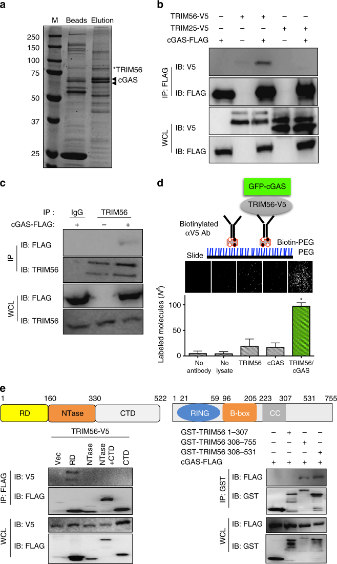
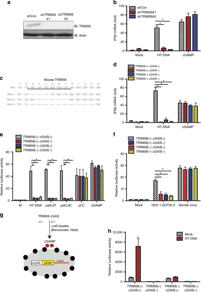
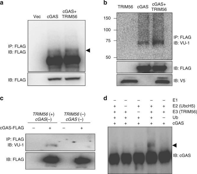
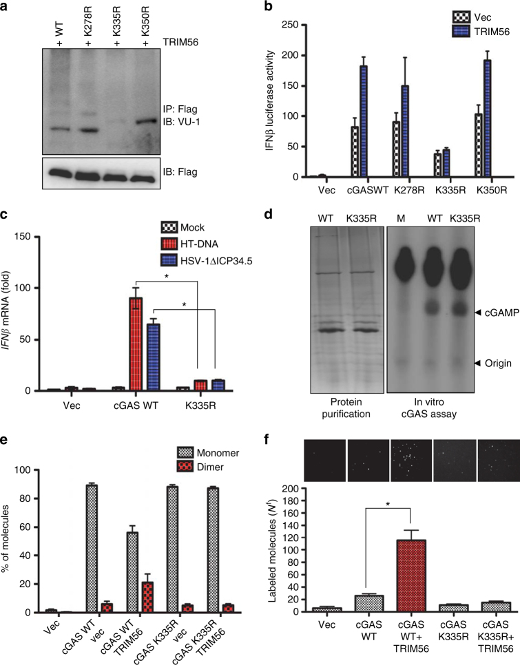
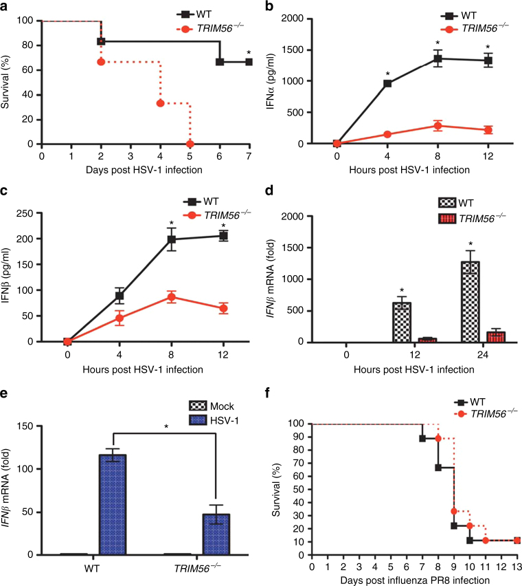
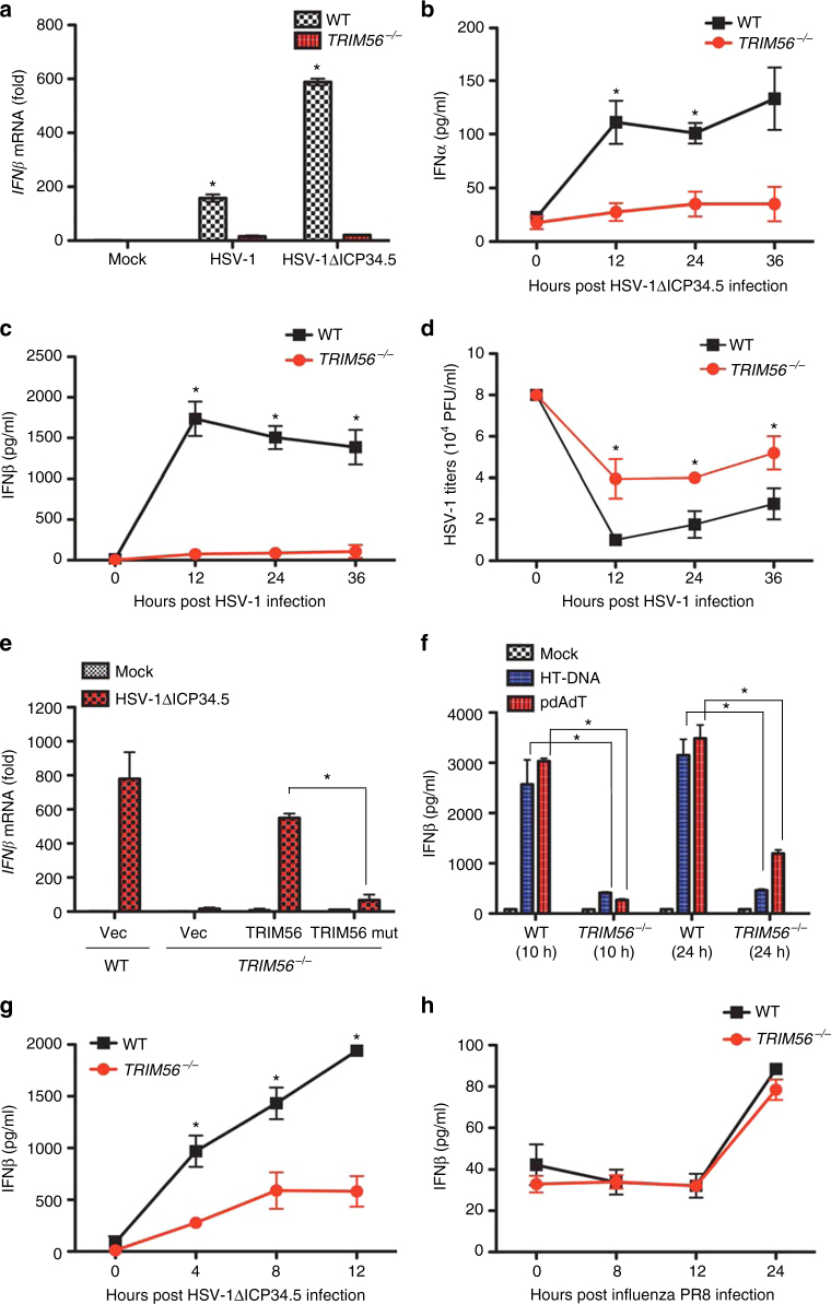
Intracellular nucleic acid sensors often undergo sophisticated modifications that are critical for the regulation of antimicrobial responses. Upon recognition of DNA, the cytosolic sensor cyclic GMP-AMP (cGAMP) synthase (cGAS) produces the second messenger cGAMP, which subsequently initiates downstream signaling to induce interferon-αβ (IFNαβ) production. Here we report that TRIM56 E3 ligase-induced monoubiquitination of cGAS is important for cytosolic DNA sensing and IFNαβ production to induce anti-DNA viral immunity. TRIM56 induces the Lys335 monoubiquitination of cGAS, resulting in a marked increase of its dimerization, DNA-binding activity, and cGAMP production. Consequently, TRIM56-deficient cells are defective in cGAS-mediated IFNαβ production upon herpes simplex virus-1 (HSV-1) infection. Furthermore, TRIM56-deficient mice show impaired IFNαβ production and high susceptibility to lethal HSV-1 infection but not to influenza A virus infection. This adds TRIM56 as a crucial component of the cytosolic DNA sensing pathway that induces anti-DNA viral innate immunity.


Conflict of interest statement

The authors declare no competing financial interests.

Figures

Fig. 1
Fig. 1
TRIM56 interacts with cGAS. a HEK293T cells were transiently transfected with cGAS-FLAG and protein complexes were purified from cell lysates with anti-FLAG M2 beads. The immunoprecipitates were eluted with FLAG peptides and fractionated using SDS-PAGE followed by Coommassie blue staining. The bands for TRIM56 and cGAS identified by mass spectrometry are indicated on the right. b Co-immunoprecipitation analysis of the interaction between cGAS-FLAG and TRIM56-V5. HEK293T cells expressing cGAS-FLAG were co-transfected with TRIM56-V5 or TRIM25-V5. cGAS-FLAG immunoprecipitates were probed with FLAG and V5 antibody and whole-cell lysates (WCLs) were probed with FLAG and V5 antibody, as indicated. c cGAS interacts with endogenously expressed TRIM56. HEK293T cells were transfected with cGAS-FLAG. Rabbit IgG or TRIM56 immunoprecipitates were probed with FLAG antibody, and WCLs were probed with TRIM56 and FLAG, as indicated. d cGAS-FLAG was transiently coexpressed with TRIM56-V5 in HEK293T cells. TRIM56-V5 was pulled down for SiMPull analysis. Top panel: schematic depiction of cGAS SiMPull; middle panel: representative GFP fluorescence images; bottom panel: average number of molecules per imaging area (Nf). e The N-terminal regulatory domain of cGAS interacts with the C-terminal NHL homologous region of TRIM56. Left panel: the regulatory domain of cGAS binds to TRIM56. WCLs of HEK293T cells transfected with TRIM56-V5 and cGAS truncation mutants were used for immunoprecipitation and immunoblotting with the indicated antibodies. Right panel: the C-terminus domain (308–531) of TRIM56 interacts with cGAS. WCLs of HEK293T cells transfected with cGAS-FLAG and TRIM56 truncation mutants were used for immunoprecipitation and immunoblotting with the indicated antibodies. Data are representative of two independent experiments ae. Error bars indicate mean ± s.d. of n = 3. *P < 0.05 versus control using Student’s t-test (d). Full blots are shown in Supplementary Fig. 10
Fig. 2
Fig. 2
TRIM56 is required for cGAS-dependent signal transduction and acts upstream of STING. a THP-1 cells were stably transfected with control shRNA, TRIM56#1 shRNA, or TRIM56#2 shRNA. Cell lysates were detected with the indicated antibodies. b THP-1 cells used in a were stimulated with HT-DNA (2 μg/ml) and cGAMP (3 μg/ml). The expression of IFNβ mRNA was measured using real-time PCR. c Generation of TRIM56−/− L929 cells. CRISPR-Cas9 targeting: the CRISPR target site in exon 1 of TRIM56 is underlined, and the protospacer adjacent motif (PAM) is shown in red. Deletions in the three TRIM56 alleles, each of which results in a frameshift, are shown by the black dashes. d cGAS-deleted L929 cells with or without TRIM56 deletion were complemented with wild-type cGAS-FLAG and stimulated with HT-DNA (2 μg/ml) and cGAMP (3 μg/ml). The expression of IFNβ mRNA was measured using real-time PCR. e L929 cell lines used in d were stably transfected with ISREIFNβ-Lucia reporter genes. Cells were stimulated with HT-DNA (2 μg/ml), poly dA:dT (1 μg/ml), poly dG:dC (1 μg/ml) polyI:C (1 μg/ml), and cGAMP (3 μg/ml). Luciferase activity was measured 18 h after stimulation. f L929 cell lines used in e were infected with HSV-1ΔICP34.5 (MOI = 5) and Sendai virus. Luciferase activity was measured 18 h after stimulation. g Schematic representation of the cGAMP bio-assay. Relative cGAMP activity was measured using THP1-Lucia ISG cells that generate luciferase in response to cGAMP. h cGAMP bio-assay. L929 cell lines used in d were stimulated with HT-DNA (2 μg/ml) for 9 h. Extracts of the cells were prepared, DNase- and heat-treated, and incubated with permeabilized THP-1-Lucia ISG cells for 18 h. Relative luciferase activity was measured. Data in a, h are representative of two independent experiments. Data in b, df are representative of three independent experiments. Error bars in b, df, h indicate mean ± s.d. of n = 3 (b, de) and n = 2 (f, h). *P < 0.05, versus control using Student’s t-test (b, df, h). Full blots are shown in Supplementary Fig. 10
Fig. 3
Fig. 3
TRIM56 mono-ubiquitinates cGAS. a, b HEK293T cells were transfected with the indicated plasmids. Twenty-four hours after transfection, whole-cell lysates (WCLs) were used for immunoprecipitation and immunoblotting, as indicated. c WCLs from L929 cell lines were used for immunoprecipitation and immunoblotting, as indicated. d Immunoblot analysis of the in vitro ubiquitination with the indicated combinations of ubiquitin, purified cGAS, E1, UbcH5 E2 enzymes, and TRIM56 (E3). Data in a, b are representative of three independent experiments. Data in c, d are representative of two independent experiments. Full blots are shown in Supplementary Fig. 10
Fig. 4
Fig. 4
TRIM56 increases cGAS dimerization and DNA-binding ability. a HEK293T cells were transfected with cGAS WT or cGAS K→R mutants. WCLs were used for immunoprecipitation and immunoblotting, as indicated. b IFNβ promoter activity in 293T cells, which were co-transfected with TRIM56 and cGAS WT or K→R mutants. c L929 cGAS−/− cells were stably complemented with empty vector, mouse cGAS WT, or cGAS K335R. Cells were stimulated with HT-DNA or infected with HSV-1ΔICP34.5 (MOI = 5). IFNβ mRNA was analyzed by RT-PCR. d In vitro enzymatic assays were performed in the presence of P32-α-GTP with amino acids 141–507 of mouse cGAS purified from engineered E. coli BL21 strain. The left panel shows the purified cGAS WT and cGAS K335R proteins. cGAMP production was analyzed by TLC and autoradiography. The bottom arrow shows the spotted origin and the top arrow shows the migrated cGAMP. e The relative ratio of observed bleaching steps for monomeric cGAS-GFP and dimeric cGAS-GFP pulled down from cell lysates. HEK293T cells were transfected with vector, cGAS WT, cGAS WT/TRIM56, cGASK335R, or cGAS335R/TRIM56, and the ratio was calculated by counting the photobleaching steps from a GFP-cGAS pull-down. For determining the stoichiometry, traces were manually scored for the number of bleaching steps. More than 300 traces were scored to reliably identify the photobleaching step distribution. See Supplementary Fig. 6. f Rhodamine-labeled double-stranded DNA bound by cGAS on the PEG-coated surface. Top panel: representative fluorescent DNA images bound by cGAS pulled down from cell lysates from vector; cGAS, cGAS- and TRIM56-expressing cells; cGASK335R-; or cGASK335R- and TRIM56-expressing cells. Rhodamine-labeled pdA:dT (1 μg/ml) was co-transfected into vector, cGAS, TRIM56/cGAS-expressing cells, cGASK335R, or cGASK335R/TRIM56-expressing cells, respectively. Bottom panel: average number of molecules per imaging area (Nf). Error bars indicate mean ± s.d. *P < 0.05, Student’s t-test. Data in a, d are representative of two independent experiments. Data in b, c, e, f are representative of three independent experiments. Error bars b, c, e, f indicate mean ± s.d. of n = 3. *P < 0.05, versus control using Student’s t-test (c, f). Full blots are shown in Supplementary Fig. 10
Fig. 5
Fig. 5
TRIM56−/− mice are susceptible to HSV-1 infection but not to IAV infection. a HSV-1 (1.8 × 108 pfu/mouse) was injected peritoneally to WT or TRIM56−/− mice (n = 6 each). Survival rates were monitored for 7 days. b, c Sera were collected from WT or TRIM56−/− mice injected with HSV-1 at the indicated time points, and IFNα or IFNβ concentration was measured by ELISA (n = 3 each at each time point). Total peritoneal cavity cells (d) and peritoneal macrophages (e) were isolated from WT or TRIM56−/− HSV-1-infected mice (n = 3 for each time point). IFNβ mRNA levels in these cells were measured using RT-PCR. f Influenza PR8 (1000 pfu/mouse) was administered intranasally to WT or TRIM56−/− mice (n = 9 each). Survival rates were monitored for 13 days. Error bars indicate mean ± s.d. *P < 0.05, Student’s t-test. Data in af are representative of two independent experiments. Error bars in be indicate mean ± s.d. of n = 3. *P < 0.05, versus control using Student’s t-test (ae)
Fig. 6
Fig. 6
TRIM56 is required for IFN induction in response to HSV-1 infection but not to IAV infection in primary cells. a RT-PCR analysis of IFNβ mRNA in BMDMs from WT and TRIM56−/− mice at 18 h after mock, HSV-1, or HSV-1ΔICP34.5 infection. ELISA of IFNα (b) or IFNβ (c) in BMDMs from WT and TRIM56−/− mice at the indicated time points after HSV-1ΔICP34.5 or HSV-1 infection. d WT or TRIM56−/− BMDMs were infected with HSV-1 (MOI = 0.1). Viral supernatants were collected at the indicated time points, and virus titers were determined using a plaque assay on Vero cells. e RT-PCR analysis of IFNβ mRNA in WT or TRIM56−/− BMDMs infected with lentivirus-derived vector, TRIM56 WT, or TRIM56 Mut. Results are presented relative to empty vector-expressing and mock-infected WT cells. f ELISA of IFNβ in MEFs from WT and TRIM56−/− embryos. MEFs were stimulated with HT- DNA (2 μg/ml) or polydA:dT (1 μg/ml). g ELISA of IFNβ in BMDCs from WT and TRIM56−/− mice at the indicated time points following infection with HSV-1ΔICP34.5. h ELISA of IFNβ in BMDMs from WT and TRIM56−/− mice at the indicated time points following infection with IAV PR8. Data in ag are representative of three independent experiments. Data in h are representative of two independent experiments. Error bars in ah indicate mean ± s.d. of n = 3. *P < 0.05 versus control using Student’s t-test (ah)

Similar articles

Cited by

References

    1. Medzhitov R, Janeway C., Jr. The Toll receptor family and microbial recognition. Trends Microbiol. 2000;8:452–456. doi: 10.1016/S0966-842X(00)01845-X. - DOI - PubMed
    1. Elinav E, Strowig T, Henao-Mejia J, Flavell RA. Regulation of the antimicrobial response by NLR proteins. Immunity. 2011;34:665–679. doi: 10.1016/j.immuni.2011.05.007. - DOI - PubMed
    1. Kawai T, Akira S. Toll-like receptors and their crosstalk with other innate receptors in infection and immunity. Immunity. 2011;34:637–650. doi: 10.1016/j.immuni.2011.05.006. - DOI - PubMed
    1. Paludan SR, Bowie AG. Immune sensing of DNA. Immunity. 2013;38:870–880. doi: 10.1016/j.immuni.2013.05.004. - DOI - PMC - PubMed
    1. Fernandes-Alnemri T, Yu JW, Datta P, Wu J, Alnemri ES. AIM2 activates the inflammasome and cell death in response to cytoplasmic DNA. Nature. 2009;458:509–513. doi: 10.1038/nature07710. - DOI - PMC - PubMed

Publication types

MeSH terms