Selective Tropism of Dengue Virus for Human Glycoprotein Ib
- PMID: 29426910
- PMCID: PMC5807543
- DOI: 10.1038/s41598-018-20914-z
Selective Tropism of Dengue Virus for Human Glycoprotein Ib
Erratum in
-
Publisher Correction: Selective Tropism of Dengue Virus for Human Glycoprotein Ib.Sci Rep. 2018 Apr 12;8(1):6000. doi: 10.1038/s41598-018-23724-5. Sci Rep. 2018. PMID: 29651159 Free PMC article.
Abstract
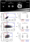
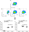
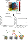
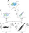
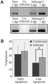
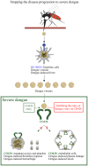
Since the hemorrhage in severe dengue seems to be primarily related to the defect of the platelet, the possibility that dengue virus (DENV) is selectively tropic for one of its surface receptors was investigated. Flow cytometric data of DENV-infected megakaryocytic cell line superficially expressing human glycoprotein Ib (CD42b) and glycoprotein IIb/IIIa (CD41 and CD41a) were analyzed by our custom-written software in MATLAB. In two-dimensional analyses, intracellular DENV was detected in CD42b+, CD41+ and CD41a+ cells. In three-dimensional analyses, the DENV was exclusively detected in CD42b+ cells but not in CD42b- cells regardless of the other expressions. In single-cell virus-protein analyses, the amount of DENV was directly correlated with those of CD42b at the Pearson correlation coefficient of 0.9. Moreover, RT- PCR and apoptosis assays showed that DENV was able to replicate itself and release its new progeny from the infected CD42b+ cells and eventually killed those cells. These results provide evidence for the involvement of CD42b in DENV infection.
Conflict of interest statement
The authors declare no competing interests.
Figures

Similar articles
-
Dengue Virus and Its Relation to Human Glycoprotein IIb/IIIa Revealed by Fluorescence Microscopy and Flow Cytometry.Viral Immunol. 2017 Nov;30(9):654-661. doi: 10.1089/vim.2017.0090. Epub 2017 Sep 25. Viral Immunol. 2017. PMID: 28945165
-
Dengue Virus Induces the Release of sCD40L and Changes in Levels of Membranal CD42b and CD40L Molecules in Human Platelets.Viruses. 2018 Jul 5;10(7):357. doi: 10.3390/v10070357. Viruses. 2018. PMID: 29976871 Free PMC article.
-
Binding of monoclonal antibodies to platelet glycoproteins Ib and IIb/IIIa in uremic patients.Nephron. 1997;75(3):283-5. doi: 10.1159/000189550. Nephron. 1997. PMID: 9069449
-
Insight into the Tropism of Dengue Virus in Humans.Viruses. 2019 Dec 9;11(12):1136. doi: 10.3390/v11121136. Viruses. 2019. PMID: 31835302 Free PMC article. Review.
-
Glycoprotein IIb-IIIa content and platelet aggregation in healthy volunteers and patients with acute coronary syndrome.Platelets. 2011;22(4):243-51. doi: 10.3109/09537104.2010.547959. Epub 2011 Feb 17. Platelets. 2011. PMID: 21329420 Review.
Cited by
-
Exposure of Platelets to Dengue Virus and Envelope Protein Domain III Induces Nlrp3 Inflammasome-Dependent Platelet Cell Death and Thrombocytopenia in Mice.Front Immunol. 2021 Apr 29;12:616394. doi: 10.3389/fimmu.2021.616394. eCollection 2021. Front Immunol. 2021. PMID: 33995345 Free PMC article.
-
Thrombocytopenia in dengue infection: mechanisms and a potential application.Expert Rev Mol Med. 2024 Oct 14;26:e26. doi: 10.1017/erm.2024.18. Expert Rev Mol Med. 2024. PMID: 39397710 Free PMC article. Review.
-
Transfusion-transmitted arboviruses: Update and systematic review.PLoS Negl Trop Dis. 2022 Oct 6;16(10):e0010843. doi: 10.1371/journal.pntd.0010843. eCollection 2022 Oct. PLoS Negl Trop Dis. 2022. PMID: 36201547 Free PMC article.
-
Sepsis - it is all about the platelets.Front Immunol. 2023 Jun 7;14:1210219. doi: 10.3389/fimmu.2023.1210219. eCollection 2023. Front Immunol. 2023. PMID: 37350961 Free PMC article. Review.
-
Role of Platelets in Detection and Regulation of Infection.Arterioscler Thromb Vasc Biol. 2021 Jan;41(1):70-78. doi: 10.1161/ATVBAHA.120.314645. Epub 2020 Oct 29. Arterioscler Thromb Vasc Biol. 2021. PMID: 33115274 Free PMC article. Review.
References
-
- Special Programme for Research and Training in Tropical Diseases., World Health Organization. Dengue : guidelines for diagnosis, treatment, prevention, and control, pp. 147 p (TDR : World Health Organization, Geneva, ed. New, 2009).
Publication types
MeSH terms
Substances
LinkOut - more resources
Full Text Sources
Other Literature Sources

