

2-Methoxyestradiol protects against pressure overload-induced left ventricular hypertrophy
- PMID: 29426916
- PMCID: PMC5807528
- DOI: 10.1038/s41598-018-20613-9
2-Methoxyestradiol protects against pressure overload-induced left ventricular hypertrophy
Abstract
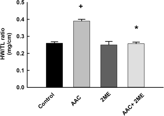
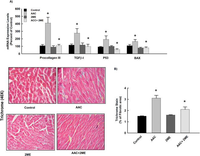
Numerous experimental studies have supported the evidence that 2-methoxyestradiol (2 ME) is a biologically active metabolite that mediates multiple effects on the cardiovascular system, largely independent of the estrogen receptor. 2 ME is a major cytochrome P450 1B1 (CYP1B1) metabolite and has been reported to have vasoprotective and anti-inflammatory actions. However, whether 2 ME would prevent cardiac hypertrophy induced by abdominal aortic constriction (AAC) has not been investigated yet. Therefore, the overall objectives of the present study were to elucidate the potential antihypertrophic effect of 2 ME and explore the mechanism(s) involved. Our results showed that 2 ME significantly inhibited AAC-induced left ventricular hypertrophy using echocardiography. The antihypertrophic effect of 2 ME was associated with a significant inhibition of CYP1B1 and mid-chain hydroxyeicosatetraenoic acids. Based on proteomics data, the protective effect of 2 ME is linked to the induction of antioxidant and anti-inflammatory proteins in addition to the modulation of proteins involved in myocardial energy metabolism. In vitro, 2 ME has shown a direct antihypertrophic effect through mitogen-activated protein kinases- and nuclear factor-κB-dependent mechanisms. The present work shows a strong evidence that 2 ME protects against left ventricular hypertrophy. Our data suggest the potential of repurposing 2 ME as a selective CYP1B1 inhibitor for the treatment of heart failure.
Conflict of interest statement
The authors declare no competing interests.
Figures


Similar articles
-
The role of cytochrome P450 1B1 and its associated mid-chain hydroxyeicosatetraenoic acid metabolites in the development of cardiac hypertrophy induced by isoproterenol.Mol Cell Biochem. 2017 May;429(1-2):151-165. doi: 10.1007/s11010-017-2943-y. Epub 2017 Mar 1. Mol Cell Biochem. 2017. PMID: 28251434
-
Inhibition of Mid-chain HETEs Protects Against Angiotensin II-induced Cardiac Hypertrophy.J Cardiovasc Pharmacol. 2017 Jul;70(1):16-24. doi: 10.1097/FJC.0000000000000494. J Cardiovasc Pharmacol. 2017. PMID: 28437282
-
Ameliorative Role of Fluconazole Against Abdominal Aortic Constriction-Induced Cardiac Hypertrophy in Rats.J Cardiovasc Pharmacol. 2022 Jun 1;79(6):833-845. doi: 10.1097/FJC.0000000000001258. J Cardiovasc Pharmacol. 2022. PMID: 35266922
-
The role of mid-chain hydroxyeicosatetraenoic acids in the pathogenesis of hypertension and cardiac hypertrophy.Arch Toxicol. 2016 Jan;90(1):119-36. doi: 10.1007/s00204-015-1620-8. Epub 2015 Nov 2. Arch Toxicol. 2016. PMID: 26525395 Review.
-
Left Ventricular Hypertrophy: Roles of Mitochondria CYP1B1 and Melatonergic Pathways in Co-Ordinating Wider Pathophysiology.Int J Mol Sci. 2019 Aug 20;20(16):4068. doi: 10.3390/ijms20164068. Int J Mol Sci. 2019. PMID: 31434333 Free PMC article. Review.
Cited by
-
Protective Effects of 2-Methoxyestradiol on Acute Isoproterenol-Induced Cardiac Injury in Rats.Saudi Pharm J. 2023 Oct;31(10):101787. doi: 10.1016/j.jsps.2023.101787. Epub 2023 Sep 13. Saudi Pharm J. 2023. PMID: 37766820 Free PMC article.
-
DPP4 Inhibition, NPY1-36, PYY1-36, SDF-1α, and a Hypertensive Genetic Background Conspire to Augment Cell Proliferation and Collagen Production: Effects That Are Abolished by Low Concentrations of 2-Methoxyestradiol.J Pharmacol Exp Ther. 2020 Apr;373(1):135-148. doi: 10.1124/jpet.119.263467. Epub 2020 Feb 3. J Pharmacol Exp Ther. 2020. PMID: 32015161 Free PMC article.
-
Estradiol Metabolism: Crossroads in Pulmonary Arterial Hypertension.Int J Mol Sci. 2019 Dec 23;21(1):116. doi: 10.3390/ijms21010116. Int J Mol Sci. 2019. PMID: 31877978 Free PMC article. Review.
-
2-Methoxyestradiol Ameliorates Angiotensin II-Induced Hypertension by Inhibiting Cytosolic Phospholipase A2α Activity in Female Mice.Hypertension. 2021 Nov;78(5):1368-1381. doi: 10.1161/HYPERTENSIONAHA.121.18181. Epub 2021 Oct 11. Hypertension. 2021. PMID: 34628937 Free PMC article.
-
Sex-dependent alterations in cardiac cytochrome P450-mediated arachidonic acid metabolism in pressure overload-induced cardiac hypertrophy in rats.Drug Metab Dispos. 2025 May;53(5):100077. doi: 10.1016/j.dmd.2025.100077. Epub 2025 Mar 31. Drug Metab Dispos. 2025. PMID: 40273825 Free PMC article.
References
-
- Morgan ET. Regulation of cytochrome p450 by inflammatory mediators: why and how? Drug metabolism and disposition: the biological fate of chemicals. 2001;29:207–212. - PubMed
Publication types
MeSH terms
Substances
Grants and funding
LinkOut - more resources
Full Text Sources
Other Literature Sources
