. 2018 Feb 9;9(1):603.
doi: 10.1038/s41467-018-03092-4.

Tridimensional visualization reveals direct communication between the embryo and glands critical for implantation

Affiliations

Tridimensional visualization reveals direct communication between the embryo and glands critical for implantation

Jia Yuan et al. Nat Commun. .

Abstract

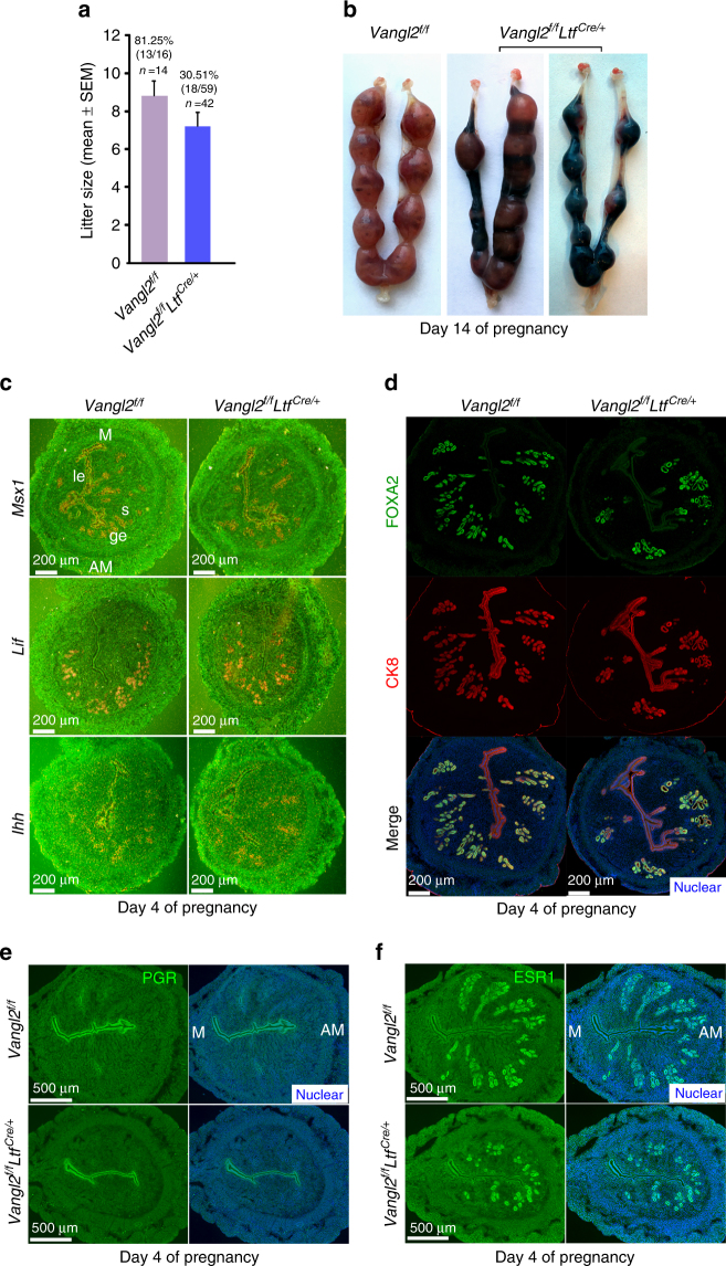
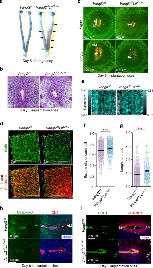
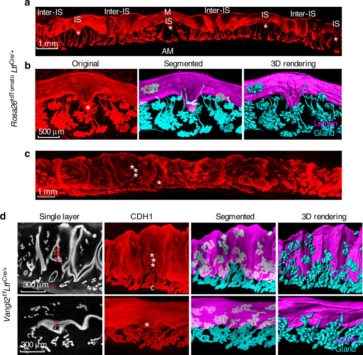
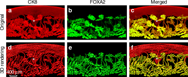
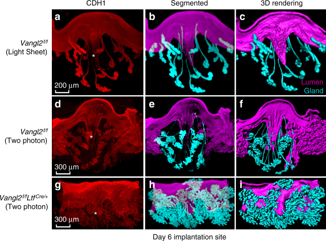
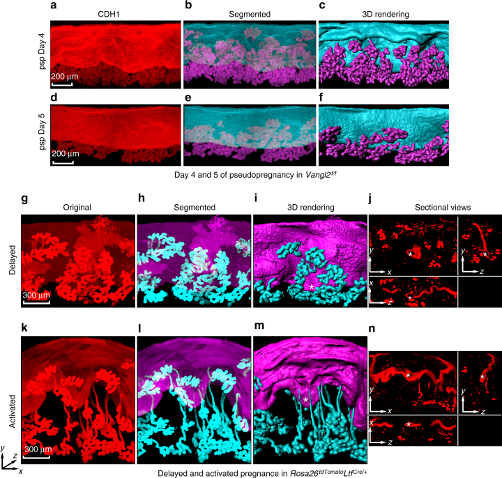
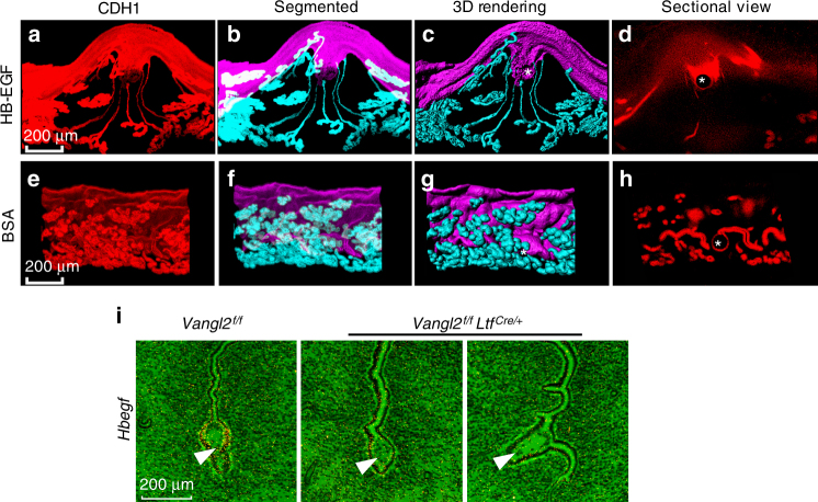
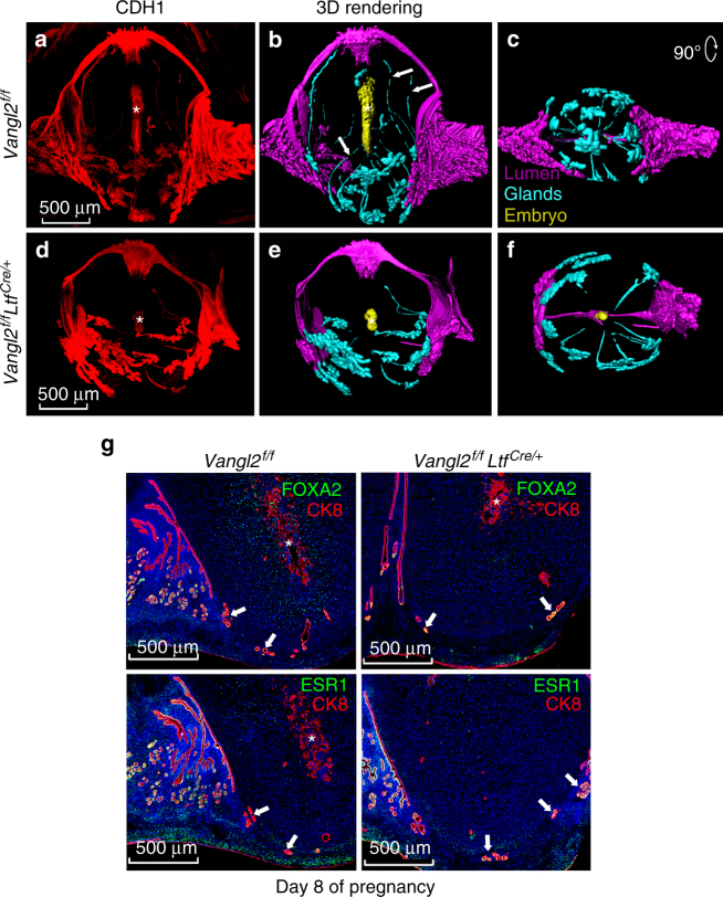
Embryo implantation is central to pregnancy success. Our previous understanding is limited by studying this phenomenon primarily in two dimensions. Here we employ 3D visualization, revealing that epithelial evaginations that form implantation chambers (crypts) consistently arise with preexisting glands, suggesting direct access of glands to embryos within the chamber. While the lobular domains of the glands become more developed, the ductal regions continue to elongate and progressively stretch following implantation. Using diapausing mice and mice with deletion of the planar cell polarity gene Vangl2 in uterine epithelial cells, we show that dynamic changes in gland topography depend on implantation-competent blastocysts and planar cell polarity. By transferring blastocyst-size beads preloaded with HB-EGF in pseudopregnant mice, we found that HB-EGF is a trigger for the communication between embryos and glands. Glands directly connecting the crypt encasing the embryo during implantation are therefore fundamental to pregnancy success.


Conflict of interest statement

The authors declare no competing financial interests.

Figures

Fig. 1
Fig. 1
Vangl2 deletion in the uterine epithelium results in severely compromised pregnancy outcome. a Percent pregnancy rate and litter size in Vangl2f/f and Vangl2f/fLtfCre/+ females. n number of females examined. The number within brackets indicate females with pups over total number of plug-positive females (Mean ± s.e.m. is derived from the indicated number of samples and analyzed by Student’s t-test). b Day 14 pregnant uteri in Vangl2f/f and Vangl2f/fLtfCre/+ females. c In situ hybridization of Msx1, Lif, and Ihh in day 4 pregnant uteri of Vangl2f/f and Vangl2f/fLtfCre/+ mice. ge gland epithelium, le luminal epithelium, s stroma, M mesometrial pole, AM antimesometrial pole. Scale bar: 200 μm. d IF of FOXA2 and CK8 in day 4 uteri of Vangl2f/f and Vangl2f/fLtfCre/+ mice. Scale bar: 200 μm. e, f IF of PGR and ESR1 in day 4 uteri of Vangl2f/f and Vangl2f/fLtfCre/+ mice. Scale bar: 500 μm. All images are representative of three independent experiments
Fig. 2
Fig. 2
Vangl2 deletion in the uterine epithelium contribute to aberrant implantation and decidualization. a Day 5 implantation sites (blue bands) in Vangl2f/f and Vangl2f/fLtfCre/+ females and arrows indicate weak blue bands. b Histology of day 5 implantation sites in Vangl2f/f and Vangl2f/fLtfCre/+ mice. * embryo. Scale bar: 200 μm. c In situ hybridization of Ptgs2 and Bmp2 in Vangl2f/f and Vangl2f/fLtfCre/+ implantation sites on day 5. Scale bar: 200 μm. Arrowheads indicate the location of embryos. d 3D reconstruction of Scrib and Phalloidin localization at the apical surface of the epithelium by IF in Vangl2f/f and Vangl2f/fLtfCre/+ uterine luminal epithelium (LE) on day 4. Scale bar: 10 μm. e Cell shape analysis of uterine epithelial cells using SEGGA software. f, g Quantification of cell eccentricity and the long-to-short axis ratio in Vangl2f/f and Vangl2f/fLtfCre epithelial cells (Mean ± s.e.m. is derived from the indicated number of samples and analyzed by Student’s t-test, ***p < 0.001). h IF of Caspase3 and CK8 in day 6 implantation sites of Vangl2f/f and Vangl2f/fLtfCre/+ mice. Scale bar: 200 μm. i IF of CDH1 and CTNNB1 in day 6 implantation sites of Vangl2f/f and Vangl2f/fLtfCre/+ mice. Scale bar: 200 μm. All images are representative of three independent experiments
Fig. 3
Fig. 3
3D images of day 5 implantation sites. a Images of one uterine horn in Rosa26tdTomatoLtfCre/+ mice on day 5. IS Implantation site, inter-IS inter-implantation site. Images were generated by Nikon A1R Multiphoton Microscope with Plan Apo 4× objective with 50 µm Z-stack. Scale bar: 1 mm. b Original images of tdTomato reporter, segmented glands and uterine lumen and 3D rendering of day 5 implantation site in Rosa26tdTomatoLtfCre/+ mice. Images were generated by a Nikon A1R Multiphoton Microscope with LWD 16× water objective with 3 µm Z-stack. Scale bar: 500 μm. c Image of one uterine horn in Vangl2f/fLtfCre/+ mice on day 5 immunostained with E-cad (CDH1). Images were generated by Nikon A1R Multiphoton Microscope; Plan Apo 4× objective with 50 µm Z-stack. Scale bar: 500 μm. d Single layer, whole-mount staining, segmented and 3D rendering of images in day 5 implantation site in two independent Vangl2f/fLtfCre/+ mice immunostained with E-cad (CDH1). Images were generated by Nikon A1R Multiphoton Microscope; LWD 16× water objective with 3 µm Z-stack. * embryos, C cyst. Scale bar: 500 μm. All images are representative of three independent experiments
Fig. 4
Fig. 4
3D visualization of day 5 implantation sites in Vangl2f/f mice stained with CK8 amd FOXA2. af Original double immunostaining with CK8 and FOXA2 and 3D rendering pictures. CK8 markes both luminal epithelial and glandular epithelial cells, while FOXA2 only marks glands. Representative images of three independent experiments, images were generated by Nikon A1R Multiphoton Microscope; LWD 16× water objective with 3 μm Z-stack. * location of embryos. Scale bar: 400 μm
Fig. 5
Fig. 5
3D visualization of day 6 implantation sites in Vangl2f/f and Vangl2f/fLtfCre/+ mice. ac 3D images of CDH1, segmented and 3D rendered images of Vangl2f/f day 6 implantation sites generated by a light sheet microscopy. Scale bar: 200 μm. di 3D immunostaining with CDH1, segmented and 3D rendered images, respectively, in Vangl2f/f and Vangl2f/fLtfCre/+ mice generated by two-photon microscopy. Images are generated by Nikon A1R Multiphoton Microscope with a LWD 16× water objective with 3 µm Z-stack. Note: aberrant crypt size and evagination in Vangl2f/fLtfCre/+ mice with derailed extension and display of glands. * location of embryos. Scale bar: 300 μm. All images are representative of three independent experiments
Fig. 6
Fig. 6
Implantation-competent blastocysts are required for crypt formation. af IF of E-Cad (CDH1), segmented and 3D rendering, respectively, in pseudopregnant days 4 and 5 uteri. psp pseudopregnancy. Scale bar: 200 μm. gn, Original images of tdTomato reporter, segmented glands, and uterine lumen, 3D rendering and sectional view of implantation sites at 24 h after terminating delayed implantation by an injection of estrogen in P4-primed Rosa26tdTomatoLtfCre/+ mice. Asterisks indicate locations of embryos in delayed and activated uterus. Scale bar: 300 μm. Sectional views (j, n) are represented images (g, k). Images are generated by Nikon A1R Multiphoton Microscope with a LWD 16× water objective with 3 µm Z-stack. All images are representative of three independent experiments
Fig. 7
Fig. 7
3D analysis of day 6 crypt induced by transfer of Affi-gel blue beads presoaked in HB-EGF or BSA. ad IF of E-Cad (CDH1), segmented glands, and uterine lumen, 3D rendering and sectional view of day 6 crypts at the site of beads carrying HB-EGF transferred into uteri of Vangl2f/f mice. eh Original image, segmented glands, and uterine lumen, 3D rendering and sectional view of day 6 at the site of beads carrying BSA uteri of Vangl2f/f mice. Asterisks indicate the location of beads. Scale bar: 200 μm. Images are generated by Nikon A1R Multiphoton Microscope with a LWD 16× water objective with 3 µm Z-stack. i In situ hybridization of Hbegf in Vangl2f/f and Vangl2f/fLtfCre/+ implantation sites on day 5. Arrowhead indicates embryo. Note: absence of Hbegf expression in the crypt epithelium. Scale bar: 200 μm. All images are representative of three independent experiments
Fig. 8
Fig. 8
3D analysis of day 8 implantation sites in Vangl2f/f and Vangl2f/fLtfCre/+ mice. a, b, d, and e Images of CDH1 immunostained and 3D rendered images of day 8 implantation sites in Vangl2f/f and Vangl2f/fLtfCre/+ mice, respectively, generated by two-photon microscopy. Arrows show the sheared ducts from the gland lobules. c, f Images correspond to a clockwise rotation of 90° from the bottom view of Vangl2f/f and Vangl2f/fLtfCre/+ implantation site. Images were generated by a Nikon A1R Multiphoton Microscope with Plan Apo λ 10× objective with 12 µm Z-stack. g IF of FOXA2 with CK8 and IF of ESR1 with CK8 on day 8 implantation sites in Vangl2f/f and Vangl2f/fLtfCre/+ mice. Arrows indicate glands at the implantation site. * location of embryos. Scale bar: 500 μm. All images are representative of three independent experiments

References

    1. Cha J, Sun X, Dey SK. Mechanisms of implantation: strategies for successful pregnancy. Nat. Med. 2012;18:1754–1767. doi: 10.1038/nm.3012. - DOI - PMC - PubMed
    1. Wang H, Dey SK. Roadmap to embryo implantation: clues from mouse models. Nat. Rev. Genet. 2006;7:185–199. doi: 10.1038/nrg1808. - DOI - PubMed
    1. Daikoku T, et al. Conditional deletion of Msx homeobox genes in the uterus inhibits blastocyst implantation by altering uterine receptivity. Dev. Cell. 2011;21:1014–1025. doi: 10.1016/j.devcel.2011.09.010. - DOI - PMC - PubMed
    1. Song H, et al. Cytosolic phospholipase A2alpha is crucial [correction of A2alpha deficiency is crucial] for ‘on-time’ embryo implantation that directs subsequent development. Development. 2002;129:2879–2889. - PubMed
    1. Hirota Y, et al. Uterine-specific p53 deficiency confers premature uterine senescence and promotes preterm birth in mice. J. Clin. Invest. 2010;120:803–815. doi: 10.1172/JCI40051. - DOI - PMC - PubMed

Publication types

MeSH terms

Substances

LinkOut - more resources