. 2018 Feb 9;9(1):612.
doi: 10.1038/s41467-018-03072-8.

Ultraconserved element uc.372 drives hepatic lipid accumulation by suppressing miR-195/miR4668 maturation

Affiliations

Ultraconserved element uc.372 drives hepatic lipid accumulation by suppressing miR-195/miR4668 maturation

Jun Guo et al. Nat Commun. .

Abstract

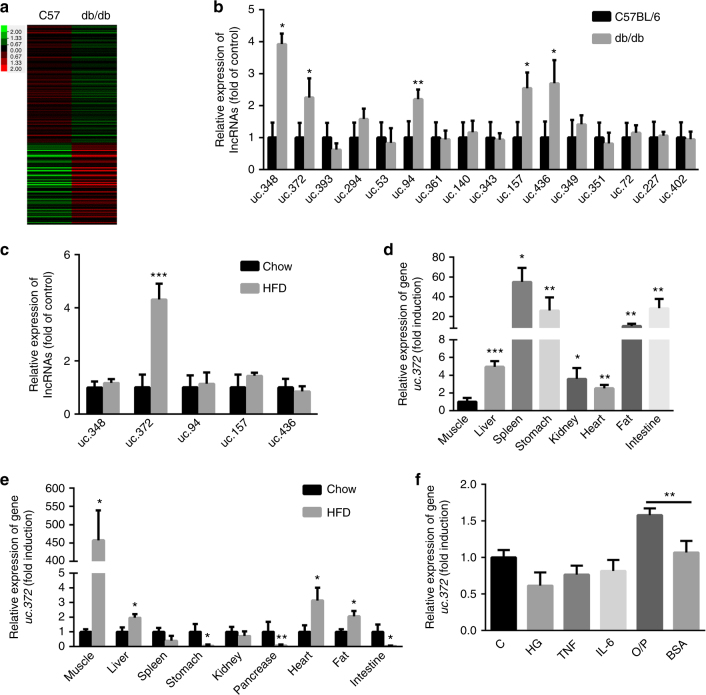
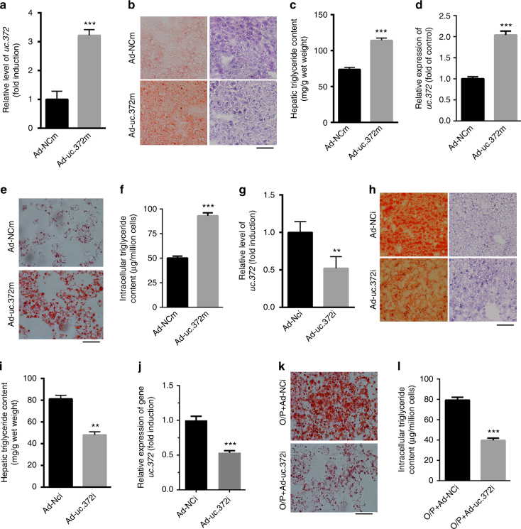
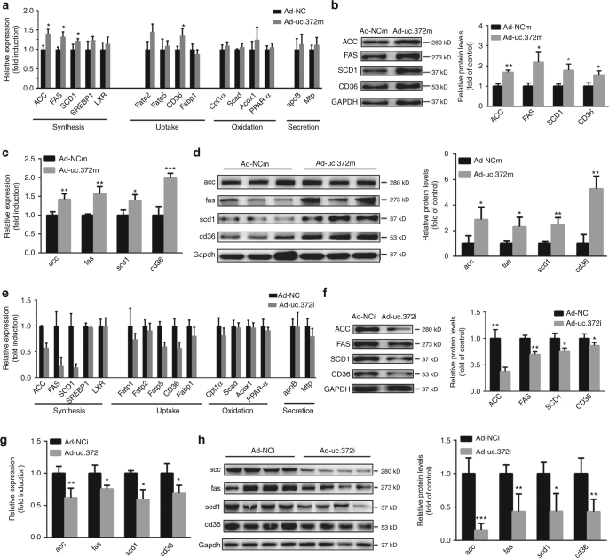
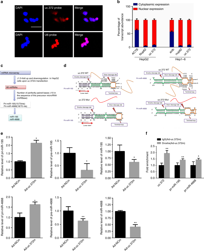
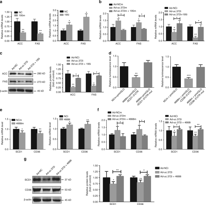
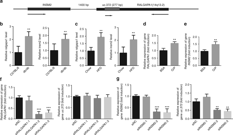
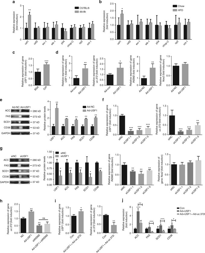
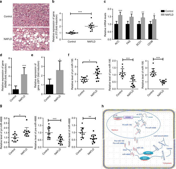
Ultraconserved (uc) RNAs, a class of long non-coding RNAs (lncRNAs), are conserved across humans, mice, and rats, but the physiological significance and pathological role of ucRNAs is largely unknown. Here we show that uc.372 is upregulated in the livers of db/db mice, HFD-fed mice, and NAFLD patients. Gain-of-function and loss-of-function studies indicate that uc.372 drives hepatic lipid accumulation in mice by promoting lipogenesis. We further demonstrate that uc.372 binds to pri-miR-195/pri-miR-4668 and suppresses maturation of miR-195/miR-4668 to regulate expression of genes related to lipid synthesis and uptake, including ACC, FAS, SCD1, and CD36. Finally, we identify that uc.372 is located downstream of the insulinoma-associated 2 (INSM2) gene that is transcriptionally activated by upstream transcription factor 1 (USF1). Our findings reveal a novel mechanism by which uc.372 drives hepatic steatosis through inhibition of miR-195/miR-4668 maturation to relieve miR-195/miR-4668-mediated suppression of functional target gene expression.


Conflict of interest statement

The authors declare no competing financial interests.

Figures

Fig. 1
Fig. 1
Identification of uc.372 upregulated in the livers of db/db mice and HFD-fed mice. a LncRNA-wide expression profiling in the liver of 8-week-old male db/db mice (n = 5). b Expression levels of ucRNAs in the liver of 8-week-old male db/db mice analyzed by real-time PCR (n = 5). c Expression levels of ucRNAs in the liver of 10-week-HFD-fed mice analyzed by real-time PCR (n = 5). d Distribution of uc.372 in various tissues of 6- to 8-week-old C57BL/6J mice (n = 3). e Distribution of uc.372 in various tissues of 10-week-HFD-fed mice (n = 5). f Expression level of uc.372 in HepG2 cells treated with 33.3 mM high glucose (HG), 20 nM tumor necrosis factor (TNF), 20 nM IL-6, or 300 μM O/P mixture (n = 3). Data are mean ± SEM; *P < 0.05; **P < 0.01; ***P < 0.001 vs. control group. (b, c, e Student’s t-test; d, f analysis of variance (ANOVA))
Fig. 2
Fig. 2
Uc.372 drives hepatic lipid accumulation in mice. a Hepatic expression level of uc.372 in 6- to 8-week-old C57BL/6J mice administrated with Ad-uc.372m for 7 days by tail vein injection, as analyzed by real-time PCR (n = 5). b Representative image from three similar experiments of H&E and Oil Red O staining in liver frozen sections of 6- to 8-week-old C57BL/6J mice after tail vein injection with Ad-uc.372 or Ad-NC for 7 days were shown. Scale bar, 400 μm. c Hepatic triglyceride content in 6- to 8-week-old C57BL/6J mice after tail vein injection with Ad-uc.372 or Ad-NC for 7 days (n = 5). d Expression level of uc.372 in HepG2 cells transfected with Ad-uc.372m or Ad-NC for 48 h (n = 3). e Representative image from three similar experiments of Oil Red O staining in HepG2 cells transfected with Ad-uc.372m or Ad-NC for 48 h. Scale bar, 40 μm. f Intracellular triglyceride content in HepG2 cells transfected with Ad-uc.372m or Ad-NC for 48 h (n = 3). g Hepatic expression level of uc.372 in 10-week-HFD-fed mice administrated with Ad-uc.372i or Ad-NC for 7 days by tail vein injection (n = 5). h Representative image from three similar experiments of H&E and Oil Red O staining in liver frozen sections of 10-week-HFD-fed mice after tail vein injection of Ad-uc.372i or Ad-NC for 7 days. Scale bar, 50 μm. i Hepatic triglyceride content in 10-week-HFD-fed mice after tail vein injection of Ad-uc.372i or Ad-NC for 7 days (n = 5). j Expression level of uc.372 in HepG2 cells transfected with Ad-uc.372i or Ad-NC for 48 h (n = 3). k Representative image from three similar experiments of Oil Red O staining in HepG2 cells transfected with Ad-uc.372i or Ad-NC for 48 h. Scale bar, 40 μm. l Intracellular triglyceride content in HepG2 cells transfected with Ad-uc.372i or Ad-NC for 48 h (n = 3). Data are mean ± SEM; **P < 0.01; ***P < 0.001 vs. control group (a, c, g, i, j, l Student’s t-test)
Fig. 3
Fig. 3
Uc.372 promotes the expression of genes related to lipid synthesis and uptake in hepatocytes. a Expression levels of genes related to lipid synthesis (ACC, FAS, SCD, SREBP1, and LXR), uptake (Fatp1, Fatp2, Fatp5, Fabp1, and CD36), oxidation (Cpt1a, Scad, Acox1, and PPAR-a), and secretion (apoB and Mtp) in HepG2 cells transfected with Ad-uc.372m or Ad-NCm for 48 h (n = 3). b Protein levels of ACC, FAS, SCD1, and CD36 in HepG2 cells transfected with Ad-uc.372m or Ad-NCm for 48 h, as analyzed by western blot (representative blots from three similar experiments) (n = 3). c Expression levels of acc, fas, scd1, and cd36 in the liver of 6- to 8-week-old C57BL/6J mice injected with Ad-uc.372m or Ad-NCm for 7 days (n = 3). d Protein levels of acc, fas, scd1, and cd36 in the liver of 6- to 8-week-old C57BL/6J mice injected with Ad-uc.372m or Ad-NCm for 7 days (n = 3). e Expression levels of genes related to lipid synthesis, uptake, oxidation, and secretion in HepG2 cells transfected with Ad-uc.372i or Ad-NCi for 48 h (n = 3). f Protein levels of ACC, FAS, SCD1, and CD36 in HepG2 cells transfected with Ad-uc.372i or Ad-NCi for 48 h in the presence of an O/P mixture (representative blots from three similar experiments) (n = 3). g Expression levels of acc, fas, scd1, and cd36 in the liver of 10-week-HFD-fed mice after tail vein injection of Ad-uc.372i or Ad-NC for 7 days (n = 3). h Protein levels of acc, fas, scd1, and cd36 in the liver of 10-week-HFD-fed mice after tail vein injection of Ad-uc.372i or Ad-NC for 7 days (n = 3). Data are mean ± SEM; *P < 0.05; **P < 0.01; ***P < 0.001 vs. control group (Student’s t-test)
Fig. 4
Fig. 4
uc.372 binds to pri-miR-195/pri-miR-4668. a Representative image from three similar experiments showed the distribution of uc.372 in HepG2 cells as analyzed by in situ hybridization. Scale bar, 25 μm. b Cellular fractionation assay in HepG2 and Hep1-6 cells was performed by quantitative real-time PCR using a specific cytosol control (gene ACTB) and a specific nuclear control (gene Nup62) (n = 3). c Schematic flowchart depicting the strategy. d The stem-loop sequence of pri-miR-195 (left panel) and pri-miR-4668 (right panel), and their partial complementarity with uc.372. e The levels of pri-miR-195/pri-miR-4668, pre-miR-195/pre-miR-4668, and mature miR-195/miR-4668 in the HepG2 cells transfected with Ad-uc.372m or Ad-NCm for 48 h (n = 3). f RIP assay showed that uc.372 could bind pri-miR-195/pri-miR-4668 in HepG2 cells transfected with Ad-uc.372m or Ad-NCm for 48 h (n = 3). Data are mean ± SEM; *P < 0.05; **P < 0.01 vs. control group (Student’s t-test)
Fig. 5
Fig. 5
uc.372 inhibits the maturation of miR-195/miR-4668 to regulate expression of ACC, FAS, SCD1, and CD36. a Expression levels of ACC and FAS in the HepG2 cells transfected with miR-195 mimic and inhibitor at a final concentration of 20 nM for 48 h (n = 3). b Expression levels of ACC and FAS in the HepG2 cells transfected with miR-195 mimic and inhibitor for 24 h in the presence with Ad-uc.372m and Ad-uc.372i (n = 3). c Protein levels of ACC and FAS in Ad-uc.372i-infected and miR-195 inhibitor-transfected HepG2 cells in the presence with with 300 μM O/P mixture for 48 h (representative blots from three similar experiments) (n = 3). d The relative luciferase units (RLU) in the HepG2 cells transfected with pmirGLO-SCD1-3′UTR and pmirGLO-SCD1-3′UTR mutant or pmirGLO-CD36-3′UTR and pmirGLO-CD36-3′UTR mutant under overexpression of miR-4668 or NC for 48 h (n = 3). e Expression levels of SCD1 and CD36 in the HepG2 cells transfected with miR-4668 mimic and inhibitor or NC for 48 h (n = 3). f Expression levels of SCD1 and CD36 in the HepG2 cells transfected with miR-4668 mimic and inhibitor for 24 h in the presence with Ad-uc.372m and Ad-uc.372i (n = 3). g Protein levels of ACC and FAS in Ad-uc.372i-infected and miR-4668 inhibitor-transfected HepG2 cells in the presence with 300 μM O/P mixture for 48 h (representative blots from three similar experiments) (n = 3). Data are mean ± SEM; *P < 0.05; **P < 0.01; ***P < 0.001 vs. control group (a, e Student’s t-test; b, c, d, f, g analysis of variance (ANOVA))
Fig. 6
Fig. 6
uc.372 expression is dependent on transcription of INSM2. a Schematic analysis of uc.372 location. b Ralgapa1 and insm2 expression in the liver of 8-week-old male db/db mice (n = 5). c Ralgapa1 and insm2 expression in the liver of 10-week-HFD-fed mice (n = 5). d The mRNA level of RALGAPA1 in the HepG2 cells treated with 300 μM O/P mixture for 48 h (n = 3). e The mRNA level of INSM2 in the HepG2 cells treated with 300 μM O/P mixture for 48 h (n = 3). f The levels of RALGAPA1 and uc.372 after silencing RALGAPA1 in HepG2 cells for 48 h (n = 3). g The levels of INSM2 and uc.372 after silencing INSM2 in HepG2 cells for 48 h (n = 3). Data are mean ± SEM; **P < 0.01; ***P < 0.001 vs. control group (b, c, d, e Student’s t-test; f, g analysis of variance (ANOVA))
Fig. 7
Fig. 7
Upstream transcription factor 1 (USF1) transcriptionally regulates expression of INSM2 and uc.372. a Identification of usf1 elevated in the liver of 8-week-old male db/db mice (n = 5). b Identification of usf1 elevated in the liver of 10-week-HFD-fed mice (n = 5). c Expression of USF1 in the HepG2 cells treated with 300 μM O/P mixture for 48 h (n = 3). d Expression levels of USF1, uc.372, INSM2, and Nox2 in the HepG2 cells infected with Ad-USF1 or Ad-NC for 48 h (n = 3). e Protein levels of ACC, FAS, SCD1, and CD36 in the HepG2 cells infected with Ad-USF1 or Ad-NC for 48 h (representative blots from three similar experiments) (n = 3). f Real-time PCR analysis of USF1, uc.372, INSM2, and Nox2 in the HepG2 cells transfected with specific siRNAs targeting USF1 or NC for 48 h (n = 3). g Western blot analysis of ACC, FAS, SCD1, and CD36 expression in the HepG2 cells transfected with specific siRNAs targeting USF1 (representative blots from three similar experiments) (n = 3). h The mRNA level of uc.372 in the HepG2 cells infected with Ad-USF1 in the presence or absence of siINSM2 or NC for 48 h (n = 3). i The mRNA levels of USF1 and uc.372 in the HepG2 cells infected with Ad-USF1 and Ad-uc.372i for 48 h (n = 3). j The mRNA levels of ACC, FAS, SCD1, and CD36 in the HepG2 cells infected with Ad-USF1 and Ad-uc.372i or Ad-NC for 48 h (n = 3). Data are mean ± SEM; *P < 0.05; **P < 0.01; ***P < 0.001 vs. control group. (a, b, c, d, e, g, i Student’s t-test; f, h, j analysis of variance (ANOVA))
Fig. 8
Fig. 8
Effect of uc.372 on abnormal lipid accumulation is subjected to regulation of miR-195/ miR-4668 in the liver of NAFLD patients. a Oil Red O staining of liver frozen sections from NAFLD patients or healthy control (representative image from three similar experiments). Scale bar, 100 μm. b The level of uc.372 in the liver samples of NAFLD patients or healthy control (n = 11). c The mRNA levels of ACC, FAS, SCD1, and CD36 in the liver samples of NAFLD patients or healthy control (n = 3). d The mRNA level of INSM2 in the liver samples of NAFLD patients or healthy control (n = 11). e The mRNA level of USF1 in the liver samples of NAFLD patients or healthy control (n = 11). f The levels of pri-miR-195, pre-miR-195, and mature miR-195 in the liver samples of NAFLD patients or healthy control (n = 11). g The levels of pri-miR-4668, pre-miR-4668, and mature miR-4668 in the liver samples of NAFLD patients or healthy control (n = 11). h Schematic depicting our proposed model that us.372 drives hepatic lipid accumulation. Data are mean ± SEM; Student’s t-test: *P < 0.05; **P < 0.01; ***P < 0.001 vs. control group

References

    1. Haga Y, et al. Nonalcoholic fatty liver disease and hepatic cirrhosis: comparison with viral hepatitis-associated steatosis. World J. Gastroenterol. 2015;21:12989–12995. doi: 10.3748/wjg.v21.i46.12989. - DOI - PMC - PubMed
    1. Farrell GC, Larter CZ. Nonalcoholic fatty liver disease: from steatosis to cirrhosis. Hepatology. 2006;43:S99–S112. doi: 10.1002/hep.20973. - DOI - PubMed
    1. Kwon H, et al. Inhibition of hedgehog signaling ameliorates hepatic inflammation in mice with nonalcoholic fatty liver disease (nafld) Hepatology. 2015;63:1155–1169. doi: 10.1002/hep.28289. - DOI - PMC - PubMed
    1. Goffredo M, et al. Role of tm6sf2 rs58542926 in the pathogenesis of nonalcoholic pediatric fatty liver disease: a multiethnic study. Hepatology. 2016;63:117–125. doi: 10.1002/hep.28283. - DOI - PMC - PubMed
    1. Klair JS, et al. A longer duration of estrogen deficiency increases fibrosis risk among postmenopausal women with nonalcoholic fatty liver disease. Hepatology. 2016;64:85–91. doi: 10.1002/hep.28514. - DOI - PMC - PubMed

Publication types

MeSH terms