. 2018 Feb 9;8(1):2701.
doi: 10.1038/s41598-018-21063-z.

Cerebral ischemia induces the aggregation of proteins linked to neurodegenerative diseases

Affiliations

Cerebral ischemia induces the aggregation of proteins linked to neurodegenerative diseases

Anja Kahl et al. Sci Rep. .

Erratum in

Abstract

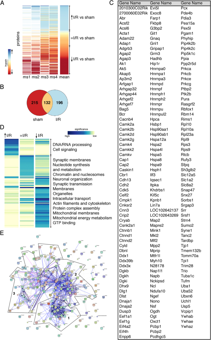
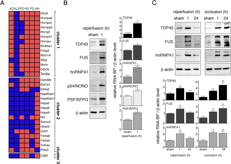
Protein aggregation critically affects cell viability in neurodegenerative diseases, but whether this also occurs in ischemic brain injury remains elusive. Prior studies report the post-ischemic aggregation of ubiquitin, small ubiquitin-related modifier (SUMO) and ribosomes, however whether other proteins are also affected is unknown. Here we employed a proteomic approach to identify the insoluble, aggregated proteome after cerebral ischemia. Mice underwent transient middle cerebral artery occlusion or sham-surgery. After 1-hour reperfusion, prior to apparent brain injury, mice were sacrificed and detergent-insoluble proteins were obtained and identified by nanoLC-MS/MS. Naturally existing insoluble proteins were determined in sham controls and aggregated proteins after cerebral ischemia/reperfusion were identified. Selected aggregated proteins found by proteomics were biochemically verified and aggregation propensities were studied during ischemia with or without reperfusion. We found that ischemia/reperfusion induces the aggregation of RNA-binding and heat-shock proteins, ubiquitin, SUMO and other proteins involved in cell signalling. RNA-binding proteins constitute the largest group of aggregating proteins in ischemia. These include TDP43, FUS, hnRNPA1, PSF/SFPQ and p54/NONO, all of which have been linked to neurodegeneration associated with amyotrophic lateral sclerosis and frontotemporal dementia. The aggregation of neurodegeneration-related disease proteins in cerebral ischemia unveils a previously unappreciated molecular overlap between neurodegenerative diseases and ischemic stroke.


Conflict of interest statement

The authors declare no competing interests.

Figures

Figure 1
Figure 1
Ischemic stroke leads to the aggregation of 196 highly interconnected proteins that are mainly associated with RNA processing, stress response and cell signaling. (A) Ipsilateral neocortical Triton-insoluble proteins were identified after sham-surgery or MCAO and 1-hour reperfusion (I/R). The heat map depicts relative levels of Triton-insoluble proteins found in I/R compared to sham-controls. Each horizontal line represents a single protein, the vertical strips depict four independent mass spectrometry runs (ms1-4) and the mean value of the 4 runs. Trypsin levels served as internal standard for the calculation of fold changes of protein levels in I/R versus sham conditions. Proteins were clustered into three groups dependent on their solubility before and after I/R (increased, ↑, blue shaded), (unchanged, = , yellow/light orange shaded), (reduced, ↓, dark orange/red shaded). Upward and downward pointing arrows indicate an increase or decrease in relative I/R versus sham insoluble protein levels, respectively. ms, mass-spectrometry. (B) A Venn diagram showing the distribution of Triton-insoluble proteins in sham conditions and after I/R is shown. (C) An alphabetical list of the 196 aggregated proteins (↑) is provided. (D) Proteins from each induction group identified in Fig. 1A were examined for GO term enrichment through the DAVID database. Similar enrichment terms were clustered and the natural logarithm of corrected P values was plotted. Only terms with an enrichment score of P < 10–5 in at least one group were considered for analysis. Blue and yellow shades represent high and low significance, respectively. (E) Aggregated proteins in I/R (↑) were analyzed for functional association networks via the STRING database. Strong association networks were compiled into three main clusters (clusters 1, 2, 3). A high-resolution image of Fig. 1E is included as Supplementary Figure 1.
Figure 2
Figure 2
Ischemic stroke induces the aggregation of the ALS- and FTD-related RNABPs TDP43, FUS, hnRNPA1, PSF/SFPQ and p54/NONO. (A) I/R-derived aggregated proteins were analyzed for their known ability to form aggregates in neurodegeneration. Identified proteins were clustered according to analysis in Fig. 1D. The heatmap points out disease affiliation of aggregate-prone proteins in ND, where blue boxes indicate positive and red boxes negative hits. AD, Alzheimer’s disease; ALS, amyotrophic lateral sclerosis; FD, frontotemporal dementia; HD, Huntington’s disease; oth, other; PD, Parkinson’s disease. (B) Triton-insoluble fractions were obtained from ipsilateral neocortical tissue from mice subjected to sham surgery or MCAO followed by 1-hour reperfusion. The presence of the RNABPs TDP43, FUS, hnRNPA1, p54/NONO and PSF/SFPQ in Triton-insoluble fractions was detected by Western Blotting with respective antibodies. Optical densities of RNABP bands were measured and normalized to β-actin. Changes in RNABPs were expressed relative to sham controls. *P < 0.05 from sham; n = 9/group. (C) Ipsilateral neocortices from sham animals, after MCAO followed by 1 and 24 hours reperfusion, or after 1 and 24 hours permanent MCAO were harvested. Triton-insoluble proteins were isolated and examined for the presence of TDP43, FUS and hnRNPA1 by Western Blotting. Quantification of results was carried out as in Fig. 2B. Full-size blots are presented in Supplementary Figures S2 and S3. *P < 0.05 from sham; n = 3–6/group. h, hours.

References

    1. Masters CL, et al. Amyloid plaque core protein in Alzheimer disease and Down syndrome. Proc Natl Acad Sci USA. 1985;82:4245–4249. doi: 10.1073/pnas.82.12.4245. - DOI - PMC - PubMed
    1. Mattson MP, et al. Beta-Amyloid peptides destabilize calcium homeostasis and render human cortical neurons vulnerable to excitotoxicity. J Neurosci. 1992;12:376–389. doi: 10.1523/JNEUROSCI.12-02-00376.1992. - DOI - PMC - PubMed
    1. Garcia-Reitbock P, et al. SNARE protein redistribution and synaptic failure in a transgenic mouse model of Parkinson’s disease. Brain. 2010;133:2032–2044. doi: 10.1093/brain/awq132. - DOI - PMC - PubMed
    1. Spillantini MG, et al. Alpha-synuclein in Lewy bodies. Nature. 1997;388:839–840. doi: 10.1038/42166. - DOI - PubMed
    1. Cui L, et al. Transcriptional repression of PGC-1alpha by mutant huntingtin leads to mitochondrial dysfunction and neurodegeneration. Cell. 2006;127:59–69. doi: 10.1016/j.cell.2006.09.015. - DOI - PubMed

Publication types

MeSH terms

Substances

LinkOut - more resources