

Programmed death ligand 1 expression and tumor infiltrating lymphocytes in neurofibromatosis type 1 and 2 associated tumors
- PMID: 29427150
- PMCID: PMC5930071
- DOI: 10.1007/s11060-018-2788-6
Programmed death ligand 1 expression and tumor infiltrating lymphocytes in neurofibromatosis type 1 and 2 associated tumors
Abstract
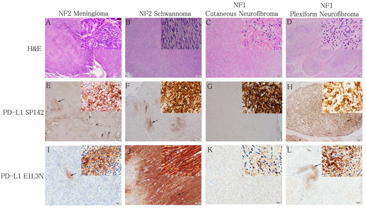
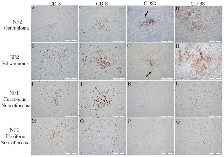
Immune checkpoint inhibitors targeting programmed cell death 1 (PD-1) or its ligand (PD-L1) have been shown to be effective in treating patients with a variety of cancers. Biomarker studies have found positive associations between clinical response rates and PD-L1 expression on tumor cells, as well as the presence of tumor infiltrating lymphocytes (TILs). It is currently unknown whether tumors associated with neurofibromatosis types 1 and 2 (NF1 and NF2) express PD-L1. We performed immunohistochemistry for PD-L1 (clones SP142 and E1L3N), CD3, CD20, CD8, and CD68 in NF1-related tumors (ten dermal and six plexiform neurofibromas) and NF2-related tumors (ten meningiomas and ten schwannomas) using archival formalin-fixed paraffin-embedded tissues. Expression of PD-L1 was considered positive in cases with > 5% membranous staining of tumor cells, in accordance with previously published biomarker studies. PD-L1 expression in tumor cells (using the SP142 and E1L3N clones, respectively) was assessed as positive in plexiform neurofibromas (6/6 and 5/6) dermal neurofibromas (8/10 and 6/10), schwannomas (7/10 and 10/10), and meningiomas (4/10 and 2/10). Sparse to moderate presence of CD68, CD3, or CD8 positive TILs was found in 36 (100%) of tumor specimens. Our findings indicate that adaptive resistance to cell-mediated immunity may play a major role in the tumor immune microenvironment of NF1 and NF2-associated tumors. Expression of PD-L1 on tumor cells and the presence of TILs suggest that these tumors might be responsive to immunotherapy with immune checkpoint inhibitors, which should be explored in clinical trials for NF patients.
Keywords: Meningioma; Neurofibroma; Neurofibromatosis type 1; Neurofibromatosis type 2; Programmed death-ligand 1; Schwannoma; Tumor infiltrating lymphocytes.
Conflict of interest statement
Figures
References
MeSH terms
Substances
Grants and funding
LinkOut - more resources
Full Text Sources
Other Literature Sources
Research Materials
Miscellaneous
