. 2018 Apr:152:26-35.
doi: 10.1016/j.antiviral.2018.02.003. Epub 2018 Feb 8.

The molecular tweezer CLR01 inhibits Ebola and Zika virus infection

Affiliations

The molecular tweezer CLR01 inhibits Ebola and Zika virus infection

Annika E Röcker et al. Antiviral Res. 2018 Apr.

Abstract

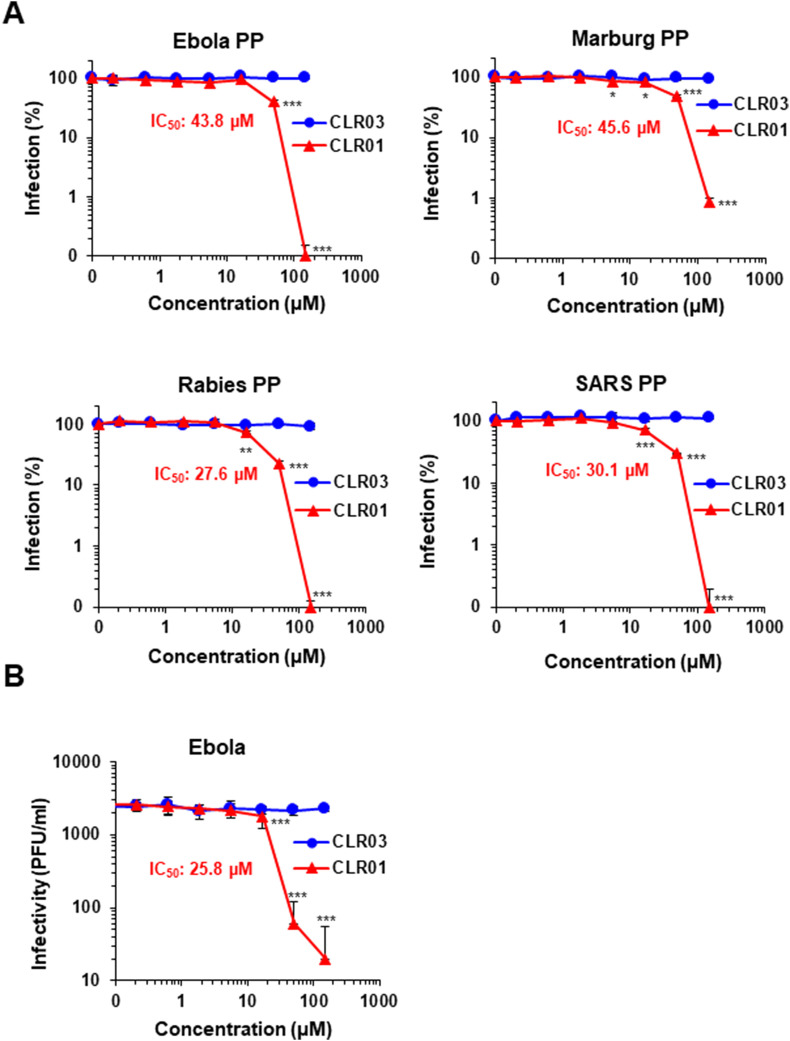
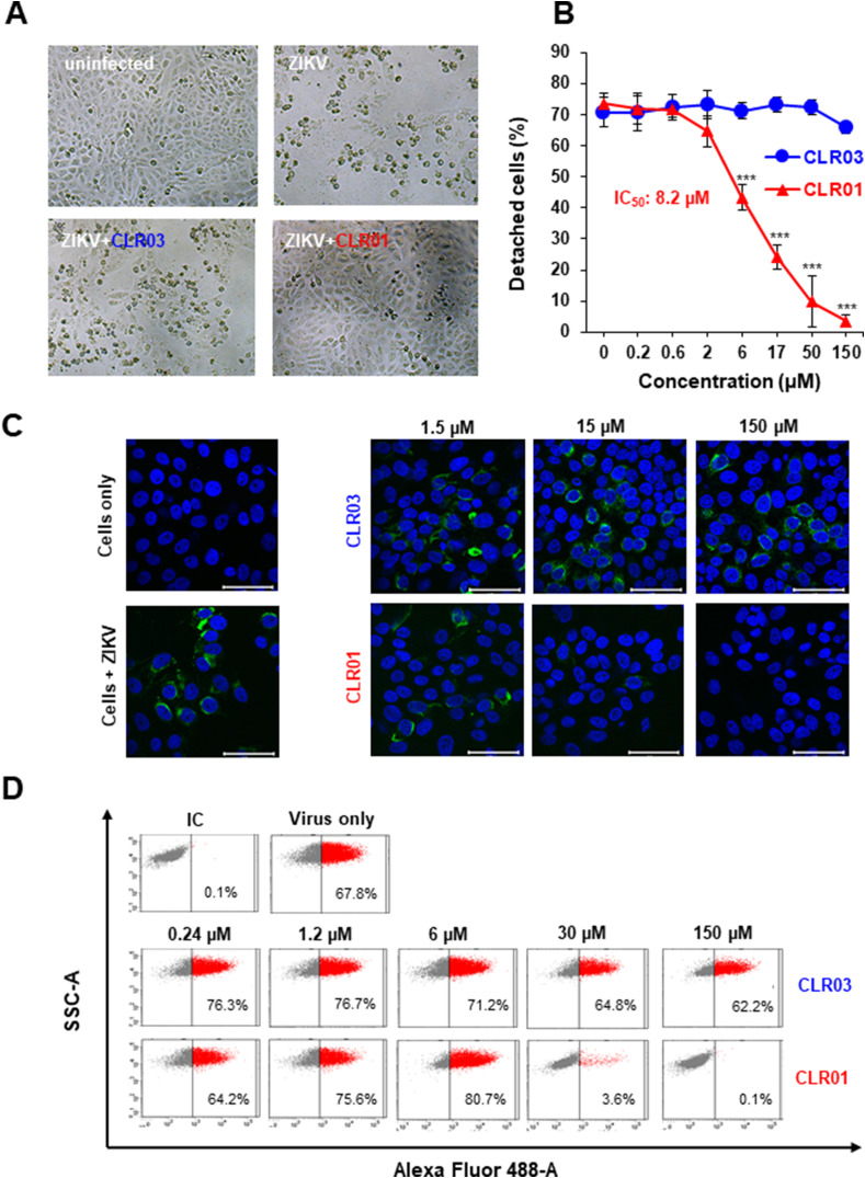
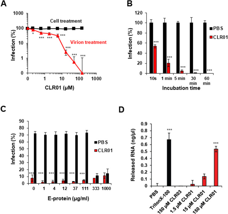
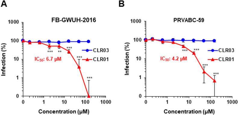
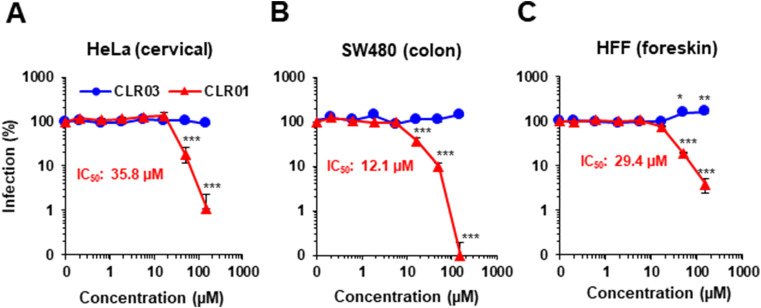
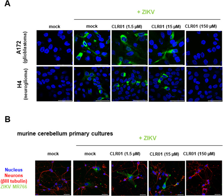
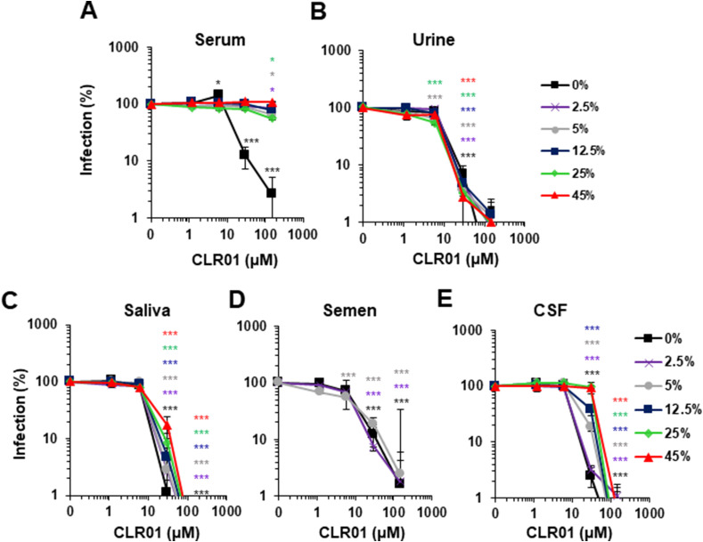
Ebola (EBOV) and Zika viruses (ZIKV) are responsible for recent global health threats. As no preventive vaccines or antiviral drugs against these two re-emerging pathogens are available, we evaluated whether the molecular tweezer CLR01 may inhibit EBOV and ZIKV infection. This small molecule has previously been shown to inactivate HIV-1 and herpes viruses through a selective interaction with lipid-raft-rich regions in the viral envelope, which results in membrane disruption and loss of infectivity. We found that CLR01 indeed blocked infection of EBOV and ZIKV in a dose-dependent manner. The tweezer inhibited infection of epidemic ZIKV strains in cells derived from the anogenital tract and the central nervous system, and remained antivirally active in the presence of semen, saliva, urine and cerebrospinal fluid. Our findings show that CLR01 is a broad-spectrum inhibitor of enveloped viruses with prospects as a preventative microbicide or antiviral agent.

Keywords: Broadly active antiviral agents; Ebola virus; Lipid rafts; Virus inactivation; Zika virus.


Figures

Fig. 1
Fig. 1
CLR01 inhibits lentiviral infection independently of the viral glycoprotein and prevents EBOV plaque formation. (A) Luciferase-encoding lentiviral particles pseudotyped with glycoproteins of viruses of the Filoviridae (EBOV, Marburg virus),Rhabdoviridae (Rabies virus) or the Coronaviridae family (SARS-CoV), were exposed to indicated concentrations of CLR01 or CLR03 and then used to infect Huh-7 cells. After three days, infection rates were determined by quantifying firefly luciferase activity and subtracting background activity derived from uninfected cells. Values represent % reporter gene activities ± SD derived from triplicate infections, normalized to values obtained for cells infected in the absence of tweezers. (B) Analysis of replication competent EBOV was performed using confluent Vero E6 cells in 24-well plates. rgEBOV-eGFP was preincubated with CLR01 or CLR03 and mixtures were added to the cells. After 7 days, samples were analyzed by counting the number of plaques per well and calculating the corresponding plaque forming units per milliliter (PFU/ml) for each inhibitor and dilution. Displayed values represent the mean of three independent experiments ± SD. IC50 values were calculated by GraphPad Prism. One-way ANOVA (non-parametric, grouped), followed by Bonferroni's multiple comparison tests were applied to compare the different CLR01/CLR03 concentrations to cells infected in the absence of compound (* denotes p < 0.01; ** denotes p < 0.001; *** denotes p < 0.0001).
Fig. 2
Fig. 2
CLR01 inhibits ZIKV infection of Vero E6 cells. (A) Light microscopy images of Vero E6 cells infected with ZIKV MR766 in the absence or presence of 150 μM CLR01 or CLR03. Images were taken 4 days post infection (dpi). (B) ZIKV MR766 was incubated with 0.2-150 μM CLR01 or CLR03 before these mixtures were used to infect Vero E6 cells. After 4 days, when significant cytopathic effects were visible, the number of adherent, viable cells were determined using the MTT assay (Müller et al., 2016). Values represent mean ± SD of percentages derived from triplicate infections. IC50 was determined using GraphPad Prism. One-way ANOVA (non-parametric, grouped), followed by Bonferroni's multiple comparison tests were applied to compare the different CLR01/CLR03 concentrations to cells infected in the absence of compound (*** denotes p < 0.0001) (C) Confocal microscopy images of Vero E6 cells infected with CLR01- or CLR03-treated ZIKV at day 3 post infection. Cells were stained for ZIKV E protein (green) and nuclei (Hoechst, blue) and imaged using confocal microscopy. Scale bar: 50 μm. (D) Flow cytometry of infected Vero E6 cells. Virus was pretreated with PBS, CLR01 or CLR03 and added to cells. 48 h later, cells were fixed, permeabilized, and stained with an anti E protein antibody, and quantified using an Alexa 488-coupled secondary antibody. Percentages indicate the fraction of protein E positive cells. IC, isotype control.
Fig. 3
Fig. 3
Mechanism of CLR01 inhibition of ZIKV infectivity. (A) For virus treatment, ZIKV was incubated with CLR01 for 10 min at room temperature before the mixtures were added to Vero E6 cells. For cell treatment, CLR01 was added directly to the cells; after 2 h, the medium was replaced and the cells were infected with ZIKV MR766. 2 dpi, cell-based ZIKV immunodetection assay was performed. Values represent mean ± SD (n = 3). One-way ANOVA (non-parametric, grouped), followed by Bonferroni's multiple comparison tests were applied to compare the different CLR01/CLR03 concentrations to cells infected in the absence of compound (* denotes p < 0.01; *** denotes p < 0.0001) (B) ZIKV was incubated for the indicated times with PBS or 10 μM CLR01 before the mixture was added to Vero E6 cells. 2 dpi, cell-based ZIKV immunodetection assay was performed. Values represent mean ± SD (n = 3). One-way ANOVA (non-parametric, grouped), followed by Bonferroni's multiple comparison tests were applied to compare cells infected with CLR01-treated ZIKV to cells infected with ZIKV that had been incubated with PBS for the same time period (*** denotes p < 0.0001). (C) Indicated concentrations of ZIKV E protein were titrated to 100 μM CLR01 or PBS before ZIKV was added. Mixtures were used to inoculate Vero E6 cells. 3 dpi, the number of adherent, viable cells were determined using the MTT assay (Müller et al., 2016). The values shown are mean ± SD from triplicate infections. One-way ANOVA (non-parametric, grouped), followed by Bonferroni's multiple comparison tests were applied to compare cells treated with different concentrations of ZIKV E protein and CLR01 to cells that had been treated with the same concentrations of E protein and PBS (*** denotes p < 0.0001). (D) ZIKV MR766 was incubated with PBS, 150 μM Triton X-100, 1.5-150 μM CLR01 or 150 μM CLR03 for 30 min at 37 °C. Samples were inactivated by UV light of a laminar flow for 60 min. Then, 10 μl of the samples were used to determine RNA concentrations using the QuantiFluor® RNA System and a Quantus Fluorometer (Promega). Background values obtained from control samples using cell supernatant of uninfected cells were subtracted from the respective signals. RNA levels of virus stock incubated with PBS were subtracted from these values. Data points represent mean ± SD (n = 3). One-way ANOVA (non-parametric, grouped), followed by Bonferroni's multiple comparison tests were applied to compare samples treated with different CLR01 concentrations, CLR03 or Triton X-100 to PBS-treated virus (*** denotes p < 0.0001).
Fig. 4
Fig. 4
CLR01 inactivates epidemic ZIKV isolates. (A) ZIKV FB-GWUH-2016 or (B) PRVABC-59 was incubated for 10 min at 37 °C with 0.2-150 μM CLR01 or CLR03. Thereafter, Vero E6 cells were infected and 2 days later, infection rates were determined via a cell-based ZIKV immunodetection assay. Data points represent mean ± SD (n = 3). IC50 values were determined with GraphPad Prism. One-way ANOVA (non-parametric, grouped), followed by Bonferroni's multiple comparison tests were applied to compare the different CLR01 concentrations to cells infected in the absence of compound (** denotes p < 0.001; *** denotes p < 0.0001).
Fig. 5
Fig. 5
CLR01 abrogates ZIKV infection of cells derived from the anogenital region. ZIKV was incubated for 10 min at 37 °C with 0.2-150 μM CLR01 or CLR03. Next, mixtures were used to inoculate HeLa (A), SW480 (B), or HFF (C) cells. 3 days later, infection rates were determined via quantification of the viral E protein using a cell-based ZIKV immunodetection assay. Data points represent mean ± SEM (n = 6). One-way ANOVA (non-parametric, grouped), followed by Bonferroni's multiple comparison tests were applied to compare the different CLR01 concentrations to cells infected in the absence of compound (* denotes p < 0.01; ** denotes p < 0.001; *** denotes p < 0.0001).
Fig. 6
Fig. 6
CLR01 inhibits infection of human and murine brain cells. (A) Human glioblastoma (A172) or neuroglioma (H4) cells were infected with ZIKV in the presence or absence of 1.5-150 μM CLR01. Two days later, cells were stained for ZIKV E protein (green) and nuclei (with Hoechst; blue) and imaged by confocal microscopy. Scale bar: 50 μm. (B) Primary murine cerebellum cultures were infected with ZIKV MR766 that had been incubated with 1.5–150 μM CLR01 for 10 min at 37 °C. 3 dpi, cultures were fixed, permeabilized and stained for the neuronal protein βIII tubulin (red), ZIKV E protein (green) and nuclei (with Hoechst; blue). Scale bar: 20 μm.
Fig. 7
Fig. 7
CLR01 loses antiviral activity in the presence of serum but is active in other body fluids. Indicated concentrations of CLR01 were incubated with ZIKV MR766 in the presence of increasing concentrations of human (A) serum, (B) urine, (C) saliva, (D) semen and (E) cerebrospinal fluid (CSF). After 10 min of incubation, the mixtures were used to infect Vero E6 cells. 2 days later, infection rates were determined via a cell-based ZIKV immunodetection assay. Data points represent mean ± SEM (n = 6), except for CSF and serum: mean ± SD (n = 3). One-way ANOVA (non-parametric, grouped), followed by Bonferroni's multiple comparison tests were applied to compare the different CLR01 concentrations to cells infected in the absence of CLR01 but in presence of the same respective body fluid concentration (* denotes p < 0.01; *** denotes p < 0.0001).

References

    1. Atkinson B., Hearn P., Afrough B., Lumley S., Carter D., Aarons E.J., Simpson A.J., Brooks T.J., Hewson R. Detection of Zika virus in semen. Emerg. Infect. Dis. 2016;22:940. - PMC - PubMed
    1. Attar A., Chan W.-T.C., Klärner F.-G., Schrader T., Bitan G. Safety and pharmacological characterization of the molecular tweezer CLR01–a broad-spectrum inhibitor of amyloid proteins' toxicity. BMC Pharmacol. Toxicol. 2014;15:1. - PMC - PubMed
    1. Attar A., Ripoli C., Riccardi E., Maiti P., Puma D.D.L., Liu T., Hayes J., Jones M.R., Lichti-Kaiser K., Yang F. Protection of primary neurons and mouse brain from Alzheimer's pathology by molecular tweezers. Brain. 2012;135:3735–3748. - PMC - PubMed
    1. Barrows N.J., Campos R.K., Powell S.T., Prasanth K.R., Schott-Lerner G., Soto-Acosta R., Galarza-Muñoz G., McGrath E.L., Urrabaz-Garza R., Gao J. A screen of FDA-approved drugs for inhibitors of Zika virus infection. Cell Host Microbe. 2016;20:259–270. - PMC - PubMed
    1. Bavari S., Bosio C.M., Wiegand E., Ruthel G., Will A.B., Geisbert T.W., Hevey M., Schmaljohn C., Schmaljohn A., Aman M.J. Lipid raft microdomains. J. Exp. Med. 2002;195:593–602. - PMC - PubMed

Publication types