. 2018 Apr 1;436(1):14-27.
doi: 10.1016/j.ydbio.2018.02.002. Epub 2018 Feb 9.

Notum attenuates Wnt/β-catenin signaling to promote tracheal cartilage patterning

Affiliations

Notum attenuates Wnt/β-catenin signaling to promote tracheal cartilage patterning

Bradley Gerhardt et al. Dev Biol. .

Abstract

Tracheobronchomalacia (TBM) is a common congenital disorder in which the cartilaginous rings of the trachea are weakened or missing. Despite the high prevalence and clinical issues associated with TBM, the etiology is largely unknown. Our previous studies demonstrated that Wntless (Wls) and its associated Wnt pathways are critical for patterning of the upper airways. Deletion of Wls in respiratory endoderm caused TBM and ectopic trachealis muscle. To understand mechanisms by which Wls mediates tracheal patterning, we performed RNA sequencing in prechondrogenic tracheal tissue of Wlsf/f;ShhCre/wt embryos. Chondrogenic Bmp4, and Sox9 were decreased, while expression of myogenic genes was increased. We identified Notum, a deacylase that inactivates Wnt ligands, as a target of Wls induced Wnt signaling. Notum's mesenchymal ventral expression in prechondrogenic trachea overlaps with expression of Axin2, a Wnt/β-catenin target and inhibitor. Notum is induced by Wnt/β-catenin in developing trachea. Deletion of Notum activated mesenchymal Wnt/β-catenin and caused tracheal mispatterning of trachealis muscle and cartilage as well as tracheal stenosis. Notum is required for tracheal morphogenesis, influencing mesenchymal condensations critical for patterning of tracheal cartilage and muscle. We propose that Notum influences mesenchymal cell differentiation by generating a barrier for Wnt ligands produced and secreted by airway epithelial cells to attenuate Wnt signaling.


Figures

Figure 1
Figure 1
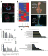
A) Epithelial deletion of Wls results in loss of Sox9 positive cells within the tracheal mesenchyme. Irregular cartilage and muscle development is shown. Sections of tracheal tissue depict the lack of ventral mesenchymal Sox9 staining within Wlsf/f;ShhCre/wt samples and increased extent of αSMA staining. B) RNA sequencing studies of Wlsf/f;ShhCre/wt trachea at E11.5 and E13.5 demonstrates differential gene regulation, Z-score normalized heat map of all differentially expressed genes is shown. C) Log2 transformed expression of select genes from E11.5 and E13.5 RNA seq analysis show differences in RNA levels. D) Representative, Toppfun functional enrichment hits. P-values were −Log2 transformed. Functional enrichment analysis was performed using all differentially expressed RNA from both E11.5 or E13.5 RNA-Seq. Specific characteristics resulting from changes in gene expression were predicted for each developmental stage.
Figure 2
Figure 2
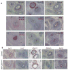
A) In-situ hybridization analysis of E13.5 transverse sections using RNA scope demonstrates differential gene regulation. Myh11 is expressed in muscle cells within the developing trachea, Wls is an epithelial marker of the developing trachea. Notum expression was complementary to Wnt7b, Wnt5a, and Myh11. B) Deletion of Wls from tracheal epithelium results in decreased Lgi3, Notum and Axin2 and increased Myh11 and Lrrc7 within the complete tracheal mesenchyme. Notum RNA was enriched in the ventro-lateral aspect of the trachea, where cartilage forms. E: Esophagus, T: Trachea.
Figure 3
Figure 3
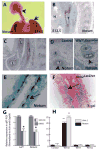
A) Notum is expressed in developing lung and trachea (arrowheads) as detected by digoxygenin labeled RNA probe. Specificity was confirmed by sense probe that yielded no staining. B and C) In-situ hybridization on E12.5 longitudinal sections of trachea (B) and lung (C) using RNAscope are shown. Notum RNA is enriched in the ventral side of the trachea and the subepithelial mesenchyme of the lung. Note expression in esophagus. D) Notum was decreased in tracheal tissue after deletion of Wls in respiratory tract epithelium as determined by in situ hybridization. E) Notum RNA is restricted to the periphery of mesenchymal condensations. F) Wnt/β-catenin activity is detected in periphery of mesenchymal condensations. G) qRT-PCR of Lef1 and Notum performed in W11.5 tracheal tissue incubated in presence of Wnt/β-catenin inhibitor XAV939 is shown (N=4). H) qRT-PCR was performed to test the ability of different Wnt ligands to induce Notum and Axin 2 RNA in mesenchymal primary cells isolated from E10.5 tracheal tissue. Wnt ligands were added at concentration 400ng/ml (N=5). T: Trachea, H: Heart, E: Esophagus. L=Lung * p<0.05, **p<0.01
Figure 4
Figure 4
A) Notum KO mouse was generated using Crispr-Cas9. The gRNAs targeted a region between exon 3 and 4 to generate a truncated protein (Stop) without active deacylase site (arrow in A). B) The wild type Notum band is detected at 250bp; mice 9103 and 9105 carry non-functional Notum alleles. C) Offspring of 9103 carry a mutation resulting in a premature stop codon. Sequence in C corresponds to exon 3,4 and corresponding intronic regions. Underlined sequences correspond to gRNAs targeted DNA regions. D) A top flash reporter assay was performed to test Notum activity. Notum conditioned media was tested as well (Supplementary Fig. 3). Notum represses Wnt/β-catenin activity as determined by the reporter, while truncated forms of Notum, that cannot be secreted ΔN Notum or do not contain the active site of the enzyme ΔC Notum did not repress the Wnt3a induced Wnt/β-catenin activity. N=4 ANOVA, Tukey’s multiple comparisons test ** p< 0.006 Top flash vs Top Flash + Wnt3a; ### p< 0.0002 Top Flash + Wnt3a vs Top Flash + Wnt3a FL Notum.
Figure 5
Figure 5. A–C) Stenosis is observed in Notum150/150 bronchi and trachea
Immunofluorescence staining of transverse sections of E16.5 trachea and bronchi and micro CT scan of E16.5 embryos show stenosis and abnormal mesenchymal patterning. Smooth muscle is reduced on the dorsal side and the luminal area of the trachea is decreased. Areas of cartilage, muscle, lumen and total area of the trachea were quantified. D,E) Alcian blue staining of E16.5 tracheal explants was performed to determine the sites of chondrogenesis. In Notum150/150 tracheas, the number of cartilaginous rings is reduced when compared to the control trachea. N=6 *p< 0.05; ***p< 0.001.
Figure 6
Figure 6. Deletion of Notum alters cell proliferation and patterning in developing trachea
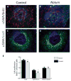
A) Notum150/Wt females were injected with BrdU at E11.5. Sections of E13.5 embryos were stained with αSMA, Sox9 and anti-BrdU antibodies. For convenience in counting the number of BrdU positive cells, the images were separated as αSMA and anti BrdU (A and B) and αSMA and Sox9 (C and D). Dotted lines in A and B represent the limits of tracheal epithelium. E) Cell proliferation was decreased in the epithelium compared to mesenchyme of Notum150/150 embryos. N=8 *p<0.05
Figure 7
Figure 7. Notum150/150 embryos lack well defined mesenchymal condensations
A: Tracheal tissue of E14.5 embryos was stained with PNA Lectin. Staining was restricted to the periphery of the trachea, demonstrating the lack of well-defined mesenchymal condensations in Notum150/150 embryos. Longitudinal sections of E14.5 sections were stained for αSMA, Sox9 and Nkx2.1. Sox9 positive cells in the trachea of Notum150/150 embryos demonstrated poorly developed mesenchymal condensations. Trachealis smooth muscle stained for αSMA on the dorsal side while tracheal epithelium stained for Nkx2.1. B: Scratch assays performed on MEF cells determined migration rates when treated with Wnt3a, Wnt5 and or Notum conditioned media. Notum inhibited Wnt5a induced cell migration. C: Cells were tracked and analyzed using Path speed within NIS Elements. Path speed was measured by calculating the total path traveled by the cells divided by time elapsed. Representative images of the morphological changes observed in migrating cells incubated with control (D), Wnt5a (E), or Wnt5a + Notum (F) conditioned media are shown.
Figure 8
Figure 8
A) X-gal staining was performed on trachea-lung tissue of E13.5 Notum 150/Wt; Axin LacZ/Wt and Notum 150150t; Axin LacZ/Wt embryos. X-gal staining was observed in trachea and lung of Notum 150/Wt; Axin LacZ/Wt. B) Intense X-gal staining was observed throughout the lungs and trachea of Notum 150/150; Axin LacZ/Wt. C) X-gal staining was primarily detected in the subepithelial mesenchyme of the airways. D) Prominent X-gal staining was observed in the mesenchyme of the lung of Notum 150/150; Axin LacZ/Wt embryos. E, F) Myh11, Col2a1 and Wnt7b RNA were unchanged while expression of Axin2 was increased after deletion of Notum. Transverse sections of E16.5 tracheas show increased β-catenin expression in mesenchyme of Notum 150150 tissue. Note the collapsed appearance of the epithelium and the reduced luminal area in the Notum 150/150 trachea (H), compared to control (G).
Figure 9
Figure 9
A) Lung and trachea tissue was isolated from Sox9GFP mice at E13.5 and incubated in air-liquid interphase for 72 hour in presence of 20 mM LiCl. Mesenchymal condensations are well formed in control samples, but are lacking in samples treated with LiCl (white arrow). B) Alcian blue staining depicted sites of cartilage in control samples. C: Note the lack of Alcian blue staining (black arrow) as well as the reduced branching of the peripheral lung of LiCl treated samples D) Increased mesenchymal expression of β-catenin in CtnnbΔ3; Dermo1Cre/wt embryos results in absence of Sox9 and αSMA staining in E12.5 respiratory tract mesenchyme, while epithelial expression of Nkx2.1 is preserved. Low and high-power magnifications of respiratory tract longitudinal sections are shown. E) E14.5 Mesenchymal deletion of Ror2 causes tracheal stenosis partially recapitulating the phenotype observed after deletion of Notum. F) ATF2 luciferase reporter assay shows that Wnt5a promotes activation of the reporter, while co-transfection of Wnt5a and Wnt3a impaired the Wnt5a induced luciferase activity. H:Heart. N=4 ANOVA, Tukey’s multiple comparisons test *** p<0.001 ATF2 vs ATF2 + Wnt5a; ###p<0.001 ATF2 + Wnt5a vs ATF2 +Wnt5a + Wnt3a.
Figure 10
Figure 10
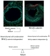
Model: Notum is a target of Wnt/β-catenin signaling in developing trachea. Wnt/β-catenin signaling is attenuated by Notum during formation of mesenchymal condensations. Notum is required for normal patterning of tracheal cartilage and muscle. In absence of Notum, Wnt/β-catenin dependent signaling is increased, leading to abnormal mesenchymal condensation formation affecting the normal tracheal development.

References

    1. Ayers KL, Mteirek R, Cervantes A, Lavenant-Staccini L, Therond PP, Gallet A. Dally and Notum regulate the switch between low and high level Hedgehog pathway signalling. Development. 2012;139:3168–3179. - PubMed
    1. Boogaard R, Huijsmans SH, Pijnenburg MW, Tiddens HA, de Jongste JC, Merkus PJ. Tracheomalacia and bronchomalacia in children: incidence and patient characteristics. Chest. 2005;128:3391–3397. - PubMed
    1. Bryja V, Andersson ER, Schambony A, Esner M, Bryjova L, Biris KK, Hall AC, Kraft B, Cajanek L, Yamaguchi TP, Buckingham M, Arenas E. The extracellular domain of Lrp5/6 inhibits noncanonical Wnt signaling in vivo. Molecular biology of the cell. 2009;20:924–936. - PMC - PubMed
    1. Cantu JA, Flowers GP, Topczewski J. Notum homolog plays a novel role in primary motor innervation. The Journal of neuroscience: the official journal of the Society for Neuroscience. 2013;33:2177–2187. - PMC - PubMed
    1. Carpenter AC, Rao S, Wells JM, Campbell K, Lang RA. Generation of mice with a conditional null allele for Wntless. Genesis. 2010:1–5. - PMC - PubMed

Publication types

MeSH terms