

Efficacy of Strain Elastography in Diagnosis and Staging of Acute Appendicitis in Pediatric Patients
- PMID: 29428963
- PMCID: PMC5817900
- DOI: 10.12659/msm.905927
Efficacy of Strain Elastography in Diagnosis and Staging of Acute Appendicitis in Pediatric Patients
Abstract
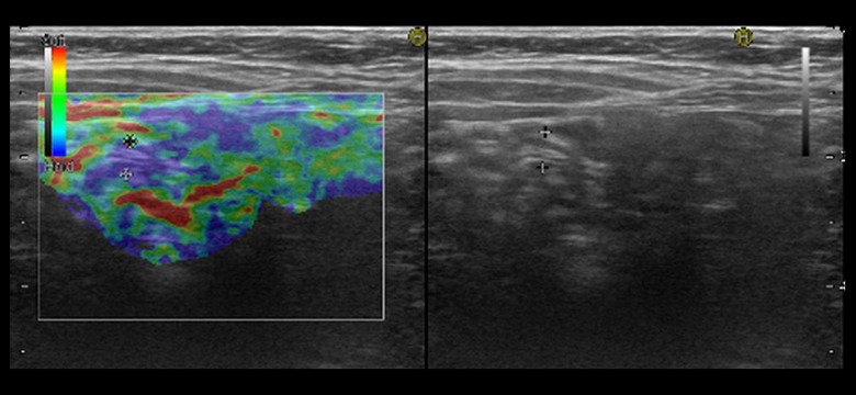
BACKGROUND In the present study, the role and efficiency of strain elastography (SE) were evaluated in diagnosis and staging of acute appendicitis in pediatric patients. MATERIAL AND METHODS We enrolled 225 pediatric patients with suspected clinical and laboratory findings of acute appendicitis. Gray-scale sonographic findings were recorded and staging was made by the colorization method of SE imaging. Appendectomy was performed in all patients and the results of the surgical pathology were compared with the imaging findings. The sensitivity, specificity, and accuracy of SE imaging were determined in terms of evaluating the "acute appendicitis". RESULTS Sonographic evaluation revealed acute appendicitis in 100 patients. Regarding the SE analysis, cases with appendicitis were classified into 3 groups as: mild (n=17), moderate (n=39), and severe (n=44). The pathological evaluation revealed 95 different stages of appendicitis and normal appendix in 5 cases: acute focal (n=10), acute suppurative (n=46), phlegmonous (n=27), and perforated (n=12), regarding the results of surgical pathology. Five patients with pathologically proven "normal" appendix were noted as "mild stage appendicitis" based on gray scale and SE analysis. In total, when gray-scale and SE results were compared with pathology results regardless of the stage of appendicitis, sensitivity, specificity, positive predictive value, negative predictive value, and accuracy rates were 96%, 96%, 95%, 96.8%, and 96%, respectively. No statistically significant difference was detected between other groups (P<0.05). CONCLUSIONS In acute appendicitis, the use of SE imaging as a supportive method for the clinical approach can be useful in diagnosis, and its results are closely correlated with the histopathologic stage of appendix inflammation.
Figures


References
-
- Birnbaum BA, Wilson SR. Appendicitis at the millennium. Radiology. 2000;215:337–48. - PubMed
-
- Lane MJ, Liu DM, Huynh MD, et al. Suspected acute appendicitis: Nonenhanced helical CT in 300 consecutive patients. Radiology. 1999;213:341–46. - PubMed
-
- Velanovich V, Satava R. Balancing the normal appendectomy rate with the perforated appendicitis rate: implications for quality assurance. Am Surg. 1992;58:264–69. - PubMed
-
- Rettenbacher T, Hollerweger A, Macheiner P, et al. Ovoid shape of the vermiform appendix: A criterion to exclude acute appendicitis – evaluation with US. Radiology. 2003;226:95–100. - PubMed
-
- Puig S, Hormann M, Rebhandl W, et al. US as a primary diagnostic tool in relation to negative appendectomy: six years experience. Radiology. 2003;226:101–4. - PubMed
MeSH terms
LinkOut - more resources
Full Text Sources
Medical
