Histologic processing artifacts and inter-pathologist variation in measurement of inked margins of canine mast cell tumors
- PMID: 29429400
- PMCID: PMC6505814
- DOI: 10.1177/1040638718757582
Histologic processing artifacts and inter-pathologist variation in measurement of inked margins of canine mast cell tumors
Abstract
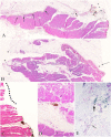
Although quantitative assessment of margins is recommended for describing excision of cutaneous malignancies, there is poor understanding of limitations associated with this technique. We described and quantified histologic artifacts in inked margins and determined the association between artifacts and variance in histologic tumor-free margin (HTFM) measurements based on a novel grading scheme applied to 50 sections of normal canine skin and 56 radial margins taken from 15 different canine mast cell tumors (MCTs). Three broad categories of artifact were 1) tissue deformation at inked edges, 2) ink-associated artifacts, and 3) sectioning-associated artifacts. The most common artifacts in MCT margins were ink-associated artifacts, specifically ink absent from an edge (mean prevalence: 50%) and inappropriate ink coloring (mean: 45%). The prevalence of other artifacts in MCT skin was 4-50%. In MCT margins, frequency-adjusted kappa statistics found fair or better inter-rater reliability for 9 of 10 artifacts; intra-rater reliability was moderate or better in 9 of 10 artifacts. Digital HTFM measurements by 5 blinded pathologists had a median standard deviation (SD) of 1.9 mm (interquartile range: 0.8-3.6 mm; range: 0-6.2 mm). Intraclass correlation coefficients demonstrated good inter-pathologist reliability in HTFM measurement (κ = 0.81). Spearman rank correlation coefficients found negligible correlation between artifacts and HTFM SDs ( r ≤ 0.3). These data confirm that although histologic artifacts commonly occur in inked margin specimens, artifacts are not meaningfully associated with variation in HTFM measurements. Investigators can use the grading scheme presented herein to identify artifacts associated with tissue processing.
Keywords: Canine; histologic tumor-free margin; mast cell tumors.
Conflict of interest statement
Figures


References
-
- Byrt T. Problems with kappa. J Clin Epidemiol 1992;45:1452–1452. - PubMed
-
- Donnelly L, et al. Evaluation of histological grade and histologically tumour-free margins as predictors of local recurrence in completely excised canine mast cell tumours. Vet Comp Oncol 2015;13:70–76. - PubMed
-
- Grizzle WE, et al. Fixation of tissues. In: Bancroft JD, et al., eds. Theory and Practice of Histological Techniques. 6th ed. Philadelphia, PA: Churchill Livingstone Elsevier, 2008:53–74.
-
- Jeyakumar S, et al. Effect of histologic processing on dimensions of skin samples obtained from cat cadavers. Am J Vet Res 2015;76:939–945. - PubMed
-
- Kamstock DA, et al. Recommended guidelines for submission, trimming, margin evaluation, and reporting of tumor biopsy specimens in veterinary surgical pathology. Vet Pathol 2011;48:19–31. - PubMed
Publication types
MeSH terms
LinkOut - more resources
Full Text Sources
Other Literature Sources
Medical

