. 2018 Mar 1;102(3):468-479.
doi: 10.1016/j.ajhg.2018.01.014. Epub 2018 Feb 8.

Mutations in the BAF-Complex Subunit DPF2 Are Associated with Coffin-Siris Syndrome

Affiliations

Mutations in the BAF-Complex Subunit DPF2 Are Associated with Coffin-Siris Syndrome

Georgia Vasileiou et al. Am J Hum Genet. .

Abstract

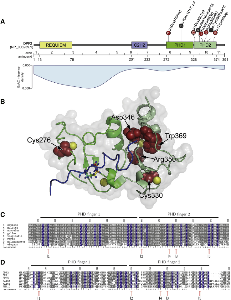
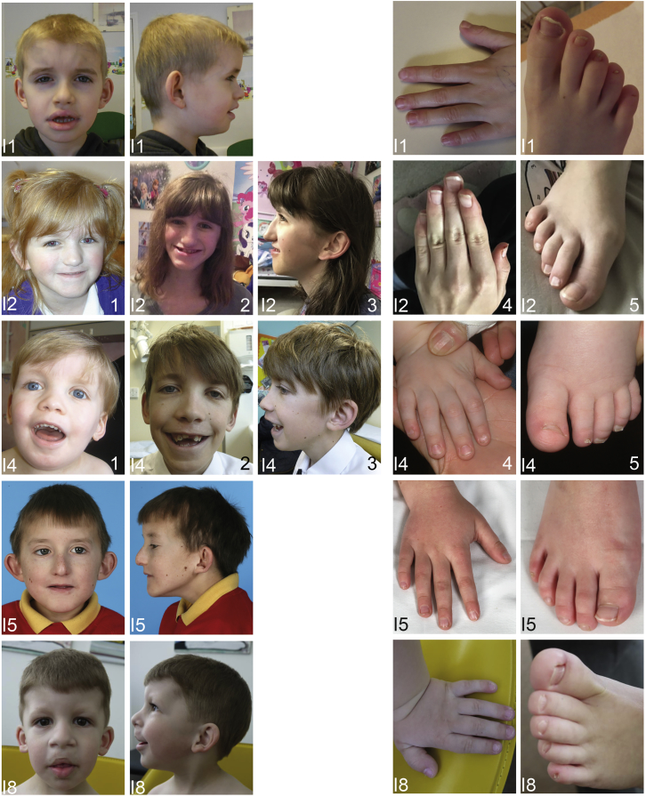
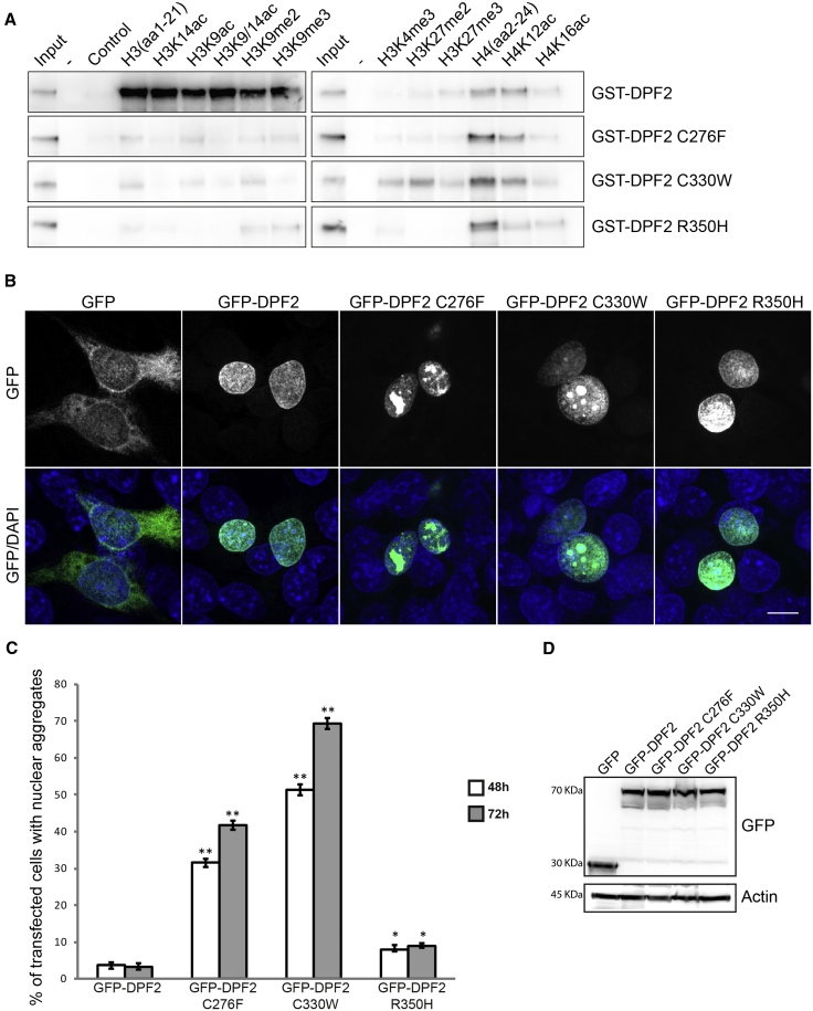
Variants affecting the function of different subunits of the BAF chromatin-remodelling complex lead to various neurodevelopmental syndromes, including Coffin-Siris syndrome. Furthermore, variants in proteins containing PHD fingers, motifs recognizing specific histone tail modifications, have been associated with several neurological and developmental-delay disorders. Here, we report eight heterozygous de novo variants (one frameshift, two splice site, and five missense) in the gene encoding the BAF complex subunit double plant homeodomain finger 2 (DPF2). Affected individuals share common clinical features described in individuals with Coffin-Siris syndrome, including coarse facial features, global developmental delay, intellectual disability, speech impairment, and hypoplasia of fingernails and toenails. All variants occur within the highly conserved PHD1 and PHD2 motifs. Moreover, missense variants are situated close to zinc binding sites and are predicted to disrupt these sites. Pull-down assays of recombinant proteins and histone peptides revealed that a subset of the identified missense variants abolish or impaire DPF2 binding to unmodified and modified H3 histone tails. These results suggest an impairment of PHD finger structural integrity and cohesion and most likely an aberrant recognition of histone modifications. Furthermore, the overexpression of these variants in HEK293 and COS7 cell lines was associated with the formation of nuclear aggregates and the recruitment of both wild-type DPF2 and BRG1 to these aggregates. Expression analysis of truncating variants found in the affected individuals indicated that the aberrant transcripts escape nonsense-mediated decay. Altogether, we provide compelling evidence that de novo variants in DPF2 cause Coffin-Siris syndrome and propose a dominant-negative mechanism of pathogenicity.

Keywords: BAF complex; Coffin-Siris syndrome; DPF2; PHD finger; autism spectrum disorder; dominant negative; histone modification; intellectual disability; nail hypoplasia; nuclear aggregates.


Figures

Figure 1
Figure 1
Clustering of DPF2 Variants in PHD Fingers (A) Schematic representation of DPF2, its domains (based on GenBank: NP_006259.1), the encoding exons (numbering based on GenBank: NM_006268.4), and the localization of DPF2 variants. Missense variants are presented in red, and truncating variants are in black. Note that the premature termination codons of the two truncating variants p.Asp340Glufs12 and p.Cys356Profs5 reside within 50 nucleotides upstream of the most 3′ exon-exon junction. The numbers in the circles indicate the affected individuals. For individual 6, c.904+1G>T is described at the genomic level because no RT-PCR could be performed. In light blue, a density blot of all missense variants reported in ExAC Browser version 0.3.1 shows markedly low frequency of variants in the PHD zinc fingers. (B) The crystal structure of the DPF2 double PHD finger bound to a histone peptide containing acetylation at lysine 14 (H3K14ac) (PDB: 5B79 and 2KWJ19) shows the clustering of the herein described missense variants in the tandem PHD finger domain (PHD finger 1 is colored in bright green, and PHD finger 2 is colored in pale green). The histone H3 backbone in the DPF2 binding pocket is shown in blue with an acetylated lysine residue at position 14 in stick representation. Zinc ions are represented as yellow spheres. The five affected amino acid residues are colored in red. The electrostatic surface is represented in gray with 80% opacity. Cys276 and Asp346 reside at the protein surface, whereas Cys330, Arg350, and Trp369 are buried in the PHD2 domain. (C) Multiple-sequence alignment of DPF2 orthologs at the de novo missense variant positions in the tandem PHD fingers shows high evolutionary sequence conservation. Residues from the conserved C4HC3 signature are marked in blue (see also Figure S5A and Data S1 sheet “DPF2_orthologs”). Numbers I1–I5 indicate the individual with the respective variant. Positions with de novo missense variants are indicated with a red arrow. Gray shading represents conservation. (D) Amino acid sequence alignment of DPF2 and putative human paralog proteins with similar tandem PHD fingers shows conservation of the C4HC3 signature (see also Figure S5B and Data S1 sheet “PHD_finger_proteins_(PHF)”). Protein sequences were obtained from NCBI and ClustalW, and the msa package within R was used for alignment. References for the orthologs are as follows: H. sapiens (DPF2), GenBank: NP_006259.1; M. mulatta (LOC721967), GenBank: XP_002808108.1; M. musculus (Dpf2), GenBank: NP_035392.1; G. gallus (DPF2), GenBank: NP_989662.1; D. rerio (dpf2), GenBank: NP_001007153.1; and X. tropicalis (dpf2), GenBank: NP_001184101.1. References for the paralogs are as follows: DPF1, GenBank: NP_001128627.1; DPF2, GenBank: NP_006259.1; DPF3, GenBank: NP_001267471.1; KAT6A, GenBank: NP_006757.2; KAT6B, GenBank: NP_036462.2; and PHF10, GenBank: NP_060758.2.
Figure 2
Figure 2
Facial Phenotype and Images from the Hands and Feet of Individuals with Variants in DPF2 (I1) Individual 1 at 9 years. (I2) Individual 2 at 5 years, 5 months (image 1) and 16 years (images 2–5). (I4) Individual 4 at 2 years (images 1, 4, and 5) and 12 years (images 2 and 3). (I5) Individual 5 at 10 years, 5 months. (I8) Individual 8 at 3 years, 9 months. Note the hypoplasia of the fifth toenails (all individuals), hypoplasia of further toenails (I1, I4, and I8), hypoplasia of fingernails (I4 and I8), generalized brachydactyly (I4 and I8) or fifth-finger brachydactyly (I1 and I5), and clinodactyly (I2, I5, and I8). Facial dysmorphisms include sparse hair (I1, I4, and I8), a prominent forehead (I1, I4, and I8), hypertelorismus (I4), macrotia (I1, I2), prominent or low-set ears (I2, I4, and I8), a broad nose (I4 and I8), thick alae nasi (I1, I4, and I8), a thin upper lip (I2, I4, I5, and I8), a thick lower vermillion (I1 and I8), cleft lip palate (I8), and a broad and short philtrum (I1, I2, I4, and I8).
Figure 3
Figure 3
Functional Consequences of the DPF2 Missense Variants on Histone Binding and Sub-nuclear Localization (A) Western blot analysis of histone peptide pull-downs shows the absence or residual binding of the GST-DPF2 recombinant proteins harboring the three missense variants to unmodified, acetylated, or methylated H3 peptides (wild-type GST-DPF2 is shown for comparison). Note that the interaction with unmodified and acetylated H4 peptides was not affected. Purified GST-fusion proteins and histone peptides are indicated. Input represents 5% of the purified GST proteins used in the pull-downs. As a control, no peptide was added in the pull-down assay. GST-tagged wild-type DPF2 and mutants were generated by N-terminal subcloning from the pEGFP-C3 into a pGEX4T1(GST) vector via BglII and SalI restriction sites (Table S4). Expression of GST-DPF2 fusion proteins in BL21 bacteria was induced with 0.1 mM IPTG. Collected cells were resuspended in 25 mM HEPES (pH 7.5), 500 mM KCl, 0.2 mM DTT, 1 mM EDTA, 10% glycerol, and proteinase inhibitor (Roche) and lysed with a French pressure cell press and then sonicated. Proteins were purified with Glutathione Sepharose 4B (GE Healthcare) according to the manufacturer’s instructions and eluted with 10 mM glutathione (pH 8.5). The eluted protein was dialysed with the Slide-A-Lyzer MINI Dialysis Device (ThermoFisher Scientific) against 25 mM HEPES (pH 7.5), 500 mM KCl, 0.2 mM DTT, 1 mM EDTA, and 10% glycerol for 2 hr and the same buffer with 60% glycerol overnight. Histone-peptide binding assays were performed as described in Lange et al. Biotinylated histone peptides were purchased from Millipore (unmodified H3, 12-403; H3K9ac, 12-431; H3K14ac, 12-425; H3K9/14ac, 12-402; H3K9me2, 12-430; H3k9me3, 12-568; H3K4me3, 12-460, H3K27me2, 12-566; H3K27me3, 12-565; unmodified H4, 12-372) and Tebu-bio (H4K12ac, 12-0032; H4K16ac, 12-0033). (B) Representative confocal immunofluorescence microscopy images of HEK293 cells overexpressing empty GFP vector (column 1) and wild-type DPF2 (column 2) and mutant (columns 3–5) GFP-tagged constructs. GFP was homogeneously distributed throughout the cell, whereas wild-type DPF2 and mutants exhibited exclusively nuclear localization. Note that C276F and C330W mutants led to the formation of nuclear protein aggregates, whereas R350H had a minor effect. The top panel shows GFP, and the lower panel shows merged pictures with DAPI. Scale bar, 10 μm. Wild-type DPF2 cDNA was amplified by PCR. The product was N-terminally inserted into the pEGFP-C3 vector with the In-Fusion HD Cloning Plus Kit (Clontech, Takara) according to the manufacturer’s instructions. The variants in DPF2 were generated by mutagenesis PCR. Primers used for cloning and mutagenesis are described in Table S4. HEK293 cells were grown as described in Vasileiou et al. 3 × 105 HEK293 cells were cultured on coverslips and transfected with 1 μg GFP and GFP-fusion plasmids with the use of polyethylenimine. After 48 hr, cells were fixed with 100% methanol at −20°C and stained with DAPI. Fluorescence images were acquired on an LSM 800 confocal laser scanning microscope (Carl Zeiss) with a 63× lens. (C) HEK293 cells were examined 48 and 72 hr after transfection, and graphs indicate the percentage of cells exhibiting nuclear aggregates. Error bars represent the mean ± SEM, and significant values are indicated with p < 0.05 or ∗∗p < 0.01 (unpaired Student’s t test). 100 random cells were used per experiment (n = 3). (D) Western blot analysis from lysates of HEK293 cells transfected with the indicated plasmids with the use of anti-GPF (top) and anti-panActin (bottom). Molecular weight (kDa) is indicated on the left of the western blotting panels.

References

    1. Kosho T., Okamoto N., Ohashi H., Tsurusaki Y., Imai Y., Hibi-Ko Y., Kawame H., Homma T., Tanabe S., Kato M. Clinical correlations of mutations affecting six components of the SWI/SNF complex: detailed description of 21 patients and a review of the literature. Am. J. Med. Genet. A. 2013;161A:1221–1237. - PubMed
    1. Coffin G.S., Siris E. Mental retardation with absent fifth fingernail and terminal phalanx. Am. J. Dis. Child. 1970;119:433–439. - PubMed
    1. Kosho T., Miyake N., Carey J.C. Coffin-Siris syndrome and related disorders involving components of the BAF (mSWI/SNF) complex: historical review and recent advances using next generation sequencing. Am. J. Med. Genet. C. Semin. Med. Genet. 2014;166C:241–251. - PubMed
    1. Hoyer J., Ekici A.B., Endele S., Popp B., Zweier C., Wiesener A., Wohlleber E., Dufke A., Rossier E., Petsch C. Haploinsufficiency of ARID1B, a member of the SWI/SNF-a chromatin-remodeling complex, is a frequent cause of intellectual disability. Am. J. Hum. Genet. 2012;90:565–572. - PMC - PubMed
    1. Kosho T., Okamoto N., Coffin-Siris Syndrome International Collaborators Genotype-phenotype correlation of Coffin-Siris syndrome caused by mutations in SMARCB1, SMARCA4, SMARCE1, and ARID1A. Am. J. Med. Genet. C. Semin. Med. Genet. 2014;166C:262–275. - PubMed

Publication types

MeSH terms

Supplementary concepts