Review
. 2018 Aug:330:27-42.
doi: 10.1016/j.cellimm.2018.01.020. Epub 2018 Feb 2.

Macrophages and lipid metabolism

Affiliations
Review

Macrophages and lipid metabolism

Anneleen Remmerie et al. Cell Immunol. 2018 Aug.

Abstract

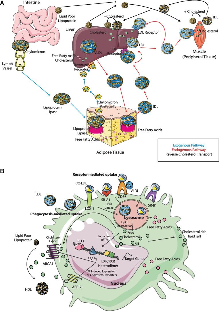
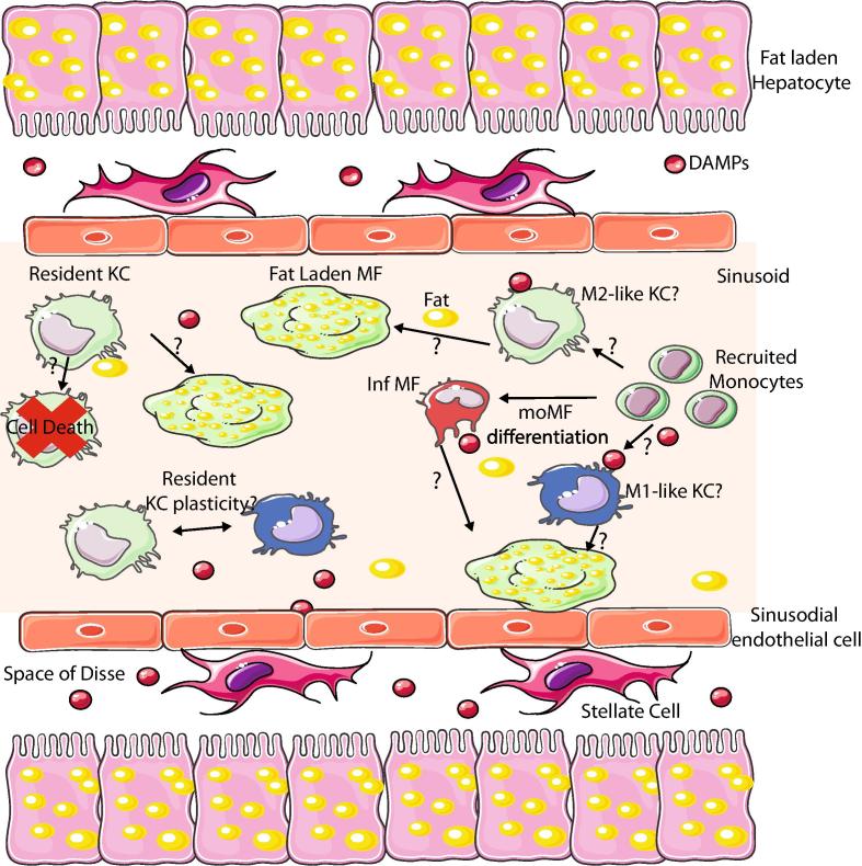
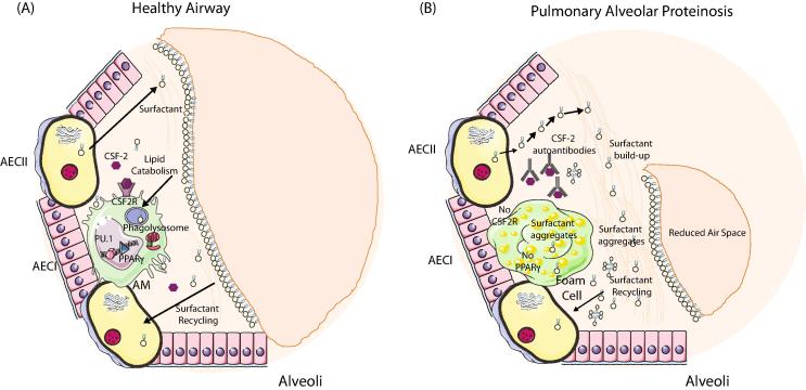
Distinct macrophage populations throughout the body display highly heterogeneous transcriptional and epigenetic programs. Recent research has highlighted that these profiles enable the different macrophage populations to perform distinct functions as required in their tissue of residence, in addition to the prototypical macrophage functions such as in innate immunity. These 'extra' tissue-specific functions have been termed accessory functions. One such putative accessory function is lipid metabolism, with macrophages in the lung and liver in particular being associated with this function. As it is now appreciated that cell metabolism not only provides energy but also greatly influences the phenotype and function of the cell, here we review how lipid metabolism affects macrophage phenotype and function and the specific roles played by macrophages in the pathogenesis of lipid-related diseases. In addition, we highlight the current questions limiting our understanding of the role of macrophages in lipid metabolism.

Keywords: AMD; Atherosclerosis; Lipid metabolism; Macrophages; NAFLD; PAP.


Figures

Fig. 1
Fig. 1
Overview of Lipid Metabolism and Macrophages. (A) The 3 pathways of Lipid Metabolism are the exogenous pathway (blue), the endogenous pathway (red) and reverse cholesterol transport (black). In the exogenous pathway, chylomicrons from the intestine are released into lymph and enter the bloodstream and subsequently adipose tissue. Here, through the action of lipoprotein lipase these are degraded into free fatty acids and chylomicron remnants which go back into circulation and then enter the liver through remnant receptors to be subsequently degraded into free fatty acids and cholesterol. In the endogenous pathway, VLDLs are exported from the liver to the circulation and adipose tissue where they are degraded again through the action of lipoprotein lipase into free fatty acids and IDL, which then binds the IDL receptor converting IDL to LDL. LDLs then bind to the LDL receptor delivering cholesterol to peripheral tissues as well as returning it to the liver. In the reverse cholesterol pathway, excess cholesterol is returned via HDL to the liver to be excreted in the bile. (B) Macrophages take up LDL, VLDL and oxidised lipoproteins via macropinocytosis, phagocytosis and scavenger receptor-mediated pathways including LOX-1, SR-A1, CD36 and SR-B1. Free cholesterol and fatty acids are generated following degradation of ingested lipids in the lysosome. Such cholesterol can be utilised to form lipid rafts. Accumulation of cellular cholesterol leads to activation of several transcription factors, including PPARγ, LXRs and RXRs which subsequently regulate expression of their target genes including transporters such as ABCA1 and ABCG1 which regulate the efflux of free cholesterol and scavenger receptors. Alternatively, passive efflux of free cholesterol can also occur. (For interpretation of the references to colour in this figure legend, the reader is referred to the web version of this article.)
Fig. 2
Fig. 2
Macrophage Niches in Atherosclerosis. (A) The onset of atherosclerosis is associated with plaque development. This generates new macrophage niches which need to be filled. This is done predominantly through the recruitment of monocytes to the developing plaque and their subsequent differentiation into macrophages. These macrophages then take up the excess fat and differentiate into lipid laden foam cells. (B) As the plaque grows, likely so too does the number of macrophage niches. These are then populated through 2 main mechanisms. The predominating mechanism is proliferation. Foam cells already present in the plaque sense the empty niches and start to proliferate thus filling up the available niches. Alternatively, new monocytes can also be recruited to the growing plaque where they will engraft and then differentiate into macrophages. (C) During plaque regression, the local environment is changed dramatically. This can have two consequences, the foam cells which are adapted to the fat-rich environment cannot adjust to the new environment and subsequently die or possibly emigrate out, resulting in niche availability which is filled through monocyte recruitment and subsequent differentiation into macrophages that are distinct from those generated in the fat-rich environment being more M2-like. Alternatively, if plastic enough to do so, the foam cell may alter its transcriptional profile rendering it more suited to this new fat-poor environment. Additionally, as the plaque regresses it is likely that the number of Mϕ niches decreases.
Fig. 3
Fig. 3
Role of Mϕs in NAFLD. There are several questions remaining regarding the role of Mϕs in NAFLD. One of the main questions relates to whether there are multiple subsets of KCs and other hepatic Mϕs present or not. Monocytes are recruited to the liver and develop into infiltrating Mϕs (Inf Mϕs) during NAFLD, but do they also contribute to the bona fide KC pool? If so are these moKCs more M1 or M2-like? Another question is where do the foam cells originate from? KCs that have become overloaded with lipid, or can these cells given their lipid metabolism enriched transcriptional profile efficiently process lipids meaning the foam cells are generated from the infiltrating Mϕs or is it a combination of both? Another key question is whether KCs are plastic and can change from a more M2 profile to that of an M1-like Mϕs and vice versa. If this is the case, how does this work and can it be manipulated to enhance M2-like cell numbers given that this appears beneficial for the patient?
Fig. 4
Fig. 4
Role of Mϕs in Alveolar Proteinosis. (A) Normal alveolar macrophage development occurs within the first week of life. Local CSF-2 signalling in the alveoli induces expression of the TF Pparg in the CSF-2R expressing fetal liver monocytes generating pre-alveolar macrophages, which subsequently differentiate into AMs. Type II alveolar epithelial cells (AECII) produce surfactant that is crucial for normal lung function. Excess or old surfactant must be recycled and this occurs primarily via the AECII but also via AMs. Surfactant is taken up via the AMs and catabolised in the phagolysosome. (B) In the absence CSF2 signalling as a result of deficiency in CSF2, CSF2R or the presence of auto-antibodies directed against CSF2, Pparg expression is not induced and as a result AMs do not develop correctly and cannot process the excess lipids. This leads to a build-up of surfactant in the alveoli and macrophages, the latter of which results in the generation of foam cells. This build up leads to a reduction in the air space in the alveoli making it difficult to breathe.

References

    1. van Furth R., Cohn Z.A., Hirsch J.G., Humphrey J.H., Spector W.G., Langevoort H.L. The mononuclear phagocyte system: a new classification of macrophages, monocytes, and their precursor cells. Bull. World Health Organ. 1972;46:845–852. - PMC - PubMed
    1. Ginhoux F., Guilliams M. Tissue-resident macrophage ontogeny and homeostasis. Immunity. 2016;44:439–449. - PubMed
    1. Hoeffel G., Chen J., Lavin Y., Low D., Almeida F.F., See P. C-myb(+) erythro-myeloid progenitor-derived fetal monocytes give rise to adult tissue-resident macrophages. Immunity. 2015;42:665–678. - PMC - PubMed
    1. Gomez Perdiguero E., Klapproth K., Schulz C., Busch K., Azzoni E., Crozet L. Tissue-resident macrophages originate from yolk-sac-derived erythro-myeloid progenitors. Nature. 2015;518:547–551. - PMC - PubMed
    1. Schulz C., Gomez Perdiguero E., Chorro L., Szabo-Rogers H., Cagnard N., Kierdorf K. A lineage of myeloid cells independent of Myb and hematopoietic stem cells. Science. 2012;336:86–90. - PubMed

Publication types

MeSH terms