. 2018 Feb 21;97(4):806-822.e10.
doi: 10.1016/j.neuron.2018.01.033. Epub 2018 Feb 8.

Immune or Genetic-Mediated Disruption of CASPR2 Causes Pain Hypersensitivity Due to Enhanced Primary Afferent Excitability

Affiliations

Immune or Genetic-Mediated Disruption of CASPR2 Causes Pain Hypersensitivity Due to Enhanced Primary Afferent Excitability

John M Dawes et al. Neuron. .

Abstract

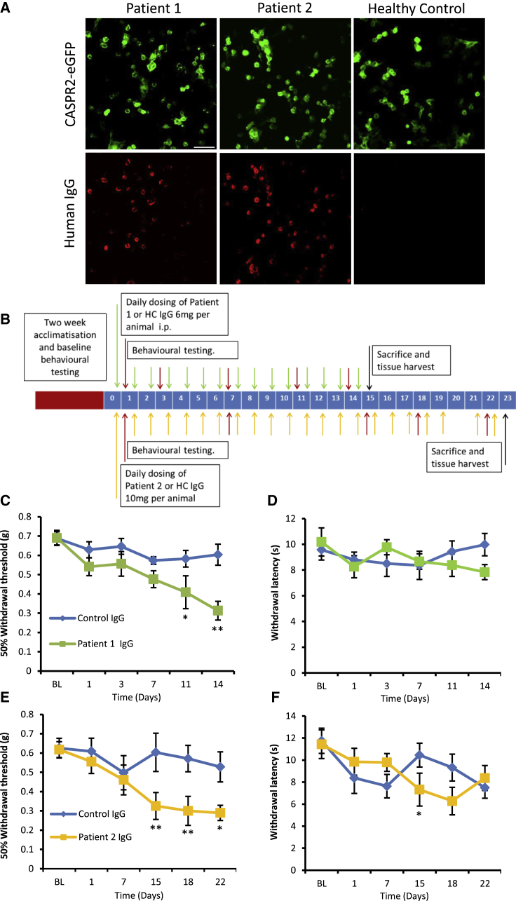
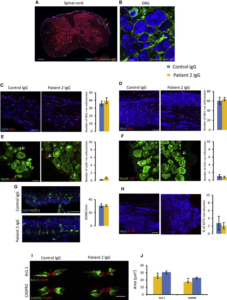
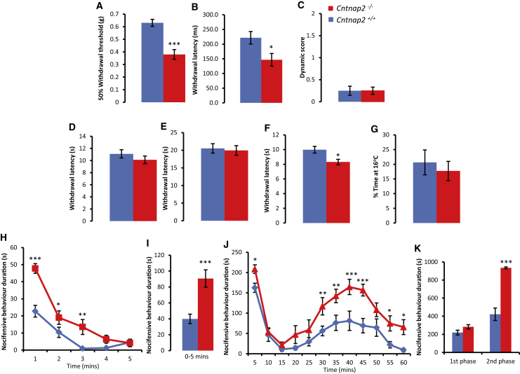
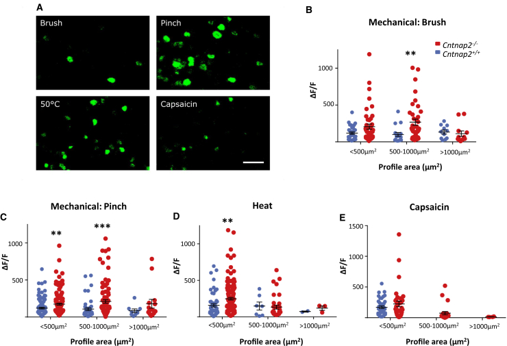
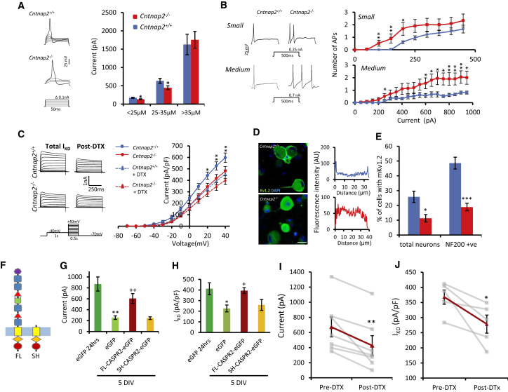
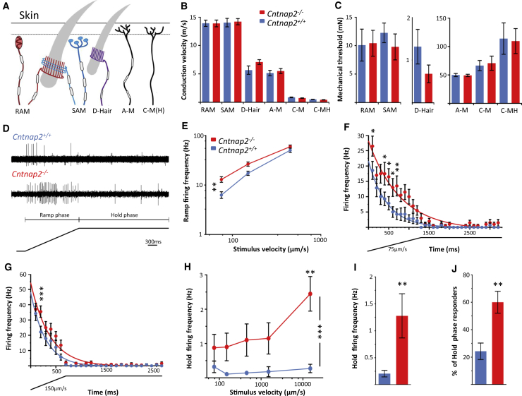
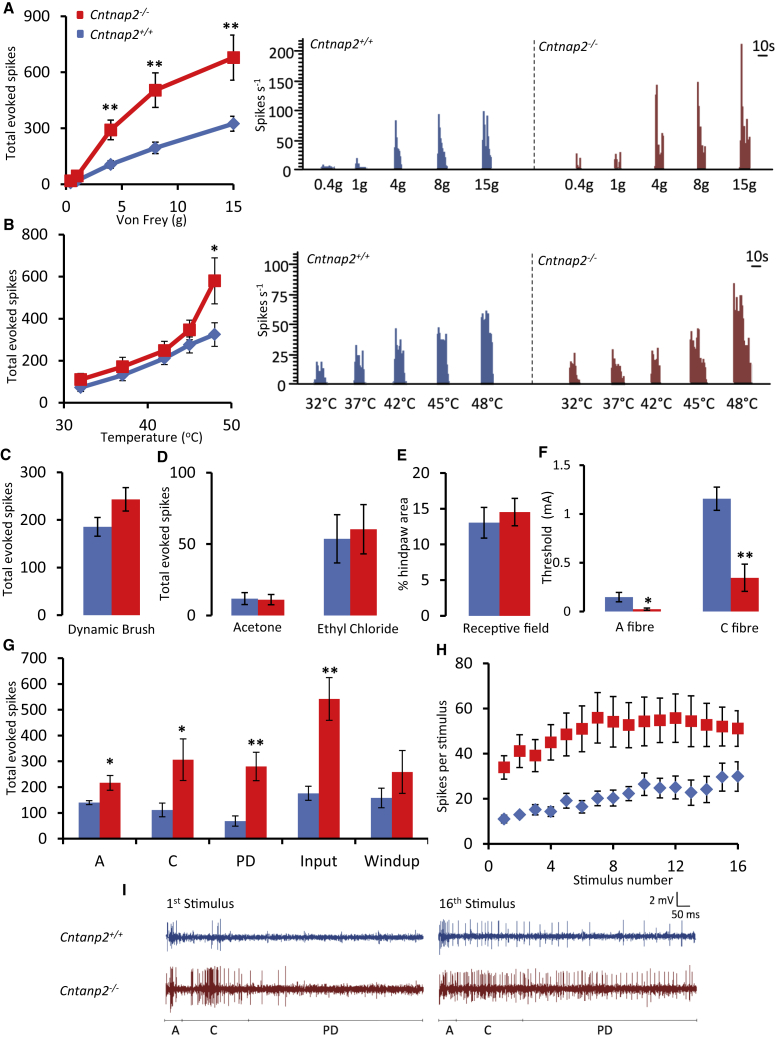
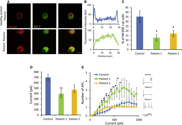
Human autoantibodies to contactin-associated protein-like 2 (CASPR2) are often associated with neuropathic pain, and CASPR2 mutations have been linked to autism spectrum disorders, in which sensory dysfunction is increasingly recognized. Human CASPR2 autoantibodies, when injected into mice, were peripherally restricted and resulted in mechanical pain-related hypersensitivity in the absence of neural injury. We therefore investigated the mechanism by which CASPR2 modulates nociceptive function. Mice lacking CASPR2 (Cntnap2-/-) demonstrated enhanced pain-related hypersensitivity to noxious mechanical stimuli, heat, and algogens. Both primary afferent excitability and subsequent nociceptive transmission within the dorsal horn were increased in Cntnap2-/- mice. Either immune or genetic-mediated ablation of CASPR2 enhanced the excitability of DRG neurons in a cell-autonomous fashion through regulation of Kv1 channel expression at the soma membrane. This is the first example of passive transfer of an autoimmune peripheral neuropathic pain disorder and demonstrates that CASPR2 has a key role in regulating cell-intrinsic dorsal root ganglion (DRG) neuron excitability.

Keywords: CASPR2; CNTNAP2; DRG; Kv1; autism; autoantibody; mechanosensation; pain; sensory neuron; voltage-gated potassium channel.


Figures

Figure 1
Figure 1
Passive Transfer of Human CASPR2-Abs Causes Pain-Related Hypersensitivity in Mice (A) CBA showing binding of antibodies from patient plasma using an anti-human IgG secondary antibody (red) to HEK cells transfected with human CASPR2-EGFP. No binding is seen using plasma from a healthy control subject. Scale bar, 50 μm. (B) Dosing regime and behavioral time course for passive transfer of WT mice with purified IgG from CASPR2-Ab-positive patients. (C–F) Using von Frey hairs, mice treated with patient 1 and 2 IgG develop a significant mechanical pain-related hypersensitivity (C and E, respectively) when compared to mice treated with IgG from a healthy control subject. Mice did not, however, develop a clear thermal hypersensitivity using the Hargreaves testing method (D and F). For (C) and (D), n = 8, and for (E) and (F), n = 9. Data shown as mean ± SEM, p < 0.05, ∗∗p < 0.01 versus control IgG group. See also Figure S1.
Figure 2
Figure 2
Patient CASPR2-Abs Bind In Vivo but Do Not Cause Gross Inflammation or Nerve Damage (A and B) Representative image of a spinal cord (A) and DRG (B) section from a mouse treated with patient IgG. No deposition of human IgG (green) seen in the spinal cord (A). In the DRG, human IgG binds to sensory neurons (B). Scale bars, 200 μm (A), 50 μm (B). (C and D) Representative images of sciatic nerve (C) and DRG (D) sections from mice treated with either control or patient 2 IgG stained for IBA1 (red). Quantification shows no difference between treatment groups; n = 4 mice. Scale bar, 50 μm. (E and F) Representative images of DRG sections from mice treated with either control or patient 2 IgG stained for Ly6G (red, E) and CD3 (red, F). The number of positive cells was very low, and no difference was found between groups; n = 4 mice. Scale bar, 25 μm. (G) Representative images of mouse glabrous skin. PGP9.5 (green) was used to mark nerve fibers. No difference was seen in the IENFD between treatment groups; n = 5 mice. Scale bar, 25 μm. (H) Representative images of mouse DRG sections stained for the injury marker ATF3 (red). Quantification showed no difference between treatment groups; n = 4 mice. Scale bar, 50 μm. (I and J) High-power representative images of single nodes (marked by CASPR [red]) from mouse sciatic nerve (I). Kv1.1 (green, top) and CASPR2 (green, bottom) staining is reduced in mice treated with patient 2 IgG. Quantification shows a significant reduction in the area of both Kv1.1 and CASPR2 staining in the patient IgG group versus control (J); n = 4 mice. Data shown as mean ± SEM, p < 0.05 versus control IgG group. See also Figures S2–S4.
Figure 3
Figure 3
CASPR2 Regulates Pain-Related Hypersensitivity in Mice (A) Using von Frey hairs, Cntnap2−/− (n = 15) mice display hypersensitivity to mechanical stimuli when compared to WT littermates (Cntnap2+/+, n = 20). (B) Withdrawal latency to pinprick application is significantly reduced in Cntnap2−/− (n = 14) compared to WT littermates (Cntnap2+/+, n = 12). (C) No difference in dynamic allodynia measured following brush application to the hindpaw between genotypes (Cntnap2−/−, n = 8; Cntnap2+/+, n = 6). (D–F) Cntnap2−/− mice (n = 8) do not display heat hypersensitivity to threshold stimuli as measured by the Hargreaves test (D) or to the hot plate set at 50°C (E). However, when using the hot plate set at 53°C (F), Cntnap2−/− mice have a reduced latency to withdrawal compared to Cntnap2+/+ mice (n = 13). (G) In comparison to WT littermates, Cntnap2−/− mice do not display cold hypersensitivity as measured by the thermal preference test; n = 7 in both groups. (H and I) In response to an intraplantar injection of capsaicin (1.5 μg), the duration of pain-related behavior is significantly greater in Cntnap2−/− (n = 11) versus Cntnap2+/+ (n = 12) mice over a 5-min period (I), but particularly in the first minute (H). (J and K) In comparison to control mice, Cntnap2−/− mice have an increased response to 5% formalin. This difference is significant in the first 5 min after injection of formalin (J), as well as in the second phase of the behavioral response (K); n = 8 for both groups. Data shown as mean ± SEM, p < 0.05, ∗∗p < 0.01, ∗∗∗p < 0.001 versus Cntnap2+/+ group. See also Figures S5 and S6.
Figure 4
Figure 4
In Vivo Calcium Imaging Shows that DRG Neurons from Cntnap2−/− Mice Are Hyper-Responsive to Mechanical and Chemical Stimuli (A) Representative images of GCaMP6 fluorescence as a measure of intracellular calcium following stimulation to the hindpaw. Scale bar, 100 μm. (B) In comparison to control, DRG neurons from Cntnap2−/− mice had a significantly greater response to brush stimulation applied to the glabrous skin. This was particularly true of medium-sized cells (500–1,000 μm2; Cntnap2+/+, n = 33 cells; Cntnap2−/−, n = 46 cells). (C) In response to noxious pinch stimulation, there was also a significant increase in the response of DRG neurons from Cntnap2−/− mice in both small- and medium-sized neurons (<500 μm2Cntnap2+/+, n = 106 cells; Cntnap2−/−, n = 132 cells; 500–1,000 μm2Cntnap2+/+, n = 46 cells; Cntnap2−/−, n = 93 cells). (D) In response to noxious heat stimulation (50°C), there was a significant increase in the response of DRG neurons from Cntnap2−/− mice in small DRG neurons (<500 μm2Cntnap2+/+, n = 54 cells; Cntnap2−/−, n = 143 cells). (E) No statistically significant difference was seen in the response of small cells to capsaicin application (<500 μm2; Cntnap2+/+, n = 41 cells; Cntnap2−/−, n = 36 cells). Note the number of medium-sized responders: Cntnap2+/+ mice = 0, Cntnap2−/− mice = 23. Cells analyzed from four Cntnap2+/+ and five Cntnap2−/− mice. Data shown as mean ± SEM, ∗∗p < 0.01, ∗∗∗p < 0.001 versus Cntnap2+/+ group.
Figure 5
Figure 5
CASPR2 Regulates the Excitability of DRG Neurons (A) Representative traces showing action potential firing to short incremental current injection in medium (25–35 μm) diameter neurons. Small (<25 μm; Cntnap2+/+, n = 47; Cntnap2−/−,n = 45 cells) and medium (25–35 μm; Cntnap2+/+, n = 20; Cntnap2−/−, n = 21 cells) diameter DRG neurons cultured from Cntnap2−/− mice have a significantly reduced rheobase when compared to neurons from control mice. There were no differences between genotype in large diameter neurons (Cntnap2+/+, n = 11; Cntnap2−/−, n = 13 cells). (B) Representative traces showing action potential firing to supra-threshold prolonged current injection in small (<25 μm) and medium (25–35 μm) diameter neurons. Quantification across a range of current steps showed that both small (Cntnap2+/+, n = 23 cells; Cntnap2−/−, n = 21 cells) and medium (Cntnap2+/+, n = 20; Cntnap2−/−, n = 21 cells) diameter Cntnap2−/− neurons display increased firing frequency in comparison to Cntnap2+/+ neurons. (C) Example traces from medium diameter neurons of outward currents evoked by depolarizing pulses. IKD was measured pre- and post-application of 100 nM DTX. Current voltage relationships for IKD demonstrating increased current in Cntnap2+/+neurons (n = 12 cells) compared to Cntnap2−/−neurons (n = 14 cells) that was not present following DTX treatment. (D) Representative images showing Kv1.2 membrane staining in DRG neurons from Cntnap2−/− and Cntnap2+/+ mice. Scale bar, 25 μm. Profile plots were used to define membrane staining. (E) Cultured DRG neurons from Cntnap2−/− mice have less Kv1.2 membrane staining when compared to control neurons. β-III-tubulin used to mark all neurons (n = 4 coverslips from two independent experiments). (F) Diagram highlighting the difference in the extracellular domain between the full-length (FL) and the short (SH) CASPR2 isoform. (G) After 5 DIV, there is a significant reduction in rheobase (EGFP 1 DIV [n = 12 cells] versus EGFP 5 DIV [n = 16 cells]) for WT neurons transfected with a plasmid containing EGFP only. FL-CASPR2-EGFP (n = 25 cells) overexpression at 5 DIV restores the rheobase to 1DIV measurements. However, overexpression of the short isoform (SH-CASPR2-EGFP) does not affect rheobase values (n = 9 cells). (H) After 5 DIV, there is a significant reduction in IKD (EGFP 1 DIV versus EGFP 5 DIV) for WT neurons transfected with a plasmid containing EGFP only (EGFP 1 DIV, n = 16 cells; EGFP 5 DIV, n = 13 cells). FL-CASPR2-EGFP overexpression at 5 DIV (n = 12 cells) restores the IKD to 1 DIV levels. Overexpression of SH-CASPR2-EGFP did not affect IKD (n = 12 cells). (I and J) The restoration of rheobase (I) and IKD (J), due to the overexpression of FL-CASPR2-EGFP at 5 DIV is reduced by the application of DTX. Gray lines show individual cells before and 5 min after the application of 100 nM DTX. Red lines show the average (rheobase, n = 7 cells; IKD, n = 5 cells). Data shown as mean ± SEM. For (A)–(C), p < 0.05, ∗∗p < 0.01, ∗∗∗p < 0.001 versus Cntnap2+/+ group. For (G) and (H), p < 0.05, ∗∗p < 0.01 versus EGFP 24 hr and +p < 0.05, ++p < 0.01 versus EGFP 5 DIV. For (I) and (J), p < 0.05, ∗∗p < 0.01 versus pre-DTX. See also Figure S7.
Figure 6
Figure 6
Genetic Deletion of FL-CASPR2 Results in Hyper-Excitable D-Hair Primary Afferents (A) Diagram illustrating the cutaneous mechanoreceptor sensory endings that were identified and recorded from using the ex vivo skin nerve preparation (B and C) Conduction velocity (B) and mechanical thresholds (C) were recorded from mechanoreceptors and nociceptors. No differences were observed between genotypes. See Table S6 for number of recorded units. (D) Example trace of evoked AP response of both Cntnap2+/+ (top) and Cntnap2−/− (bottom) D-hairs following a mechanical stimulus consisting of a ramp phase and hold phase. (E) The stimulus response curve showing that Cntnap2−/− D-hairs have a significantly higher ramp firing frequency compared to Cntnap2+/+ D-hairs at slow stimulus velocities. (F and G) Whole-stimulus (ramp and hold) firing frequency was analyzed every 100 ms to assess D-hair adaptation. At stimulus velocities 75 μm/s (F) and 150 μm/s (G), Cntnap2−/− D-hairs elicited increased firing frequencies and significantly less firing adaptation compared to Cntnap2+/+ D-hairs. Note ramp hold stimulus below x axis. (H) D-hair firing frequency was analyzed during the hold phase of each stimulus only. Cntnap2−/− D-hairs have a significantly higher firing frequency during the hold phase than control D-hairs. (I) The average hold firing frequency to a stimulus (independent of velocity) was significantly increased in Cntnap2−/− D-hairs. (J) The average percentage of D-hairs that responded to a stimulus during the hold phase was significantly higher in Cntnap2−/− mice compared to controls. For (E)–(J), Cntnap2+/+ n = 14, Cntnap2−/− n = 20 units recorded from 16 Cntnap2+/+ and 15 Cntnap2−/− mice. Data shown as mean ± SEM. p < 0.05, ∗∗p < 0.01, ∗∗∗p < 0.001 versus Cntnap2+/+ group. See also Figure S9.
Figure 7
Figure 7
Increased Activity of Lamina V/VI Dorsal Horn Neurons to Sensory Stimuli Cntnap2−/− Mice (A and B) Evoked neuronal responses to punctate mechanical stimuli (A) are significantly greater in Cntnap2−/− (n = 10 cells) compared to Cntnap2+/+ (n = 11 cells) mice. Cntnap2−/− mice also display increased neuronal responses to heat stimuli (B). Histogram traces represent typical single unit responses. (C and D) No significant differences were seen between evoked neuronal responses to dynamic brush (C) or innocuous (acetone) and noxious (ethyl chloride) evaporative cooling (D). (E) No differences were seen in the size of the receptive field. (F) WDR neurons in Cntnap2−/− mice display a significantly reduced threshold for both A- and C-fibers following electric stimulation. (G) Cntnap2−/− display an increased neuronal response following electrical stimulation. (H and I) No difference in the degree of windup was seen between genotypes (H). Representative single-unit traces also shown for the first and last stimulus for both Cntnap2−/− and Cntnap2+/+ (I). Data shown as mean ± SEM, p < 0.05, ∗∗p < 0.01 versus Cntnap2+/+ group. Cells recorded from 7 mice per genotype. See also Figure S10.
Figure 8
Figure 8
Patient CASPR2-Abs Reduce Kv1 Membrane Expression on DRG Neurons and Increase Their Excitability (A) Representative images showing that Kv1.2 (green) membrane staining is decreased in NF200-positive (red) DRG neurons treated with plasma from CASPR2-Ab-positive patients when compared to control. Scale bar, 25 μm. (B) Profile plots showing fluorescent intensity for Kv1.2 immunostaining across the cell. The profile suggests that most of the Kv1.2 is internal following treatment with patient plasma. (C) Quantification of Kv1.2 membrane staining showing a significant reduction in those cells treated with CASPR2-Ab-positive patient plasma (n = 4 coverslips from two independent experiments). (D) Medium-sized DRG neurons treated with patient plasma (patient 1, n = 12; patient 2, n = 16 cells) had a significantly reduced rheobase when compared to cells treated with plasma from healthy control (n = 20 cells). (E) DRG neurons treated with patient 1 (n = 10 cells), but not patient 2 (n = 15 cells), plasma display increased firing frequency in response to prolonged (500 ms) graded inputs of current (0–950 pA) in comparison to controls (n = 20 cells). Representative traces are show for each group. Data shown as mean ± SEM,p < 0.05, ∗∗p < 0.01 versus control group. See also Figure S8.

Comment in

References

    1. Arcourt A., Gorham L., Dhandapani R., Prato V., Taberner F.J., Wende H., Gangadharan V., Birchmeier C., Heppenstall P.A., Lechner S.G. Touch receptor-derived sensory information alleviates acute pain signaling and fine-tunes nociceptive reflex coordination. Neuron. 2017;93:179–193. - PubMed
    1. Calvo M., Richards N., Schmid A.B., Barroso A., Zhu L., Ivulic D., Zhu N., Anwandter P., Bhat M.A., Court F.A. Altered potassium channel distribution and composition in myelinated axons suppresses hyperexcitability following injury. eLife. 2016;5:e12661. - PMC - PubMed
    1. Carter R.J., Morton J., Dunnett S.B. Motor coordination and balance in rodents. Curr. Protoc. Neurosci. 2001;Chapter 8 Unit 8.12. - PubMed
    1. Cascio C.J. Somatosensory processing in neurodevelopmental disorders. J. Neurodev. Disord. 2010;2:62–69. - PMC - PubMed
    1. Chaplan S.R., Bach F.W., Pogrel J.W., Chung J.M., Yaksh T.L. Quantitative assessment of tactile allodynia in the rat paw. J. Neurosci. Methods. 1994;53:55–63. - PubMed

Publication types

MeSH terms