. 2018 Mar 13;10(3):766-779.
doi: 10.1016/j.stemcr.2018.01.008. Epub 2018 Feb 8.

Generation of Functioning Nephrons by Implanting Human Pluripotent Stem Cell-Derived Kidney Progenitors

Affiliations

Generation of Functioning Nephrons by Implanting Human Pluripotent Stem Cell-Derived Kidney Progenitors

Ioannis Bantounas et al. Stem Cell Reports. .

Abstract

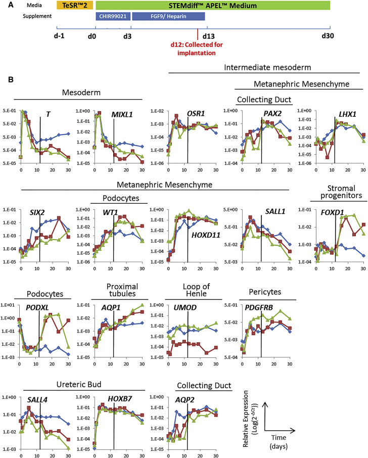
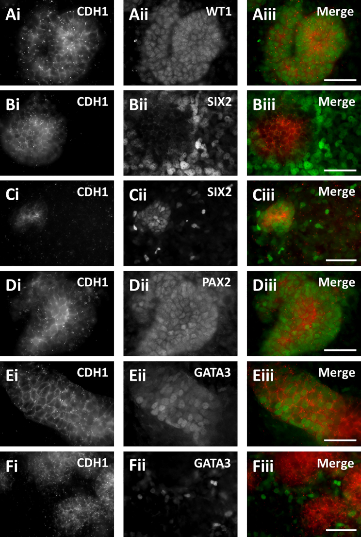
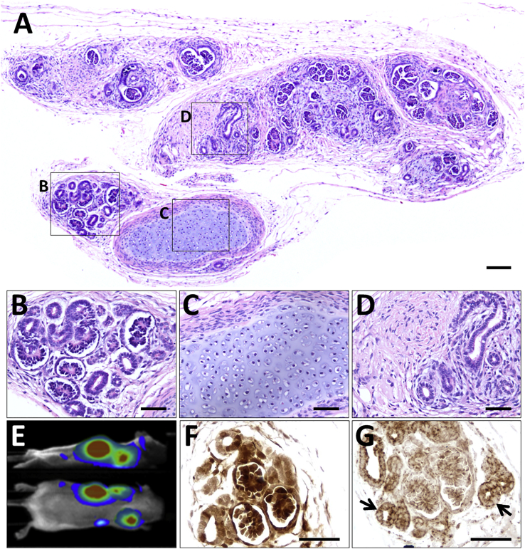
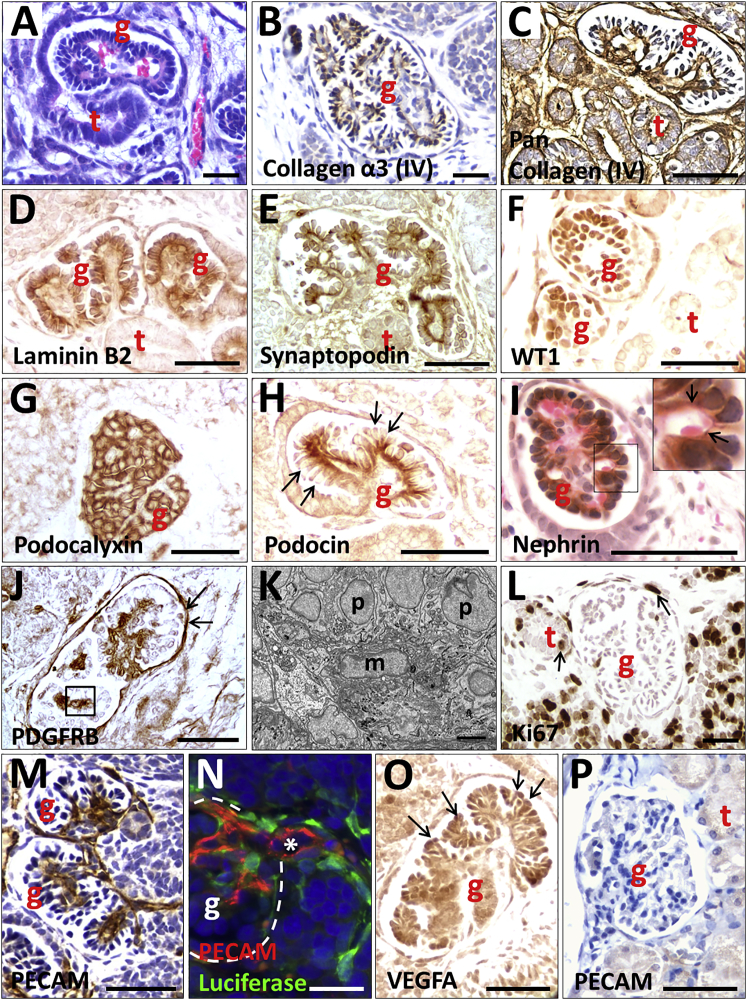
Human pluripotent stem cells (hPSCs) hold great promise for understanding kidney development and disease. We reproducibly differentiated three genetically distinct wild-type hPSC lines to kidney precursors that underwent rudimentary morphogenesis in vitro. They expressed nephron and collecting duct lineage marker genes, several of which are mutated in human kidney disease. Lentiviral-transduced hPSCs expressing reporter genes differentiated similarly to controls in vitro. Kidney progenitors were subcutaneously implanted into immunodeficient mice. By 12 weeks, they formed organ-like masses detectable by bioluminescence imaging. Implants included perfused glomeruli containing human capillaries, podocytes with regions of mature basement membrane, and mesangial cells. After intravenous injection of fluorescent low-molecular-weight dextran, signal was detected in tubules, demonstrating uptake from glomerular filtrate. Thus, we have developed methods to trace hPSC-derived kidney precursors that formed functioning nephrons in vivo. These advances beyond in vitro culture are critical steps toward using hPSCs to model and treat kidney diseases.

Keywords: cell therapy; glomerulus; human embryonic stem cells; kidney; kidney progenitors; lentivirus; metanephric mesenchyme; nephron; ureteric epithelium; vascularization.


Figures

Figure 1
Figure 1
Differentiation of MAN13 hPSC to Kidney Lineages in 2D Culture (A) Schematic of the 30 day differentiation protocol depicting the timing of application of CHIR99021 and FGF9/heparin. The time point of cell harvest for implantation into mice is also indicated (d12, in red). (B) qPCR profiling of 17 transcripts at 11 time points over 30 days. The results for three independent experiments are shown in blue, red, and green, with levels of target transcripts normalized to GAPDH expression. The characteristic tissue/lineage that expresses each gene in vivo is indicated above the graph for each transcript. The black vertical line in each graph indicates the time of collection of cells for implantation into mice.
Figure 2
Figure 2
Immunocytochemistry of hPSC Cultures MAN13 cells were differentiated for 12 days in vitro, when rudimentary morphogenesis had begun. Cultures were co-stained with the epithelial cell-cell adhesion protein CDH1 (left frames) and transcription factors (middle frames) to detect: MM-derived cells (WT1 or SIX2); UB-derived tubules (GATA3); and PAX2, expressed in both lineages. Right-hand frames show merged images (red CDH1 and green nuclear proteins). (A) A WT1+ cell cluster with a central zone expressing CDH1. (B) Typically, loose SIX2+ cells surrounded CDH1+ zones. (C) Occasionally, subsets of SIX2+ cells appeared to co-express CDH1. (D) A PAX2+ cluster with a central zone expressing CDH1. (E) A CDH1+ tubule-like structure containing a subset of GATA3+ nuclei. (F) In other areas, loosely packed GATA3+ cells surrounded CDH1+ structures. Scale bars, 40 μm.
Figure 3
Figure 3
Transduction of MAN13 hPSCs with a Lentiviral Vector Expressing a Bicistronic iRFP-E2A-Luciferase Cassette (A) Diagram of the expression cassette showing the two reporter genes and the EF1α promoter. (B) iRFP fluorescence in transduced cells. (C and D) Viability and cytotoxicity (mean ± SEM, n = 4) in MAN13 cultures transduced with lentivirus (LV-iRFP/Luc), with no significant difference compared with untransduced controls (LV-CTRL). As a positive “death” control, MAN13 cells were treated with 500 nM staurosporine for 24 hr. (E) Examples of transduced and untransduced differentiating MAN13 cultures assessed by immunocytochemistry for SIX2, WT1, and CDH1. (F) Similar qPCR profiles of transduced versus parent cells during 2D kidney differentiation. A representative experiment of three independent biological repeats is shown for each line. Scale bars, 200 μm in (B) and 90 μm in (E).
Figure 4
Figure 4
Subcutaneous Implantation into beige/SCID Mice of Luciferase-Labeled MAN13-Derived Kidney Precursor Cells Harvested at Day 12 in 2D Culture (A–D) Histological overview 12 weeks after transplantation. The implanted cells have formed a differentiated mass (A). Boxed areas indicate the following: (B) differentiated nephrons; (C) cartilage; and (D) poorly differentiated tubules and stroma. (E) Side and dorsal views showing bioluminescence in a living mouse that had received kidney precursor transplants 12 weeks previously. (F) Immunostaining (brown) for luciferase in area containing nephron-like structures. (G) Immunostaining (brown) for human mitochondria, arrows indicate tubules. Sections in (F) and (G) are not counterstained. Scale bars, 100 μm (A) and 50 μm in (B)–(D), (F), and (G).
Figure 5
Figure 5
Histology of Glomeruli Generated from Implanted Luciferase-Labeled MAN13-Derived Kidney Precursor Cells Harvested at Day 12 of 2D Culture Images in (A)–(O) are implants, whereas (P) is an adult mouse glomerulus. (A) and (I) were counterstained with H&E; (B), (C), (M), and (P) were counterstained with hematoxylin only; other sections were not counterstained. All frames are bright-field views apart from (K), which is TEM and (N), which is epifluorescence. g indicates a glomerulus, t indicates a tubule, p indicates a podocyte, and m indicates a mesangial cell. (A) Glomerulus with red blood cells in its tuft. Note the lumen of the tubule in continuity with the Bowman space of the glomerulus. (B) Collagen α-3 (IV) immunostaining (brown) in a GBM-like pattern. (C) Pan-collagen IV immunostaining (brown) in a glomerulus and nearby tubules. (D) Laminin B2 (brown) immunostaining in two glomeruli but negligible in the tubule. (E) Synaptopodin IHC (brown) in a linear pattern on the basal aspect of the podocytes. (F) WT1 IHC (brown) in podocyte nuclei. (G) Podocalyxin IHC (brown) in podocytes. (H) Podocin immunostaining (brown) in a linear pattern at the basal side of podocytes (arrows indicate apical sides of podocytes). (I) Nephrin immunostaining (brown) in a glomerulus. The boxed area is enlarged on the top right corner of the frame: arrows indicate nephrin in a linear pattern adjacent to a capillary loop containing a red blood cell. (J) Platelet-derived growth factor receptor B (PDGFRB) IHC (brown) in the center of a glomerular tuft where mesangial cells reside (boxed area). Arrows indicate additional immunostaining in Bowman capsule. (K) TEM of a similar area as depicted by a box in (J). m marks a mesangial-like cell and p indicates podocytes. (L) Ki67 immunostaining (brown) marking proliferation in more poorly differentiated cells near a glomerulus and a tubule; occasional positive nuclei (arrows) were detected in tubules and Bowman capsules. (M) PECAM immunostaining (brown) shows an extensive capillary network in glomerular tufts. (N) PECAM (red) and luciferase (green) double immunostaining. The white asterisk marks the lumen of a small artery that is continuous with the capillary network in the glomerular tuft; the white dotted line marks the Bowman capsule. Note that luciferase-expressing cells are closely associated with endothelia whose luminal surface is positive for PECAM. (O) VEGFA immunostaining (brown) was prominent in podocytes (arrows). (P) Mouse glomerulus is not reactive with the anti-human PECAM monoclonal antibody. Scale bars, 50 μm in (A)–(J) and (L)–(P) and 500 nm in (K).
Figure 6
Figure 6
TEM of a Glomerulus Generated from Implanted MAN13-Derived Kidney Precursor Cells (A) TEM overview of a capillary lumen (CL) containing a red blood cell (RBC) and lined by endothelial cells (E). Podocyte-like cells (PC) and endothelia abut a shared basement membrane (white arrows). (B) Diagram of ultrastructure of a mature glomerulus showing the spatial relationships between the capillary lumen (CL, red), endothelial cells (E, light blue), and podocytes (PN, dark blue, is a podocyte nucleus, and PC, green, is podocyte cytoplasm). Endothelia and podocytes rest on a shared trilaminar GBM, with the central lamina densa (black) flanked by the lamina rara interna on the endothelial side and lamina rara externa on the podocyte side (both light grey). Yellow arrows indicate electron dense slit diaphragm like structures joining podocyte foot processes that abut the GBM. Asterisks indicate spaces between the foot processes that receive glomerular ultrafiltrate. (C) High-power TEM showing mature organization of trilaminar GBM (between white arrowheads) and dark slit diaphragm-like structures (yellow arrowheads) between podocyte foot processes. Asterisks indicate urinary spaces between foot processes. (D) TEM of another zone showing a less mature appearance. This GBM has two dark lamina densae (white arrow), as occurs in nascent glomeruli. Scale bars, 1 μm in (A), 200 nm in (C), and 500 nm in (D).
Figure 7
Figure 7
Tubules Formed from Implanted MAN13-Derived Kidney Precursor Cells and Evidence of Nephron Functionality (A)–(G) show bright-field IHC, with (B), (C), (D), and (F) counterstained with hematoxylin. (H) and (I) are TEM images. (J) and (K) are epifluorescence imaging. (A) Brush border-like immunostaining (brown) with antibody to cubulin, a proximal tubule protein, in a subset of tubules. A negative tubule is marked with a red asterisk. (B) Aquaporin 1 immunostaining (brown) in a subset of tubules; another (asterisk) is negative. Note, as expected, aquaporin 1 is also present in glomerular and interstitial capillaries. (C) Immunostaining for uromodulin (brown), a protein in thick ascending limbs of loops of Henle. (D) Immunostaining for TRPV5 (brown), a protein that marks distal convoluted tubules; another tubule (asterisk) and glomeruli are negative. (E) CDH1 immunostaining (brown) in large branched tubules. (F) GATA3 immunostaining (brown) in a large tubule; the smaller tubule (asterisk) is negative. (G) Negative control: rabbit secondary antibody applied but primary antibody omitted. (H) Overview TEM image of a cross-section of a tubule, outlined in red dashes. (I) High-power TEM of central zone of tubules showing a primary cilium (red arrow); above its basal body is a zone with cross-sections of a cluster of microvillus-like structures. (J) Section from a kidney progenitor-derived mass harvested from a mouse intravenously injected with low-molecular-weight, fluorescein isothiocyanate (FITC)-labeled dextran. White dashes surround a cross-section of a tubule containing green fluorescence, most marked in its apical, central, zone. The yellow arrow indicates a small blood vessel that itself contains injected FITC-dextran. (K) An equivalent section from an implant in a mouse not injected with FITC-dextran shows background green fluorescence only. Scale bars, 50 μm in (A)–(G) and (J)–(K), 5 μm in (H), and 0.5 μm in (I).

References

    1. Barbaux S., Niaudet P., Gubler M.C., Grünfeld J.P., Jaubert F., Kuttenn F., Fékété C.N., Souleyreau-Therville N., Thibaud E., Fellous M., McElreavey K. Donor splice-site mutations in WT1 are responsible for Frasier syndrome. Nat. Genet. 1995;17:467–470. - PubMed
    1. Baxter M.A., Camarasa M.V., Bates N., Small F., Murray P., Edgar D., Kimber S.J. Analysis of the distinct functions of growth factors and tissue culture substrates necessary for the long-term self-renewal of human embryonic stem cell lines. Stem Cell Res. 2009;3:28–38. - PubMed
    1. Canham M.A., Van Deusen A., Brison D.R., De Sousa P.A., Downie J., Devito L., Hewitt Z.A., Ilic D., Kimber S.J., Moore H.D. The molecular karyotype of 25 clinical-grade human embryonic stem cell lines. Sci. Rep. 2015;5:17258. - PMC - PubMed
    1. Carpenter M.K., Frey-Vasconcells J., Rao M.S. Developing safe therapies from human pluripotent stem cells. Nat. Biotechnol. 2009;27:606–613. - PubMed
    1. Cheng A., Hardingham T.E., Kimber S.J. Generating cartilage repair from pluripotent stem cells. Tissue Eng. Part B Rev. 2014;20:257–266. - PMC - PubMed

Publication types