. 2018 Feb 20;115(8):E1926-E1935.
doi: 10.1073/pnas.1714377115. Epub 2018 Feb 2.

Endocytosis as a stabilizing mechanism for tissue homeostasis

Affiliations

Endocytosis as a stabilizing mechanism for tissue homeostasis

Miri Adler et al. Proc Natl Acad Sci U S A. .

Abstract

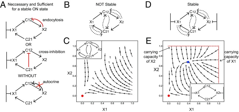
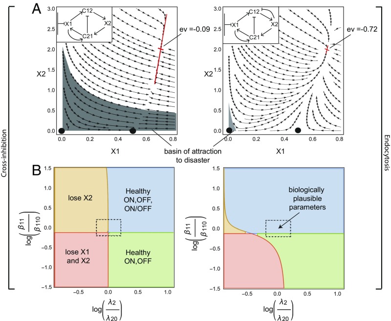
Cells in tissues communicate by secreted growth factors (GF) and other signals. An important function of cell circuits is tissue homeostasis: maintaining proper balance between the amounts of different cell types. Homeostasis requires negative feedback on the GFs, to avoid a runaway situation in which cells stimulate each other and grow without control. Feedback can be obtained in at least two ways: endocytosis in which a cell removes its cognate GF by internalization and cross-inhibition in which a GF down-regulates the production of another GF. Here we ask whether there are design principles for cell circuits to achieve tissue homeostasis. We develop an analytically solvable framework for circuits with multiple cell types and find that feedback by endocytosis is far more robust to parameter variation and has faster responses than cross-inhibition. Endocytosis, which is found ubiquitously across tissues, can even provide homeostasis to three and four communicating cell types. These design principles form a conceptual basis for how tissues maintain a healthy balance of cell types and how balance may be disrupted in diseases such as degeneration and fibrosis.

Keywords: bistability; cell circuits; mathematical models; systems medicine; tissue biology.


Conflict of interest statement

The authors declare no conflict of interest.

Figures

Fig. 1.
Fig. 1.
Homeostasis of cell numbers through circuits of communicating cells. (A) An ON state exists when cell numbers converge to the same steady-state concentration starting from a range of different initial conditions. (B) The ON state requires careful regulation, because if proliferation and removal rates are unequal cells rise to a high carrying capacity or decline to zero. (C) A schematic phase portrait of the FB–MP coculture experiment, showing its three fixed points: a stable OFF state (red), a stable ON state (blue), and an ON–OFF state (green). (D) The FB–MP circuit topology: CSF1 cross-inhibits PDGF gene expression, FB and MP endocytose their GFs, and FB secrete PDGF. (Lower) Description of the interactions in the circuits. (E) The phase portrait of the FB–MP circuit provided by the model of Eqs. 14, using the biologically plausible parameter values λ1=3.7,λ2=2.2,μ=1,β12=3.4,β11=6.8,andα12=10,α21=1. The axes are dimensionless cell numbers, with conversion factors to cell numbers shown. Examples of trajectories that go to each fixed point are shown in color. (F) Dynamics of the cell and GF concentrations for the trajectories highlighted in E as well as two other trajectories for each fixed point.
Fig. 2.
Fig. 2.
A necessary and sufficient condition for a stable ON state in cell circuits. (A) The GF for the cell that is far from carrying capacity (C12) must be inhibited either by endocytosis or by cross-regulation and must have no autocrine loop. (B) Circuits in which neither cell type is close to carrying capacity do not have a stable ON state. (C) Stream plot of one of the circuits in B shows that cell numbers either degenerate to zero (marked in red) or grow without bound. (D) Circuits in which both cells are close to carrying capacity are stable with no need for cross-regulation or feedback. (E) Stream plot of one of the circuits in D shows that even without regulation on the GFs cells reach either an OFF state (marked in red) or an ON state (marked in blue).
Fig. 3.
Fig. 3.
Endocytosis is a more robust and rapid mechanism than cross-regulation for cell circuits to stabilize their ON state. (A) A mathematically controlled comparison of endocytosis and cross-inhibition mechanisms shows that endocytosis (Right) provides a smaller basin of attraction for losing one or both cell types (gray region). The endocytosis circuit also reaches the stable ON state eightfold faster, as indicated by its more negative Jacobian eigenvalue (ev) at the fixed point. (B) The circuit with endocytosis is more robust with respect to parameter variation than the circuit with cross-inhibition, in the sense that it is less prone to lose one or both cell types. The center of the dashed rectangle corresponds to parameter values of λ1=8,λ2=8,μ=1,β11=4.4,andα21=15.0. For the circuit with cross-inhibition (Left) β12=0.35andα12=0.001. For the circuit with endocytosis (Right) β12=1.99andα12=5. Axes are log relative variation from these parameters.
Fig. 4.
Fig. 4.
The two-cell circuits can scale up to form a modular, bistable circuit made of three and four cell types. (A) A typical tissue unit made of parenchymal cells, macrophages, fibroblasts, and endothelial cells. (B) A three-cell circuit topology made of modules of the observed two-cell circuit topology. Fibroblasts (F) and parenchymal cells (P) have a carrying capacity, whereas macrophages (M) are far from carrying capacity. In this circuit, macrophages integrate the responses of the other two cell types. (C) Dynamics of cell numbers show that all cell types converge to an ON steady state from a wide range of initial conditions (blue curves). For initial cell numbers below a threshold cells decline to zero (OFF state, red lines). The parameters used are β12=β23=β32=10,β11=5,α12=α21=α23=α32=10,andλ1=λ2=λ3=10. (D) A four-cell circuit topology made of modules of the observed two-cell circuit topology with endothelial cells (E) that are far from carrying capacity. (E) The same as in C with the parameters β12=β23=β32=β24=β4=10,β11=5,α12=α21=α23=α32=α24=α4=10,andλ1=λ2=λ3=λ4=10.

References

    1. Stefater JA, 3rd, Ren S, Lang RA, Duffield JS. Metchnikoff’s policemen: Macrophages in development, homeostasis and regeneration. Trends Mol Med. 2011;17:743–752. - PMC - PubMed
    1. Wynn TA, Chawla A, Pollard JW. Macrophage biology in development, homeostasis and disease. Nature. 2013;496:445–455. - PMC - PubMed
    1. Okabe Y, Medzhitov R. Tissue biology perspective on macrophages. Nat Immunol. 2016;17:9–17. - PubMed
    1. Pierce GF, et al. Platelet-derived growth factor and transforming growth factor-beta enhance tissue repair activities by unique mechanisms. J Cell Biol. 1989;109:429–440. - PMC - PubMed
    1. Michalopoulos GK, DeFrances MC. Liver regeneration. Science. 1997;276:60–66. - PubMed

Publication types

LinkOut - more resources