Review
. 2018 Feb 12;16(3):125-142.
doi: 10.1038/nrmicro.2017.170.

Rewiring cellular networks by members of the Flaviviridae family

Affiliations
Review

Rewiring cellular networks by members of the Flaviviridae family

Christopher J Neufeldt et al. Nat Rev Microbiol. .

Abstract

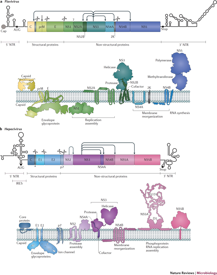
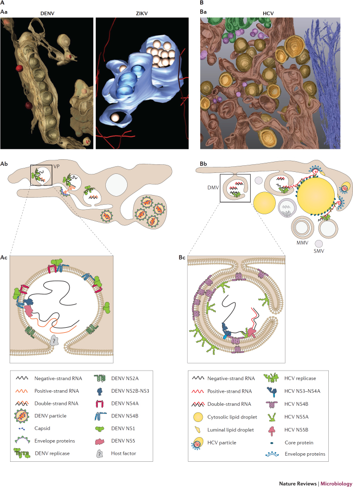
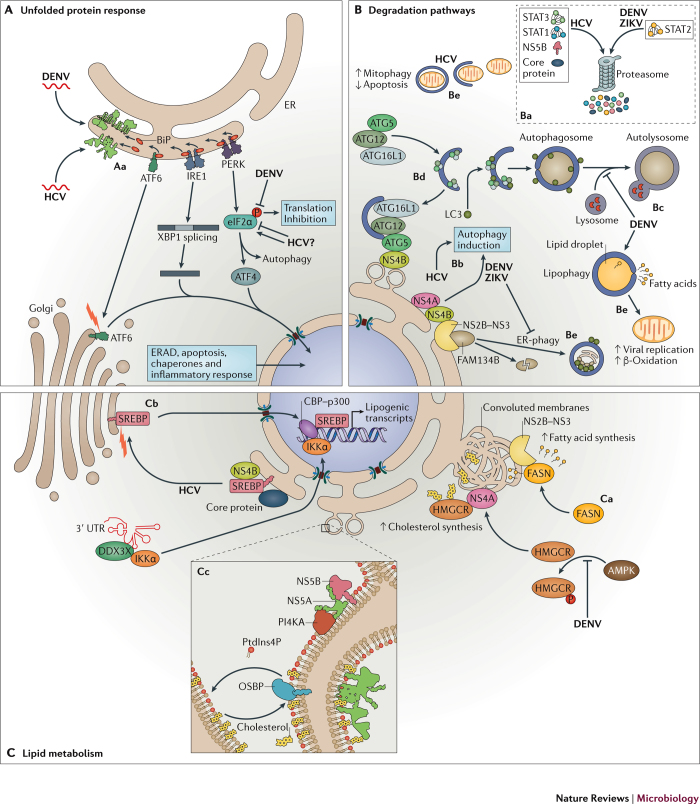
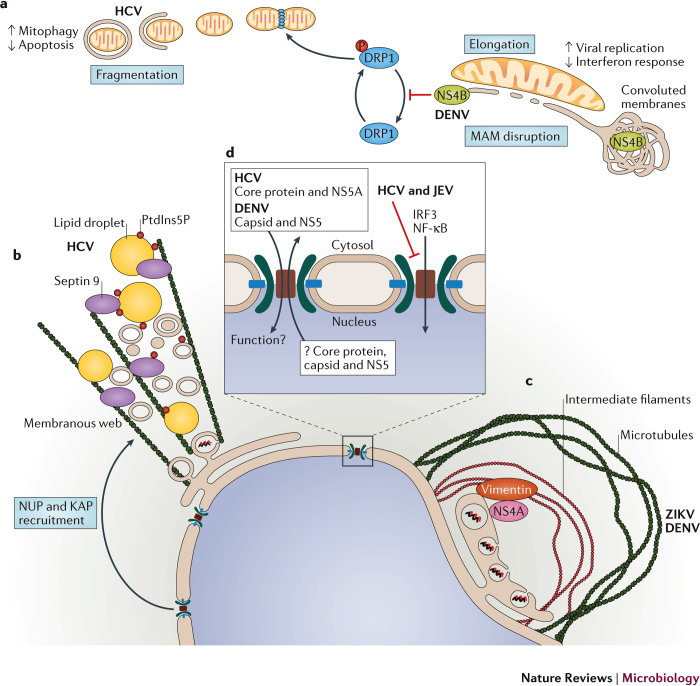
Members of the Flaviviridae virus family comprise a large group of enveloped viruses with a single-strand RNA genome of positive polarity. Several genera belong to this family, including the Hepacivirus genus, of which hepatitis C virus (HCV) is the prototype member, and the Flavivirus genus, which contains both dengue virus and Zika virus. Viruses of these genera differ in many respects, such as the mode of transmission or the course of infection, which is either predominantly persistent in the case of HCV or acutely self-limiting in the case of flaviviruses. Although the fundamental replication strategy of Flaviviridae members is similar, during the past few years, important differences have been discovered, including the way in which these viruses exploit cellular resources to facilitate viral propagation. These differences might be responsible, at least in part, for the various biological properties of these viruses, thus offering the possibility to learn from comparisons. In this Review, we discuss the current understanding of how Flaviviridae viruses manipulate and usurp cellular pathways in infected cells. Specifically, we focus on comparing strategies employed by flaviviruses with those employed by hepaciviruses, and we discuss the importance of these interactions in the context of viral replication and antiviral therapies.


Conflict of interest statement

The authors declare no competing financial interests.

Figures

Figure 1
Figure 1. Flavivirus and Hepacivirus genome organization and membrane topology of mature viral proteins.
The ORF encoding the dengue virus (DENV) (part a) or hepatitis C virus (HCV) (part b) polyprotein and the predicted secondary structures of the 5′ and 3′ non-translating regions (NTR) are depicted on the top of each panel. a | The DENV genome contains a type 1 cap structure at the 5′ end. Polyprotein cleavage by cellular signal peptidases is indicated by scissors. Arrows denote the cleavage by the viral protease, whereas the black vertical arrow indicates cleavage by the Golgi apparatus-resident protease furin. The question mark denotes a DENV polyprotein cleavage performed by an unknown protease. The DENV structural proteins capsid protein C, prM and envelope protein E are constituents of the virion; NS1, the only non-structural protein residing in the lumen of the endoplasmic reticulum (ER), and NS2A are essential for virus replication and production of infectious particles; serine protease subunit NS2B acts as a cofactor for serine protease NS3 and recruits NS3 to ER membranes; NS3 is a multifunctional protein with protease, nucleotide 5′ triphosphatase (NTPase), RNA 5′ triphosphatase and helicase activities; NS4A is an integral membrane protein with membrane curvature-inducing activity; the 2K peptide serves as a signal peptide for co-translational NS4B insertion into the ER membrane; NS4B is a protein with no reported enzymatic activity that interacts with NS3 and is absolutely required for virus replication; NS5 consists of an N-terminal domain that possesses guanylyltransferase, guanine-N7-methyltransferase and nucleoside-2′-O-methyltransferase activities involved in 5′-RNA capping and methylation of the viral genome, and a C-terminal domain with RNA-dependent RNA polymerase activity responsible for viral RNA synthesis. b | The HCV RNA genome is ∼9.6 kb long, uncapped and flanked by highly structured 5′ and 3′ NTRs. The 5′ NTR contains a type III internal ribosome entry site (IRES) that directs the cap-independent translation of the viral RNA genome. Polyprotein cleavage by the viral protease is indicated by arrows, whereas cleavage by cellular signal peptidases is indicated by scissors. The cleavage by the cellular signal peptide peptidase resulting in the removal of the HCV core carboxy- terminal region is indicated by an asterisk. The core protein and the envelope glycoproteins E1 and E2 constitute the viral particle, whereas p7 and NS2 support particle assembly yet are not incorporated into virions; the NS2 C-terminal domain contains a cysteine protease that catalyses the cleavage of the NS2–NS3 junction; NS3 contains serine protease, RNA helicase and NTPase activities; NS4A acts as cofactor for the NS3 protease and anchors NS3 to ER membranes; NS4B is involved in the formation of the HCV replication organelle; NS5A is a phosphoprotein with an intrinsically unfolded C-terminal region that mediates interactions with numerous cellular proteins; NS5B is the viral RNA-dependent RNA polymerase responsible for RNA synthesis. Note that only the DENV capsid and NS1 as well as HCV NS5A are shown as dimers, but additional viral proteins form homodimers and heterodimers or oligomeric complexes. AUG, methionine (start codon); D1, domain 1. Part b is adapted from Ref. , Macmillan Publishers Limited. PowerPoint slide
Figure 2
Figure 2. Three-dimensional structure and organization of Flavivirus and Hepacivirus replication organelles.
A | Architecture of Flavivirus replication organelles. Aa | Three-dimensional surface rendering of dengue virus (DENV) and Zika virus (ZIKV) replication compartments. The endoplasmic reticulum (ER) is shown in brown and blue for DENV and ZIKV, respectively. Virus-induced vesicles are in light brown for DENV and dark blue for ZIKV. Virus particles are depicted in red (DENV) and gold (ZIKV). Ab | Schematic representation of the Flavivirus replication and assembly compartments. Genome replication occurs within vesicle packets (VPs) formed upon ER membrane invagination. Pore-like openings connect the interior of vesicles with the cytosol to allow for metabolite exchange and trafficking of the newly synthesized RNA genome. Virions assemble through nucleocapsid budding within ER cisternae close to VPs. Particles arrange in crystalline arrays within swollen ER cisternae connected to VPs. Ac | Model of Flavivirus-induced vesicle biogenesis. Negative membrane curvatures might be induced by non-structural protein 4A (NS4A) and NS4B amphipathic α-helices that are partially embedded within the ER luminal membrane leaflet. Negative curvature might be stabilized by homo-oligomerization and hetero-oligomerization between NS4A and NS4B along with interaction with NS1 dimers associated with the luminal side of the vesicle. NS2A and other host factors might further contribute to induce membrane alterations and stabilize the pore-like opening. B | Architecture of Hepacivirus replication organelles. Ba | Hepatitis C virus (HCV) membranous web as revealed by electron tomography and three-dimensional reconstruction of infected cells. The ER is shown in dark brown. Double-membrane vesicle (DMV) outer membranes are depicted in light brown and inner membranes in orange. Cytoskeletal filaments, Golgi apparatus cisternae and single-membrane vesicles (SMVs) are shown in blue, green and violet, respectively. Bb | Schematic representation of the HCV replication and assembly compartment. DMVs form as ER protrusions and contain the non-structural proteins that are responsible for viral genome replication. Replicase activity might cease upon vesicle closure (grey shaded vesicle). Alternatively, the viral replicase might be associated with the exterior of DMVs. Newly synthetized RNA might be delivered to assembly sites by NS5A and serine protease NS3 in a process assumed to involve core protein-loaded cytosolic lipid droplets. Formation of nucleocapsids is concomitant with particle budding into the ER lumen. Bc | Model of HCV-induced DMV biogenesis. The amphipathic α-helices and the oligomerization capabilities of the replicase proteins NS4B and NS5A, perhaps along with components of the autophagy machinery (not shown), might promote positive membrane curvature and DMV formation. MMVs, multimembrane vesicles. Part Aa is adapted with permission from Refs ,, Elsevier. Part Bb is adapted from Ref. . PowerPoint slide
Figure 3
Figure 3. Cellular pathways co-opted by Flavivirus and Hepacivirus.
A | Expression of the viral proteins during hepatitis C virus (HCV) and dengue virus (DENV) infection recruits immunoglobulin heavy chain-binding protein (BiP), resulting in depletion of BiP from the endoplasmic reticulum (ER)-stress sensors cyclic AMP-dependent transcription factor ATF6-α (ATF6), serine/threonine-protein kinase/endoribonuclease IRE1 and eukaryotic translation initiation factor 2-α kinase 3 (PERK). Aa | These BiP-less sensors become activated to induce the unfolded protein response (UPR) pathway. HCV and DENV have developed strategies to manipulate UPR pathways to promote virus replication (see main text for details). B | Manipulation of the cellular degradation pathways. Ba | HCV, DENV and Zika virus (ZIKV) infection induce proteasome-mediated degradation of innate signalling proteins to dampen the antiviral immune response. Bb | Viral infection triggers autophagy and alters the autophagy flux. Bc | For instance, established DENV infection suppresses the autophagic flux, blocking the fusion between autophagosomes and lysosomes. Bd | HCV non-structural proten 4B (NS4B) interacts with autophagy protein 5 (ATG5) and recruits the autophagosome membrane elongation complex (ATG5–ATG12–ATG16L1) to the membranous web, contributing to its formation. Be | In addition to general autophagy, selective autophagic pathways are hijacked during Flaviviridae infection (see main text for details). C | Subversion of lipid homeostasis. Ca | DENV serine protease NS3 recruits fatty acid synthase (FASN) to replication compartments to stimulate lipid biosynthesis. De novo synthesized fatty acids are incorporated into replication organelles (ROs). 3-Hydroxy-3-methylglutaryl-CoA reductase (HMGCR) is recruited to DENV ROs, where it colocalizes with NS4A. Inactivation of the protein kinase 5′-AMP-activated protein kinase (AMPK) during DENV infection enhances HMGCR enzymatic activity, which results in higher cholesterol levels within ROs. Cb | During HCV infection, both viral proteins and regulatory elements within the 3′ UTR of the viral genome activate the transcription of lipogenic genes through sterol regulatory element-binding protein (SREBP)-mediated pathways (see main text for details). Cc | HCV NS5A and NS5B recruit and stimulate phosphatidylinositol 4-kinase-α (PI4KA) activity, thus increasing the local concentration of phosphatidylinositol-4- phosphate (PtdIns4P). Lipid transfer proteins such as oxysterol-binding protein 1 (OSBP) bind to PtdIns4P-rich membranes and release cholesterol in exchange for PtdIns4P. ATF4, cyclic AMP-dependent transcription factor ATF-4; ATG12, ubiquitin-like protein ATG12; ATG16L1, autophagy-related protein 16-like 1; CBP, CREB-binding protein; DDX3X, ATP-dependent RNA helicase DDX3X; eIF2α, eukaryotic translation initiation factor 2 subunit 1; ERAD, ER-associated degradation; FAM134B, reticulophagy regulator 1; IKKα, inhibitor of nuclear factor-κB kinase subunit-α; LC3, microtubule-associated proteins 1A/1B light chain 3B (also known as MAP1LC3B); p300, histone acetyltransferase p300; STAT1, signal transducer and activator of transcription 1; STAT2, signal transducer and activator of transcription 2; STAT3, signal transducer and activator of transcription 3; XBP1, X-box-binding protein 1. PowerPoint slide
Figure 4
Figure 4. Cellular organelles co-opted by Flavivirus and Hepacivirus.
a | Flaviviridae viruses alter mitochondrial morphodynamics, leading to mitochondrial elongation in the case of dengue virus (DENV) or mitochondrial fragmentation in the case of hepatitis C virus (HCV), through the manipulation of specific mitochondrial fission and fusion proteins. b | HCV infection increases the intracellular levels of septin 9 and phosphatidylinositol 5-phosphate (PtdIns5P). The septin 9 interaction with PtdIns5P modulates lipid droplet growth and recruitment to the perinuclear area in a microtubule-dependent manner, thus creating an environment that favours viral replication. HCV proteins also recruit components of the nuclear transport machinery to regions of viral replication and assembly. c | DENV and ZIKV infection causes the rearrangement of microtubules and intermediate filaments surrounding replication organelles and leads to nuclear distortion in the case of ZIKV infection. d | Several Flaviviridae virus proteins contain nuclear localization and nuclear export signal sequences and are recruited to the nuclear compartment in infected cells. However, the dynamics of nuclear import and/or export as well as the specific function of these proteins within the nuclear compartment are not clear. HCV and Japanese encephalitis virus (JEV) impair nuclear import of immune transcription factors to limit immune activation in infected cells. DRP1, dynamin 1-like protein; IRF3, interferon regulatory factor 3; KAP, keratin-associated protein; MAM, mitochondrial-associated membrane; NS, non-structural; NF-κB, nuclear factor-κB; NUP, nuclear pore complex protein. PowerPoint slide

References

    1. [No authors listed.] Taxonomy. International Committee on Taxonomy of Viruses (ICTV)https://talk.ictvonline.org/taxonomy/ (2017).
    1. [No authors listed.] Hepatitis C fact sheet. World Health Organizationhttp://www.who.int/mediacentre/factsheets/fs164/en/ (2017).
    1. Manns MP, et al. Hepatitis C virus infection. Nat. Rev. Dis. Primers. 2017;3:17006. doi: 10.1038/nrdp.2017.6. - DOI - PubMed
    1. Stanaway JD, et al. The global burden of dengue: an analysis from the Global Burden of Disease Study 2013. Lancet Infect. Dis. 2016;16:712–723. doi: 10.1016/S1473-3099(16)00026-8. - DOI - PMC - PubMed
    1. Hadinegoro SR, et al. Efficacy and long-term safety of a dengue vaccine in regions of endemic disease. N. Engl. J. Med. 2015;373:1195–1206. doi: 10.1056/NEJMoa1506223. - DOI - PubMed

Publication types

LinkOut - more resources