

Case Reports
doi: 10.4103/jips.jips_136_17.
Prosthetic rehabilitation in neurosurgical cranioplasty
Affiliations
- PMID: 29430147
- PMCID: PMC5799974
- DOI: 10.4103/jips.jips_136_17
Item in Clipboard
Case Reports
Prosthetic rehabilitation in neurosurgical cranioplasty
J Indian Prosthodont Soc.
2018 Jan-Mar.
Abstract
The defects of the skull cause mechanical vulnerability of the brain, esthetic disfigurement, and transmission of vibrations and pulsation of the brain. Subsequent cranioplasty may be required to compensate for the defect and to alleviate various signs and symptoms. When long-term outcome of biomaterial use in pediatric cases is limited, alloplastic cranioplasty in adults are supported by several large case series. This case report narrates cranioplasty using titanium alloplastic implant material.
Keywords: Cranioplasty; prosthesis; reconstruction; titanium implants; trauma.
Conflict of interest statement
There are no conflicts of interest.
Figures
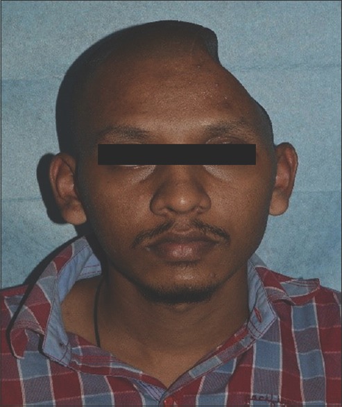
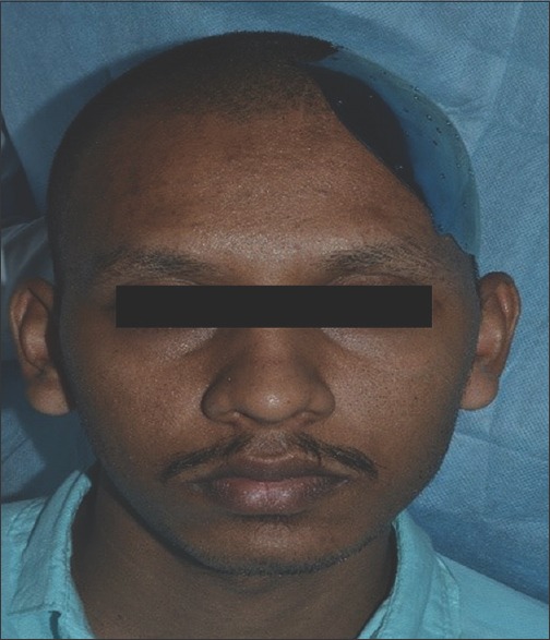
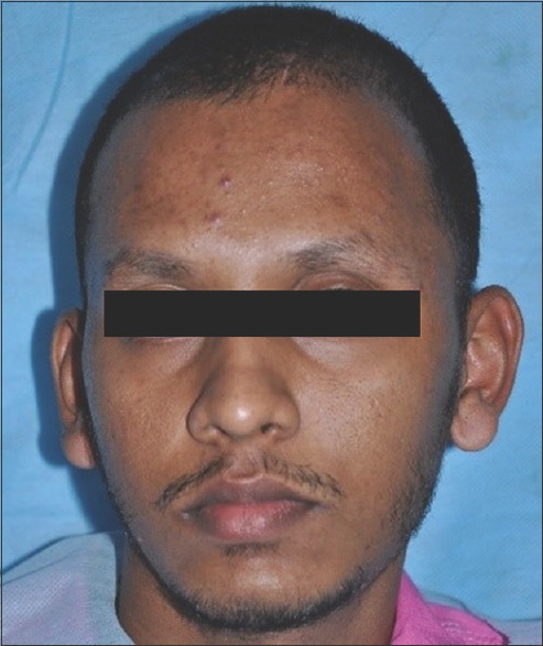
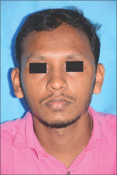
Preoperative photographs showing left cranial bone defect (frontal view)
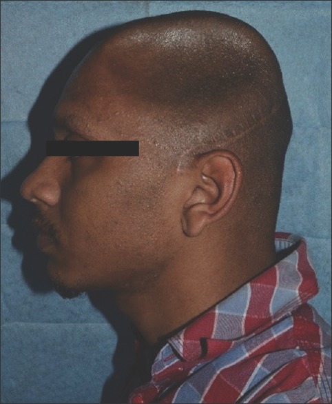
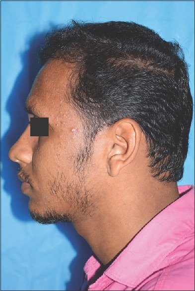
Preoperative photographs (lateral view)
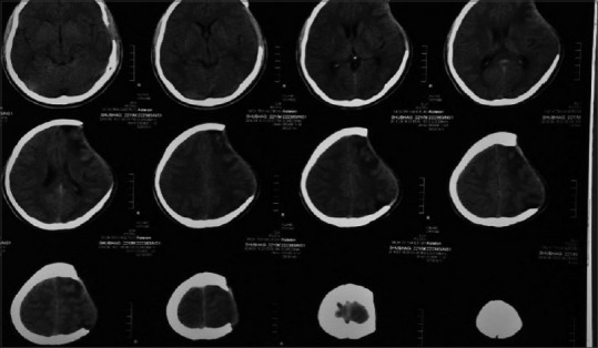
Computed tomography with skull defect
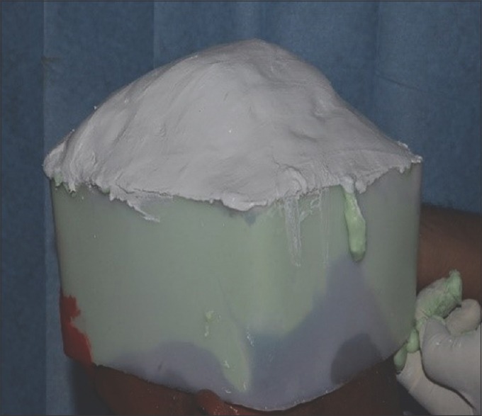
Impression of the defect
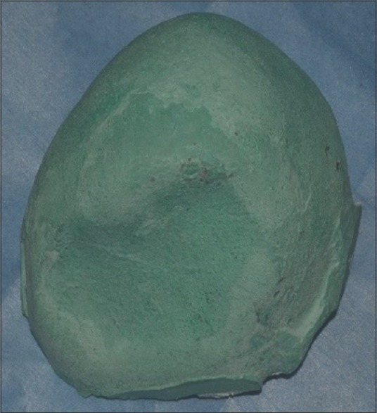
Retrieved cast
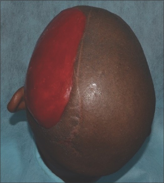
Wax pattern of the defect
Extracranial tryin of titanium plate
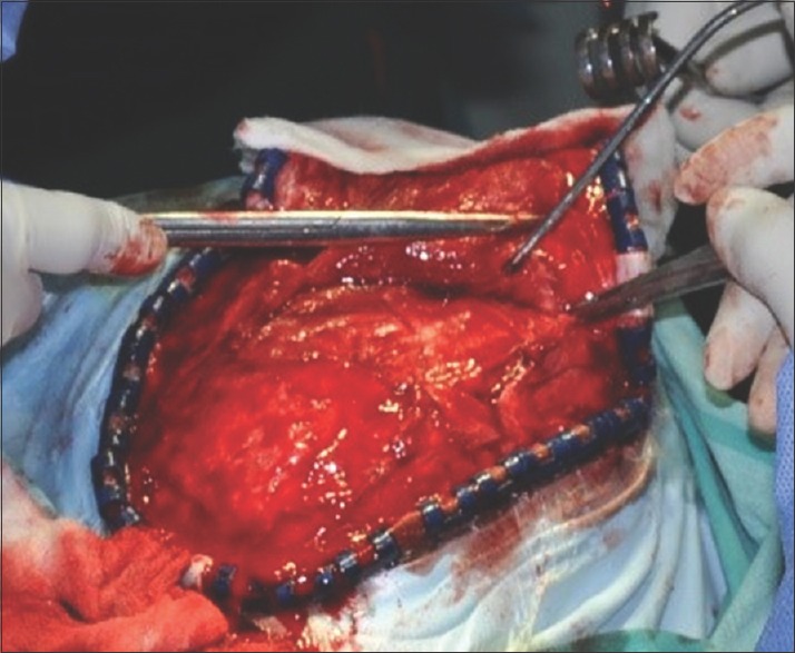
Neurosurgical procedure
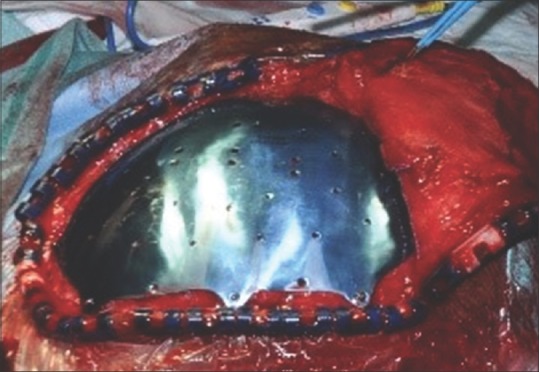
Neurosurgical procedure showing titanium implant in position
Sutures placed
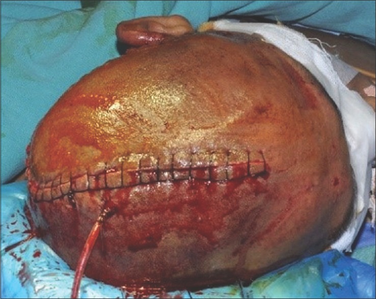
Postoperative view showing excellent adaptation and symmetry
Postoperative aerial view
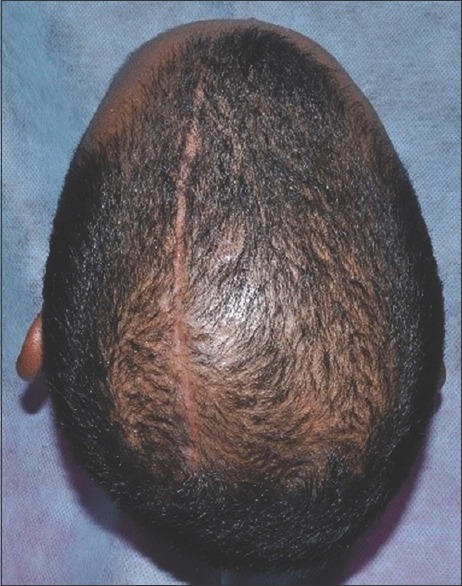
Follow-up 8 months
Follow-up 8 months
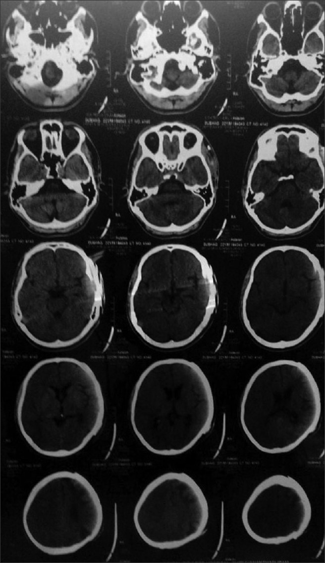
Computed tomography scan after 10 months
Similar articles
-
Cranioplasty Using Three-Dimensional-Printed Polycaprolactone Implant and Free Latissimus Dorsi Musculocutaneous Flap in a Patient with Repeated Wound Problem following Titanium Cranioplasty.Arch Plast Surg. 2022 Dec 13;49(6):740-744. doi: 10.1055/s-0042-1748656. eCollection 2022 Nov. Arch Plast Surg. 2022. PMID: 36523917 Free PMC article.
-
Deformation of a Titanium Calvarial Implant following Trauma: A Case Report.Craniomaxillofac Trauma Reconstr. 2016 Jun;9(2):158-61. doi: 10.1055/s-0035-1567810. Epub 2015 Nov 5. Craniomaxillofac Trauma Reconstr. 2016. PMID: 27162574 Free PMC article.
-
Histological Processing of CAD/CAM Titanium Scaffold after Long-Term Failure in Cranioplasty.Materials (Basel). 2022 Jan 27;15(3):982. doi: 10.3390/ma15030982. Materials (Basel). 2022. PMID: 35160928 Free PMC article.
-
Reconstruction of cranial bone defects using alloplastic implants produced from a stereolithographically-generated cranial model.Ann Acad Med Singap. 1999 Sep;28(5):692-6. Ann Acad Med Singap. 1999. PMID: 10597355 Review.
-
Implants for cranioplasty.Otolaryngol Clin North Am. 1995 Apr;28(2):381-400. Otolaryngol Clin North Am. 1995. PMID: 7596618 Review.
Cited by
-
Metal-polyphenol networks-modified tantalum plate for craniomaxillofacial reconstruction.Sci Rep. 2024 Jan 10;14(1):1023. doi: 10.1038/s41598-024-51640-4. Sci Rep. 2024. PMID: 38200230 Free PMC article.
-
Rapid prototyping technology for cranioplasty: A case series.J Indian Prosthodont Soc. 2019 Apr-Jun;19(2):184-189. doi: 10.4103/jips.jips_295_18. J Indian Prosthodont Soc. 2019. PMID: 31040554 Free PMC article.
-
Multidimensional evaluation of prosthetically rehabilitated\cranial defects using key behavior change inventory.J Indian Prosthodont Soc. 2021 Jul-Sep;21(3):311-315. doi: 10.4103/jips.jips_587_20. J Indian Prosthodont Soc. 2021. PMID: 34380821 Free PMC article.
-
Stereophotogrammetric Method for Fabrication of Cranioplast Using Digital Facial Scan Technology - A Case Report.Ann Maxillofac Surg. 2022 Jul-Dec;12(2):240-243. doi: 10.4103/ams.ams_279_21. Epub 2022 Dec 26. Ann Maxillofac Surg. 2022. PMID: 36874772 Free PMC article.
-
Diversifying the rehabilitation of calvarial defects: Rejuvenating precision: A case series.Natl J Maxillofac Surg. 2021 Sep-Dec;12(3):426-430. doi: 10.4103/njms.NJMS_288_20. Epub 2021 Dec 13. Natl J Maxillofac Surg. 2021. PMID: 35153444 Free PMC article.
References
-
- Pavaiya A, Tyagi VK, Tripathi A, Singh SV, Chand P. Cranioplasty with alloplastic cranial implant. J Indian Prosthodont Soc. 2009;9:109–11.
-
- Goiato MC, Pesqueira AA, dos Santos DM, Antenucci RM, Ribeiro Pdo P. Evaluation of dimensional change and detail reproduction in silico nes for facial prostheses. Acta Odontol Latinoam. 2008;21:85–8. - PubMed
-
- Punchak M, Chung LK, Lagman C, Bui TT, Lazareff J, Rezzadeh K, et al. Outcomes following polyetheretherketone (PEEK) cranioplasty: Systematic review and meta-analysis. J Clin Neurosci. 2017;41:30–5. - PubMed
-
- Alqutaibi AY. Materials of facial prosthesis: History and advances. Int J Contemp Dent Med Rev. 2015;2015:4.
Publication types
LinkOut - more resources
Full Text Sources
Other Literature Sources