. 2018 Feb 1:352:55-64.
doi: 10.1016/j.jphotochem.2017.10.042. Epub 2017 Oct 28.

Development of colloidally stable carbazole-based fluorescent nanoaggregates

Affiliations

Development of colloidally stable carbazole-based fluorescent nanoaggregates

Denis Svechkarev et al. J Photochem Photobiol A Chem. .

Abstract

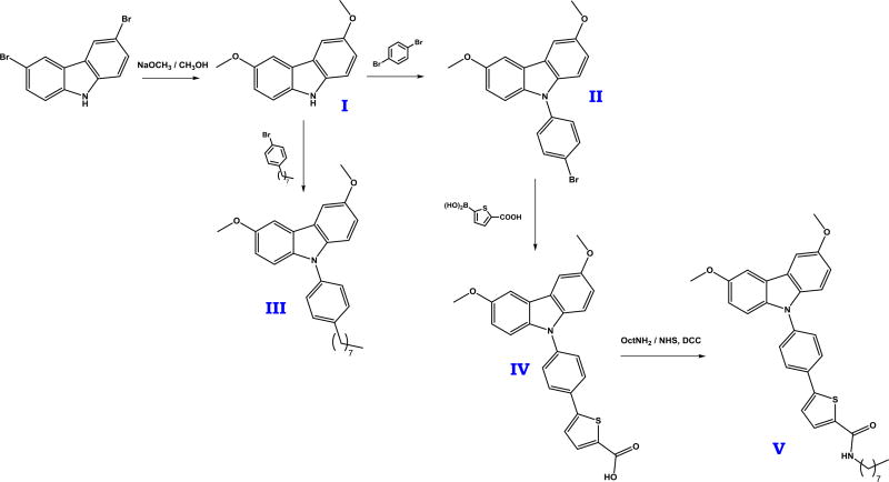
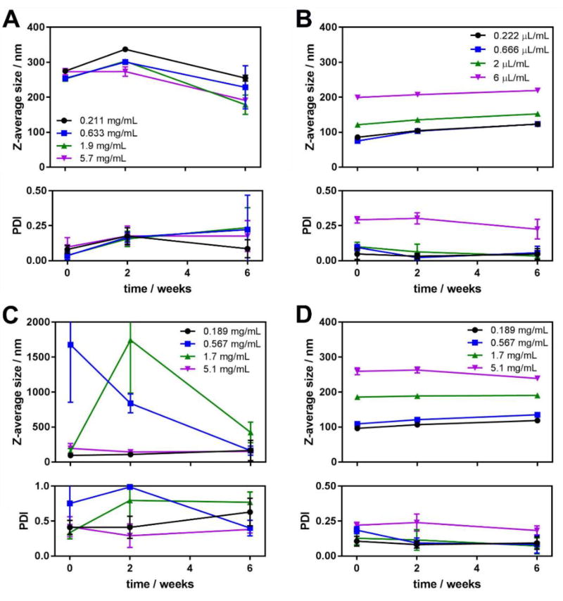
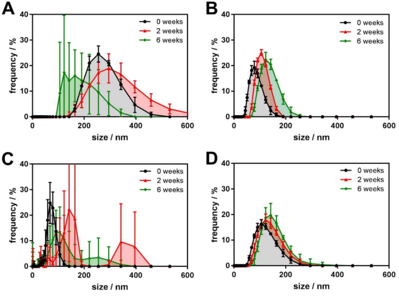
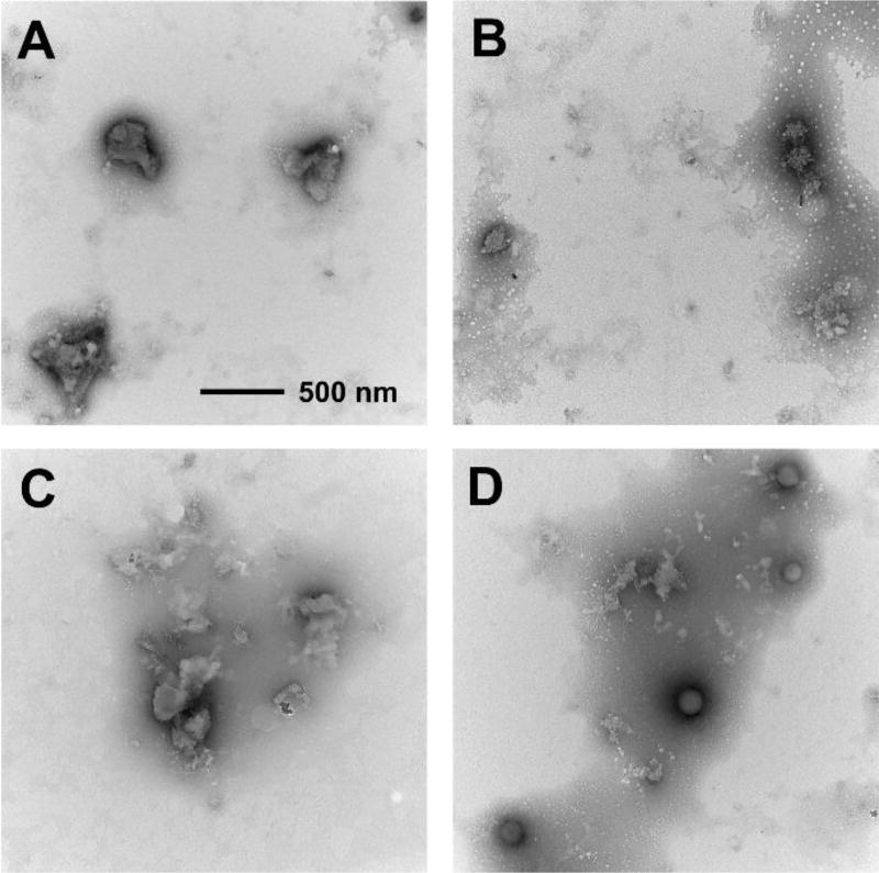
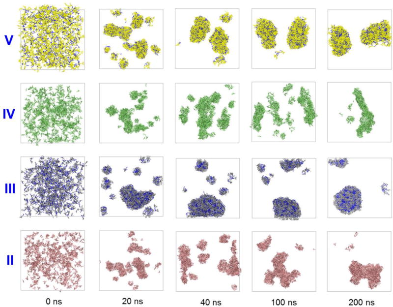
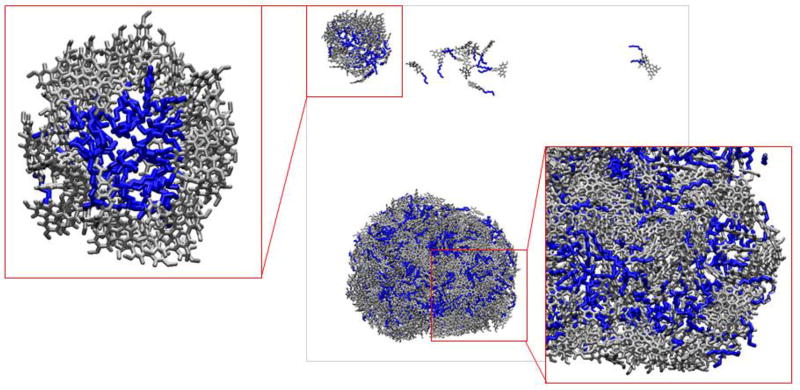
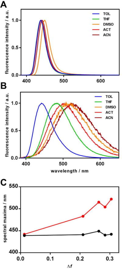
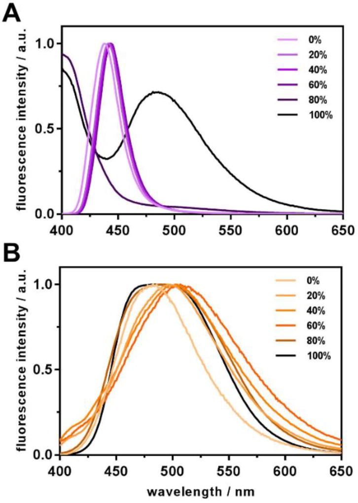
Fluorescent nanomaterials require high colloidal stability for effective use in imaging and sensing applications. We herein report the synthesis of carbazole-based organic fluorescent nanoaggregates, and demonstrate the superior colloidal stability of alkyl-substituted dye aggregates over their non-alkylated analogs. The role of alkyl chains in self-assembly and stability of such nanoaggregates is discussed based on both experimental and molecular dynamics simulation data, and spectral characteristics of the precursor dyes and their aggregates are described. The obtained results provide new insights on development of colloidally stable organic fluorescent nanomaterials with low polydispersity.

Keywords: MD simulation; alkyl chain; dynamic light scattering; fluorescent imaging; polydispersity.


Conflict of interest statement

Conflict of Interests The authors declare no conflicting financial interests.

Figures

Figure 1
Figure 1
Synthetic scheme leading to target compounds III and V.
Figure 2
Figure 2
Average aggregate size (top) and polydispersity index (bottom) obtained from cumulants analysis of the DLS experiment data for the aggregates of compounds II (A), III (B), IV (C) and V (D) in 0, 2 and 6 weeks after preparation. Each experimental point is an average of 5 independent measurements, and the error bars represent the standard deviation.
Figure 3
Figure 3
Aggregate size distributions obtained from DLS experiment for the fluorescent aggregates: A – II, 0.633 mg/mL; B – III, 0.666 µL/mL; C – IV, 0.567 mg/mL; and D – V, 0.567 mg/mL at 0, 2 and 6 weeks after preparation. Each experimental point is an average of 5 independent measurements, and the error bars represent the standard deviation.
Figure 4
Figure 4
Transmission electron micrographs of the aggregates of compounds II (A), III (B), IV (C) and V (D). All images are taken at the same magnification (note the scale bar in section A).
Figure 5
Figure 5
Dye aggregation dynamics illustrated by MD simulations snapshots of the systems consisting of 729 dye molecules solvated by ~2·105 water molecules (water molecules are omitted for clarity of representation).
Figure 6
Figure 6
Snapshot of the system of 729 molecules of III in approximately 2·105 water molecules (water molecules are omitted for clarity of representation) at 100 ns of simulation.
Figure 7
Figure 7
Intensity-normalized fluorescence spectra of target compounds III (A) and V (B) in various solvents. Spectral maxima for fluorescence are plotted against solvent polarity function Δf (C).
Figure 8
Figure 8
Intensity-normalized fluorescence spectra of target compounds III (A) and V (B) in water-THF binary solvent systems. Water content is indicated in the legends.

References

    1. Walling MA, Novak JA, Shepard JRE. Quantum Dots for Live Cell and In Vivo Imaging. Int. J. Mol. Sci. 2009;10:441–491. doi: 10.3390/ijms10020441. - DOI - PMC - PubMed
    1. Jin S, Hu Y, Gu Z, Liu L, Wu H-C. Application of Quantum Dots in Biological Imaging. J. Nanomater. 2011;2011:1–13. doi: 10.1155/2011/834139. - DOI - PubMed
    1. Li J, Zhu J-J. Quantum dots for fluorescent biosensing and bio-imaging applications. Analyst. 2013;138:2506. doi: 10.1039/c3an36705c. - DOI - PubMed
    1. Kumar P, Bohidar HB, Kumar R. Non-Functionalized Fluorescent Carbon Nanoparticles: In Vitro Imaging and Organic Solvent Sensing Applications. Sci. Adv. Mater. 2015;7:706–713. doi: 10.1166/sam.2015.1892. - DOI
    1. Ali H, Bhunia SK, Dalal C, Jana NR. Red Fluorescent Carbon Nanoparticle-Based Cell Imaging Probe. ACS Appl. Mater. Interfaces. 2016;8:9305–9313. doi: 10.1021/acsami.5b11318. - DOI - PubMed

LinkOut - more resources