

Metabolomic characterization of experimental ovarian cancer ascitic fluid
- PMID: 29430218
- PMCID: PMC5804489
- DOI: 10.1007/s11306-017-1254-3
Metabolomic characterization of experimental ovarian cancer ascitic fluid
Abstract
Introduction: Malignant ascites (MA) is a major cause of morbidity that occurs in 37% of ovarian cancer patients. The accumulation of MA in the peritoneal cavity due to cancer results in debilitating symptoms and extremely poor quality of life. There is an urgent unmet need to expand the understanding of MA to design effective treatment strategies, and to improve MA diagnosis.
Objective: Our purpose here is to contribute to a better characterization of MA metabolic composition in ovarian cancer.
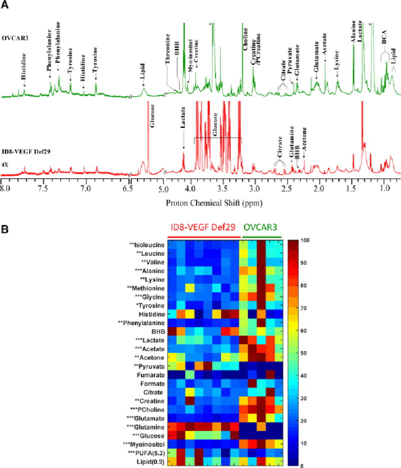
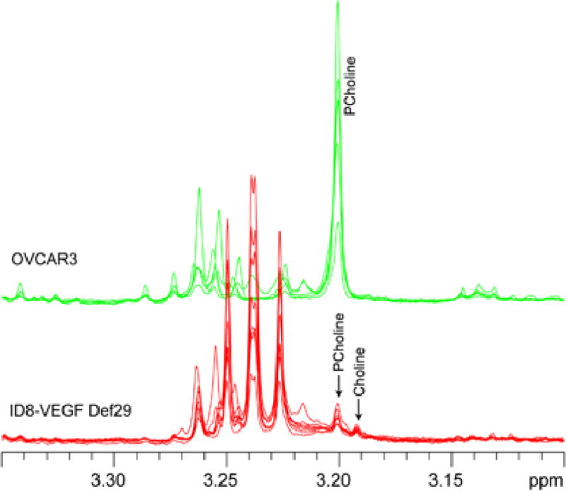
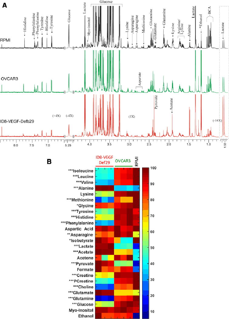
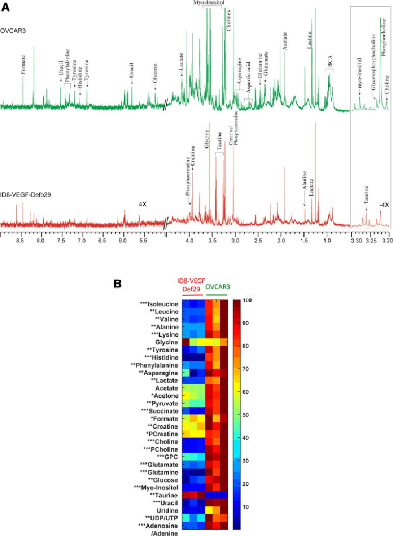
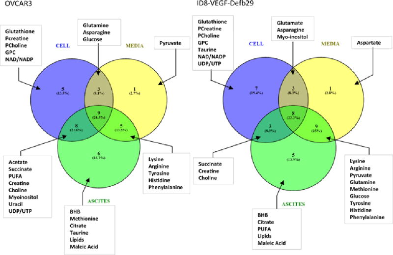
Method: We determined the metabolic composition of ascitic fluids resulting from orthotopic growth of two ovarian cancer cell lines, the mouse ID8-vascular endothelial growth factor (VEGF)-Defb29 cell line and the human OVCAR3 cell line using high-resolution 1H MRS. ID8-VEGF-Defb29 tumors induce large volumes of ascites, while OVCAR3 tumors induce ascites less frequently and at smaller volumes. To better understand the factors driving the metabolic composition of the fluid, we characterized the metabolism of these ovarian cancer cells in culture by analyzing cell lysates and conditioned culture media with 1H NMR.
Results: Distinct metabolite patterns were detected in ascitic fluid collected from OVCAR3 and ID8-VEGF-Defb29 tumor bearing mice that were not reflected in the corresponding cell culture or conditioned medium.
Conclusion: High-resolution 1H NMR metabolic markers of MA can be used to improve characterization and diagnosis of MA. Metabolic characterization of MA can provide new insights into how MA fluid supports cancer cell growth and resistance to treatment, and has the potential to identify metabolic targeting strategies to reduce or eliminate the formation of MA.
Keywords: Ovarian cancer; ascitic fluid; cancer cells and conditioned culture media; high-resolution proton NMR; metabolites; orthotopic tumor implantation.
Conflict of interest statement
CONFLICT OF INTEREST DISCLOSURE The authors declare that they have no conflict of interest.
Figures
Similar articles
-
Copper content in ascitic fluid is associated with angiogenesis and progression in ovarian cancer.J Trace Elem Med Biol. 2021 Dec;68:126865. doi: 10.1016/j.jtemb.2021.126865. Epub 2021 Sep 21. J Trace Elem Med Biol. 2021. PMID: 34601284
-
Vascular endothelial growth factor activating matrix metalloproteinase in ascitic fluid during peritoneal dissemination of ovarian cancer.Oncol Rep. 2003 Jan-Feb;10(1):89-95. Oncol Rep. 2003. PMID: 12469150
-
Ovarian cancer-derived ascitic fluids induce a senescence-dependent pro-cancerogenic phenotype in normal peritoneal mesothelial cells.Cell Oncol (Dordr). 2016 Oct;39(5):473-481. doi: 10.1007/s13402-016-0289-1. Epub 2016 Jul 21. Cell Oncol (Dordr). 2016. PMID: 27444787
-
Malignant ascites: Current therapy options and treatment prospects.Cancer Treat Rev. 2023 Oct 24;121:102646. doi: 10.1016/j.ctrv.2023.102646. Online ahead of print. Cancer Treat Rev. 2023. PMID: 39492370 Review.
-
The role of vascular endothelial growth factor and interleukins in the pathogenesis of severe ovarian hyperstimulation syndrome.Hum Reprod Update. 1997 May-Jun;3(3):255-66. doi: 10.1093/humupd/3.3.255. Hum Reprod Update. 1997. PMID: 9322101 Review.
Cited by
-
PD-L1 near Infrared Photoimmunotherapy of Ovarian Cancer Model.Cancers (Basel). 2022 Jan 26;14(3):619. doi: 10.3390/cancers14030619. Cancers (Basel). 2022. PMID: 35158887 Free PMC article.
-
Peripheral Blood Serum NMR Metabolomics Is a Powerful Tool to Discriminate Benign and Malignant Ovarian Tumors.Metabolites. 2023 Sep 1;13(9):989. doi: 10.3390/metabo13090989. Metabolites. 2023. PMID: 37755269 Free PMC article.
-
Ovarian Cancer Biomarkers: Moving Forward in Early Detection.Adv Exp Med Biol. 2020;1219:355-363. doi: 10.1007/978-3-030-34025-4_18. Adv Exp Med Biol. 2020. PMID: 32130708 Review.
-
Ascites Volumes and the Ovarian Cancer Microenvironment.Front Oncol. 2018 Dec 17;8:595. doi: 10.3389/fonc.2018.00595. eCollection 2018. Front Oncol. 2018. PMID: 30619738 Free PMC article.
-
Biguanide drugs enhance cytotoxic effects of cisplatin by depleting aspartate and NAD+ in sensitive cancer cells.Cancer Biol Ther. 2021 Dec 2;22(10-12):579-586. doi: 10.1080/15384047.2021.1982599. Epub 2021 Oct 31. Cancer Biol Ther. 2021. PMID: 34720054 Free PMC article.
References
-
- Adosraku RK, Choi GT, Constantinou-Kokotos V, Anderson MM, Gibbons WA. NMR lipid profiles of cells, tissues, and body fluids: proton NMR analysis of human erythrocyte lipids. Journal of lipid research. 1994;35(11):1925–1931. - PubMed
Grants and funding
LinkOut - more resources
Full Text Sources
Other Literature Sources