. 2018 Mar;592(6):987-998.
doi: 10.1002/1873-3468.13004. Epub 2018 Feb 19.

The role of OleA His285 in orchestration of long-chain acyl-coenzyme A substrates

Affiliations

The role of OleA His285 in orchestration of long-chain acyl-coenzyme A substrates

Matthew R Jensen et al. FEBS Lett. 2018 Mar.

Abstract

Renewable production of hydrocarbons is being pursued as a petroleum-independent source of commodity chemicals and replacement for biofuels. The bacterial biosynthesis of long-chain olefins represents one such platform. The process is initiated by OleA catalyzing the condensation of two fatty acyl-coenzyme A substrates to form a β-keto acid. Here, the mechanistic role of the conserved His285 is investigated through mutagenesis, activity assays, and X-ray crystallography. Our data demonstrate that His285 is required for product formation, influences the thiolase nucleophile Cys143 and the acyl-enzyme intermediate before and after transesterification, and orchestrates substrate coordination as a defining component of an oxyanion hole. As a consequence, His285 plays a key role in enabling a mechanistic strategy in OleA that is distinct from other thiolases.

Keywords: OleA; X-ray crystallography; thiolase.


Figures

Figure 1
Figure 1
Proposed mechanism of bacterial olefin biosynthesis. A) Representative four-gene (left) and three-gene (right) cluster arrangements for oleABCD. The cluster containing an oleBC fusion (orange) is found in olefin-producing actinobacteria. B) Mechanistic steps of olefin production catalyzed by OleA, OleD, OleC, and OleB. Typical acyl-CoA substrates are between 10 and 16 carbons in length in Xanthomonas campestris. Figure produced from data presented in [–11].
Figure 2
Figure 2
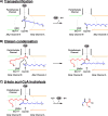
Proposed mechanism of OleA. A) Transesterification: the first acyl-CoA (blue) binds in alkyl channel A (orange) forming an acyl-enzyme intermediate after nucleophilic attack by Cys143. B) Claisen condensation: Glu117 abstracts a proton from C2 of the second substrate (red) bound in alkyl channel B (green), priming nucleophilic attack between the carbanion and the acyl-enzyme intermediate. C) β-keto acyl-CoA hydrolysis: the final CoA-SH is released through hydrolytic displacement by a hypothesized activated water.
Figure 3
Figure 3
Sequence alignment of select OleA enzymes from divergent bacteria highlighting the invariant histidine residue. Xanthomonas campestris pv. campestris str. ATCC 33913, Geobacter uraniireducens Rf4, Shewanella oneidensis MR-1, Streptomyces ambofaciens ATCC 23877, Arthrobacter aurescens TC1, Kineococcus radiotolerans SRS30216, Micrococcus luteus NCTC 2665, Stenotrophomonas maltophilia K279a, Chloroflexus aggregans DSM 9485.
Figure 4
Figure 4
Active site comparisons of OleA His285 variant structures. A) WT OleA active site residues (gray) showing water molecules (red spheres) bound in oxyanion holes 1 and 2 (blue dashed circles) [12]. B) Example simulated annealing omit mFoDFc electron density (contoured at 3σ) from H285N OleA structure (green) used to assign oxidation of Cys143. C) Monomer A active site of H285A OleA (blue). The two conformers of Cys143 are modelled at 80% and 20%. D) Monomer B active site of H285A OleA (blue). Cys143 is modelled as 55% cysteine and 45% sulfenic acid. The water molecule bound in oxyanion hole 2 is modeled at 55% occupancy to match that of the unmodified cysteine. E) Monomer A active site of H285N OleA (green). Cys143 is oxidized to sulfenic acid with the hydroxyl group bound in oxyanion hole 2. F) Monomer B of H285N OleA (green). Cys143 is modelled as two conformers of sulfenic acid each at 50% occupancy. G) Monomer A active site of H285D OleA (cyan). The two conformers of Cys143 are modelled at 60% and 40%. H) Monomer B of H285D OleA (cyan). Cys143 is modelled as 60% and 40% sulfenic acid conformers. All hydrogen bonds are depicted as black dashed lines. Equivalent stereo figures with electron density for panels C–H are in Supporting Information (Fig. S1–S6).
Figure 5
Figure 5
Asn285 side chain interactions near alkyl channel A. A) Overlay of H285N OleA (green) and C143S OleA (yellow, PDB 4KU3) [13]. Asn285 (green) forms two hydrogen bonds with the main chain of Ile345 that do not allow it to shift to the substrate-bound conformation that Ile345 (yellow) adopts when alkyl channel A is occupied. B) Overlay of H285N OleA (green) and C143S OleA (yellow, PDB 4KU3) showing the conformational changes around Ile345 needed to position myristic acid (grey) within alkyl channel A [13].
Figure 6
Figure 6
Position of Cys143 in line with the α-helix 4 dipole moment. WT OleA position of Cys143 in line with the α-helix 4 dipole moment (PDB 3ROW) [12]. His285 shares a hydrogen bond with the side chain of Asn315 and is 3.5 Å from the thiol of Cys143 (black dashed lines).
Figure 7
Figure 7
Spatial arrangement and usage of oxyanion holes in several thiolase enzymes. A) Acetylated HMG-CoA synthase (HMGS, orange) bound with acetoacetyl-CoA (AcAc-CoA, red) (PDB 1×PK) [38]. The first substrate occupies oxyanion hole 2 (red circle) while the second substrate binds in oxyanion hole 1 (blue circle). B) Acetylated FabH coordinating substrate (red) in oxyanion hole 2 (red circle) (PDB 1HNH) [35]. C) Enzyme-substrate complex between C143A OleA and myristoyl-CoA (Myr-CoA, red) (PDB 4KU2) [13]. The carbonyl oxygen of Myr-CoA occupies oxyanion hole 1 (blue circle). D) WT OleA active site and substrate modelling displaying putative oxyanion hole usage before condensation. Myristic acid (blue) is acylated to Cys143 and occupies oxyanion hole 1 and alkyl channel A. Myristoyl-CoA (red) is bound in oxyanion hole 2 and alkyl channel B. Glu117 is modelled in the position found in OleA C143S (PDB 4KU3)) and is located ~3.5 Å from the putative position of myristoyl-CoA C2 atom (red line) [13]. All models are superimposed and viewed in isolation from the same vantage point. Hydrogen bonds are shown as black dashed lines. Sa, Staphylococcus aureus; Ec, Escherichia coli; Xc, Xanthomonas campestris.

References

    1. Channon HJ, Chibnall AC. The ether-soluble substances of cabbage leaf cytoplasm: The isolation of n-nonacosane and di-n-tetradecyl ketone. Biochem J. 1929;23:168–75. - PMC - PubMed
    1. Wackett LP. Engineering microbes to produce biofuels. Curr Opin Biotechnol. 2011;22:388–93. - PubMed
    1. Rude MA, Schirmer A. New microbial fuels: a biotech perspective. Curr Opin Microbiol. 2009;12:274–81. - PubMed
    1. Steen EJ, Kang Y, Bokinsky G, Hu Z, Schirmer A, McClure A, Del Cardayre SB, Keasling JD. Microbial production of fatty-acid-derived fuels and chemicals from plant biomass. Nature. 2010;463:559–62. - PubMed
    1. Friedman L RM. Process for producing low molecular weight hydrocarbons from renewable resources. WO/2008/113014 World Patent. 2008

Publication types

MeSH terms

LinkOut - more resources