. 2018 Apr;285(7):1290-1304.
doi: 10.1111/febs.14405. Epub 2018 Feb 25.

Architecture and hydration of the arginine-binding site of neuropilin-1

Affiliations

Architecture and hydration of the arginine-binding site of neuropilin-1

Filipa Mota et al. FEBS J. 2018 Apr.

Abstract

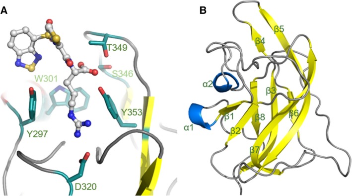
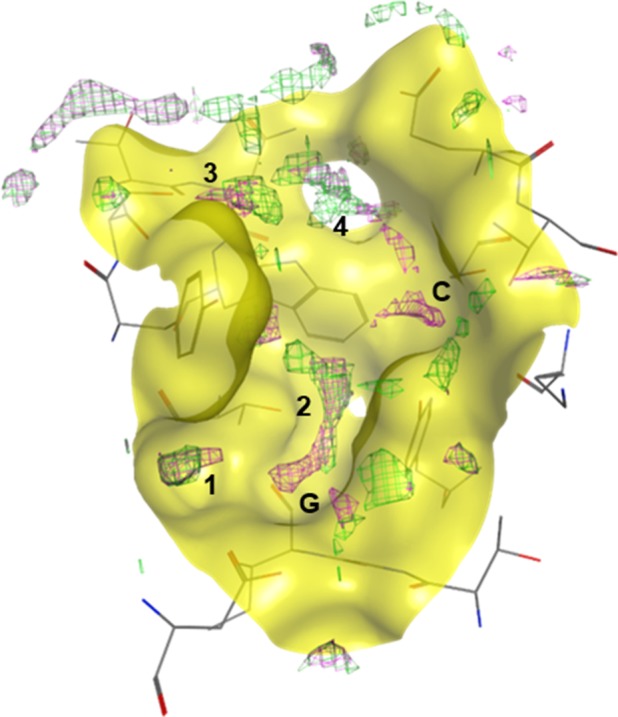
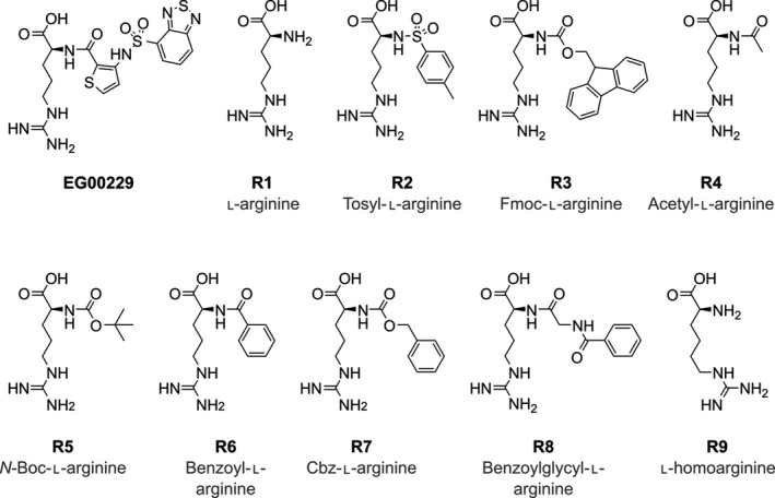
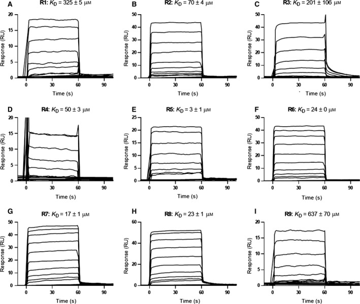
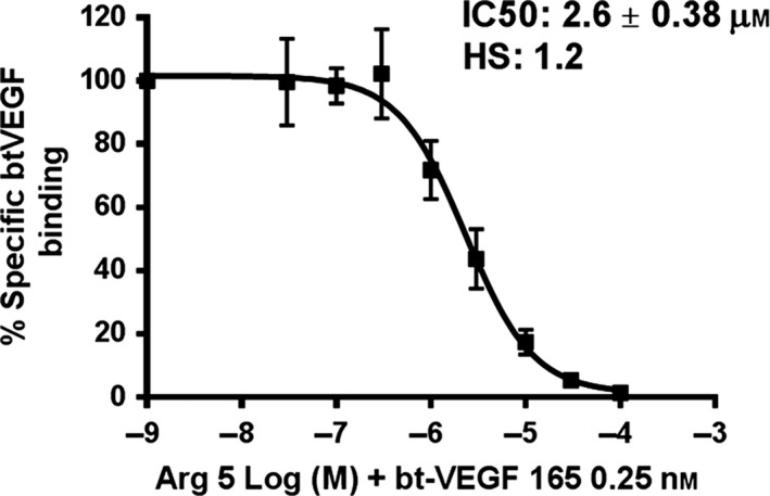
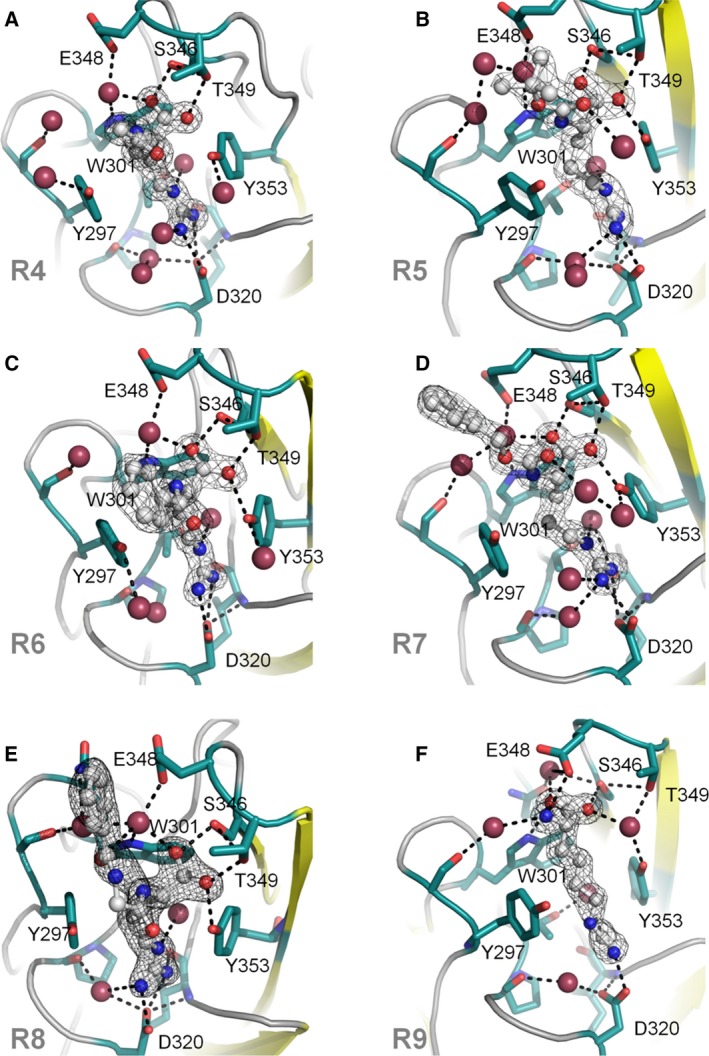
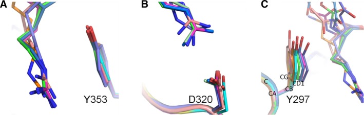
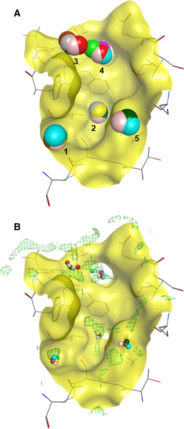
Neuropilin-1 (NRP1) is a transmembrane co-receptor involved in binding interactions with variety of ligands and receptors, including receptor tyrosine kinases. Expression of NRP1 in several cancers correlates with cancer stages and poor prognosis. Thus, NRP1 has been considered a therapeutic target and is the focus of multiple drug discovery initiatives. Vascular endothelial growth factor (VEGF) binds to the b1 domain of NRP1 through interactions between the C-terminal arginine of VEGF and residues in the NRP1-binding site including Tyr297, Tyr353, Asp320, Ser346 and Thr349. We obtained several complexes of the synthetic ligands and the NRP1-b1 domain and used X-ray crystallography and computational methods to analyse atomic details and hydration profile of this binding site. We observed side chain flexibility for Tyr297 and Asp320 in the six new high-resolution crystal structures of arginine analogues bound to NRP1. In addition, we identified conserved water molecules in binding site regions which can be targeted for drug design. The computational prediction of the VEGF ligand-binding site hydration map of NRP1 was in agreement with the experimentally derived, conserved hydration structure. Displacement of certain conserved water molecules by a ligand's functional groups may contribute to binding affinity, whilst other water molecules perform as protein-ligand bridges. Our report provides a comprehensive description of the binding site for the peptidic ligands' C-terminal arginines in the b1 domain of NRP1, highlights the importance of conserved structural waters in drug design and validates the utility of the computational hydration map prediction method in the context of neuropilin.

Database: The structures were deposited to the PDB with accession numbers PDB ID: 5IJR, 5IYY, 5JHK, 5J1X, 5JGQ, 5JGI.

Keywords: SPR; X-ray crystallography; ligand-binding protein; neuropilin; vascular endothelial growth factor (VEGF).


Figures

Figure 1
Figure 1
Structure of the binding site of NRP1‐b1 domain. (A) Ball and stick representation of EG00229 (carbon atoms are coloured grey, nitrogen blue, sulphur yellow and oxygen red) bound to NRP‐b1 (PDB entry 3I97). NRP1‐b1 domain residues involved in the non‐covalent interactions with the ligand are shown in sticks representation. (B) Ribbon diagram of NRP1‐b1 fold: β‐sheets are represented in yellow and α‐helixes in blue.
Figure 2
Figure 2
Prediction model of NRP1‐b1 binding site hydration. Computational analysis performed on PDB entry 3I97, where the green mesh represents the solvation prediction with EG00229 bound to NRP1‐b1, and the pink mesh represents the solvation prediction of NRP1‐b1 in the apo form. Labels 1–4 indicate the regions predicted to be occupied by water molecules in the binding site; G and C indicate the regions occupied by the arginine guanidine and carboxylate groups, respectively, if EG000229 is bound. In the apo form of NRP1‐b1, these regions are also predicted to be occupied by water molecules.
Figure 3
Figure 3
Chemical structures of selected arginine analogues R1–R9.
Figure 4
Figure 4
SPR measurements of arginine analogues R1–R9 binding to immobilised NRP1‐b1. Equilibrium dissociation constants (KD) were calculated using steady‐state binding levels and assuming a 1 : 1 binding model of the arginine analogues R1–R9 to immobilised NRP1‐b1. All sensorgrams are double‐referenced, using a blank surface and sample. Concentration ranges are as follows: R1 and R9 were tested at 121500 μm, R5 was tested at 0.1620 μm, all other analogues were tested at 0.6300 μm (n = 2).
Figure 5
Figure 5
Inhibition of bt‐VEGF‐A165 binding to NRP1 by R5 arginine analogue. The various concentrations of R5 compound were added to the 96‐well plates precoated with NRP1‐b1 protein, followed by addition of 0.25 nm bt‐VEGF‐A165. Non‐specific binding of bt‐VEGF‐A165 to the plates was determined in the absence of NRP1‐b1. R5 analogue competed with bt‐VEGF‐A165 for binding to plates coated with NRP1 with an IC50 of 2.6 μm. Values presented are the means ± SEM obtained from three independent experiments each performed in duplicates.
Figure 6
Figure 6
X‐ray crystal structures of arginine analogues R4R9 bound to NRP1‐b1. Side chains of key residues in the binding site are shown in green, ligands are shown in grey, nitrogen atoms are shown in blue and oxygen atoms are shown in red. Oxygen atoms corresponding to water molecules are larger and shown in raspberry colour. The figures were generated in PyMOL. The electron density of the 2Fo‐Fc map is shown as a wire mesh and contoured to 1 sigma level; hydrogen bonds are represented as black dashes. (A) R4 (PDB entry 5JGI); (B) R5 (PDB entry 5J1X); (C) R6 (PDB entry 5JGQ); (D) R7 (PDB entry 5IYY); (E) R8 (PDB entry 5JHK); (F) R9 (PDB entry 5IJR).
Figure 7
Figure 7
Side chain flexibility of Asp320 and Tyr297 in the NRP1‐b1 binding site. Effect of ligand binding on the side chain conformation of Y297, Y353 and D320 amino acids. The complexed protein structures (pdb IDs: 5JGI – dark pink, 5J1X – dark blue, 5JGQ – lilac, 5IYY – light pink, 5JHK – green, 5IJR – orange) were superimposed over the apo‐structure of NRP1‐b1 domain (pdb code: 1KEX, turquoise colour) (A) π–π stacking between the guanidino‐group of the bound small molecules and phenyl ring of Y353 residue. (B) A stick representation of the side chain rotamers of D320 residue of NRP1‐b1 protein as it has been observed in the X‐ray structures of the complexes. The interacting portion of the arginine analogues is shown as well. (C) A demonstration of the variability of the rotamer conformation of Y297 amino acid upon compound binding. As it is shown in Table 2, the differences between the values of torsion angles Chi1 and Chi2 of the ligand‐bound structures and those in the apo structure are significant and vary depending on the compound.
Figure 8
Figure 8
Analysis of hydration sites in the binding site of NRP1‐b1. (A) Superposition of nine high‐resolution X‐ray crystal structures (10 chains) (PDB entries: 4RN5, 2QQI, 1KEX, 5JGI, 5J1X, 5JGQ, 5IYY, 6JHK, 5IJR). 5 sites were identified and 4 or more structures show a water molecule conserved in that position. (B) Overlay of superposed structures showing conserved water molecules with the protein hydration prediction map. The computational model correctly predicted four sites where conserved water molecules were found.

References

    1. Pellet‐Many C, Frankel P, Jia H & Zachary I (2008) Neuropilins: structure, function and role in disease. Biochem J 411, 211–226. - PubMed
    1. Prud'homme GJ & Glinka Y (2012) Neuropilins are multifunctional coreceptors involved in tumor initiation, growth, metastasis and immunity. Oncotarget 3, 921–939. - PMC - PubMed
    1. He Z & Tessier‐Lavigne M (1997) Neuropilin is a receptor for the axonal chemorepellent Semaphorin III. Cell 90, 739–751. - PubMed
    1. Soker S, Takashima S, Miao HQ, Neufeld G & Klagsbrun M (1998) Neuropilin‐1 is expressed by endothelial and tumor cells as an isoform‐specific receptor for vascular endothelial growth factor. Cell 92, 735–745. - PubMed
    1. Migdal M, Huppertz B, Tessler S, Comforti A, Shibuya M, Reich R, Baumann H & Neufeld G (1998) Neuropilin‐1 is a placenta growth factor‐2 receptor. J Biol Chem 273, 22272–22278. - PubMed

Publication types