. 2019 Jul;39(7):1306-1313.
doi: 10.1177/0271678X18758997. Epub 2018 Feb 12.

[11C](R)-Rolipram positron emission tomography detects DISC1 inhibition of phosphodiesterase type 4 in live Disc1 locus-impaired mice

Affiliations

[11C](R)-Rolipram positron emission tomography detects DISC1 inhibition of phosphodiesterase type 4 in live Disc1 locus-impaired mice

Maarten Ooms et al. J Cereb Blood Flow Metab. 2019 Jul.

Abstract

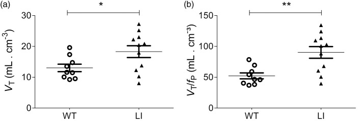
Although still a matter of controversy, disrupted in schizophrenia protein 1 (DISC1) was suggested as a potential inhibitor of phosphodiesterase 4 (PDE4). We used Disc1 locus impairment (LI) mice to investigate the interaction between PDE4 and DISC 1 in vivo and in vitro. [11C](R)-Rolipram binding was measured by PET in LI (n = 11) and C57BL/6 wild-type (WT, n = 9) mice. [11C](R)-Rolipram total distribution volumes (VT) were calculated and corrected for plasma-free fraction (fP) measured in a separate group of LI (n = 6) and WT (n = 7) mice. PDE4 enzyme activity was measured using in vitro samples of cerebral cortices from groups of LI (n = 4), heterozygote (n = 4), and WT (n = 4) mice. Disc1 LI mice showed a 41% increase in VT (18 ± 6 vs. 13±4 mL/cm3, P = 0.04) compared to WT mice. VT/fP showed a 73% significant increase (90 ± 31 vs. 52 ± 15 mL/cm3, P = 0.004) in Disc1 LI compared to WT mice. PDE4 enzymatic activity assay confirmed in vivo findings showing significant group differences (p < 0.0001). In conclusion, PDE4 activity was increased in the absence of critical DISC1 protein isoforms both in vivo and in vitro. Additionally, [11C](R)-Rolipram PET was sensitive enough to assess altered PDE4 activity caused by PDE4-DISC1 interaction.

Keywords: Positron emission tomography; [C]()-Rolipram; disrupted in schizophrenia; locus impairment mouse model; phosphodiesterase 4.


Figures

Figure 1.
Figure 1.
Time activity curves of whole brain (a) and heart (b) derived from wild-type (O) and Disc1 locus-impairment mice (▴). Mice were scanned after a bolus-infusion protocol using [11C](R)-Rolipram that resulted in stable steady state concentrations in both brain and heart. Time activity curves are presented as the normalized concentration, which is the radioactive concentration normalized for body weight and infusion rate. Data are presented as average of wild-type (n = 9) and Disc1 LI mice (n = 11) with error bars representing the standard deviation. WT: wild-type; LI: Disc1 locus impairment mouse model.
Figure 2.
Figure 2.
Comparison of VT and VT/fP in wild type (O) and Disc1 locus-impairment mice (▴). Distribution volume (VT; a) was calculated as the ratio of average brain radioactivity in PET images (60–90 min) divided by the concentration of [11C](R)-Rolipram in plasma. Distribution volume corrected for plasma-free fraction (VT/fP; b) was calculated as the ratio of average brain radioactivity in PET images (60–90 min) divided by the concentration of free [11C](R)-Rolipram in plasma. Both VT and VT/fP were significantly higher in Disc1 LI (n = 11) compared to WT mice (n = 9). *p < 0.05, **p < 0.005; unpaired t-test.
Figure 3.
Figure 3.
Parametric VT/fP images in wild type and Disc1 locus-impairment mice. These are representative coronal and transversal images showing VT/fP of [11C](R)-Rolipram in Disc1 locus-impairment mice and wild-type mice image intensity in each voxel is radioactivity normalized to free [11C](R)-Rolipram levels in plasma (i.e., VT/fp). The arrows indicate the location of the brain in the image. Note the higher uptake of [11C](R)-Rolipram in the brain of Disc1 locus-impairment.
Figure 4.
Figure 4.
Enzymatic activity assays in wild-type mice as well as homozygous and heterozygous Disc1 locus-impairment mice. The effect of genotype across the three groups was significant (p = 0.0073) based on Kruskal–Wallis test, n = 4 per genotype group. WT: wild-type; LI: Disc1 locus impairment mouse model.

Similar articles

Cited by

References

    1. Fujita M, Richards EM, Niciu MJ, et al. cAMP Signaling in brain is decreased in unmedicated depressed patients and increased by treatment with a selective serotonin reuptake inhibitor. Mol Psychiatry 2017; 22: 754–759. - PMC - PubMed
    1. Nestler EJ, Aghajanian GK. Molecular and cellular basis of addiction. Science 1997; 278: 58–63. - PubMed
    1. Duman RS. Synaptic plasticity and mood disorders. Mol Psychiatry 2002; 7: S29–S34. - PubMed
    1. Hill EV, Sheppard CL, Cheung Y-F, et al. Oxidative stress employs phosphatidyl inositol 3-kinase and ERK signalling pathways to activate cAMP phosphodiesterase-4D3 (PDE4D3) through multi-site phosphorylation at Ser239 and Ser579. Cell Signal 2006; 18: 2056–2069. - PubMed
    1. MacKenzie SJ, Baillie GS, McPhee I, et al. Long PDE4 cAMP specific phosphodiesterases are activated by protein kinase A-mediated phosphorylation of a single serine residue in Upstream Conserved Region 1 (UCR1). Br J Pharmacol 2002; 136: 421–433. - PMC - PubMed

Publication types

Substances

LinkOut - more resources