. 2018 Aug;24(15-16):1251-1261.
doi: 10.1089/ten.TEA.2017.0483. Epub 2018 May 10.

Oversized Biodegradable Arterial Grafts Promote Enhanced Neointimal Tissue Formation

Affiliations

Oversized Biodegradable Arterial Grafts Promote Enhanced Neointimal Tissue Formation

Cameron Best et al. Tissue Eng Part A. 2018 Aug.

Abstract

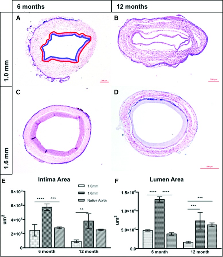
Most tissue-engineered arterial grafts are complicated by aneurysmal dilation secondary to insufficient neotissue formation after scaffold degradation. The optimal graft would form an organized multilayered structure with a robust extracellular matrix that could withstand arterial pressure. The purpose of the current study was to determine how oversizing a biodegradable arterial scaffold affects long-term neotissue formation. Size-matched (1.0 mm, n = 11) and oversized (1.6 mm, n = 9) electrospun polycaprolactone/chitosan scaffolds were implanted as abdominal aortic interposition grafts in Lewis rats. The mean lumen diameter of the 1.6 mm grafts was initially greater compared with the native vessel, but matched the native aorta by 6 months. In contrast, the 1.0 mm grafts experienced stenosis at 6 and 9 months. Total neotissue area and calponin-positive neotissue area were significantly greater in the 1.6 mm grafts by 6 months and similar to the native aorta. Late-term biomechanical testing was dominated by remaining polymer, but graft oversizing did not adversely affect the biomechanics of the adjacent vessel. Oversizing tissue-engineered arterial grafts may represent a strategy to increase the formation of organized neotissue without thrombosis or adverse remodeling of the adjacent native vessel by harnessing a previously undescribed process of adaptive vascular remodeling.

Keywords: chitosan; electrospinning; polycaprolactone; rat model; size mismatch; tissue-engineered arterial graft.


Conflict of interest statement

Dr. C.K.B. is on the Scientific Advisory board of Cook Medical (Bloomington, IN). Dr. C.K.B. receives research support from Gunze, Ltd. (Kyoto Japan) and Cook Regentec (Indianapolis, IN). None of the funding from these grants was used to support this research. Dr. J.J. is a cofounder of Nanofiber Solutions, Inc. (Hilliard, OH). Dr. C.K.B. and Mr. C.B. are cofounders of LYST Therapeutics, LLC (Columbus, OH). Dr. N.H. receives research support from Secant Medical (Telford, PA). Remaining authors have no conflicts of interest to disclose.

Figures

<b>FIG. 1.</b>
FIG. 1.
(A) Schematic overview of study design: TEVG scaffolds were implanted and followed with serial ultrasound imaging at 1, 3, 6, and 9 months postoperatively. Samples were harvested at 6 months for histologic analyses and at 12 months for μCT-angiography, biaxial mechanical testing, and histology. (B) Representative scanning electron microscope images of 1.0 mm (left) and 1.6 mm (right) TEVG scaffolds before implantation at equivalent magnification (40 × ). (C) Intraoperative photographs of TEVG explantation demonstrating the size-matched 1.0 mm (left) and size mismatched 1.6 mm (right) grafts. Adventitial sutures are placed at explant to allow determination of in vivo axial stretch for biaxial mechanical testing. Scale bar = 1.0 mm. μCT, microcomputed tomography; TEVG, tissue-engineered vascular graft. Color images available online at www.liebertpub.com/tea
<b>FIG. 2.</b>
FIG. 2.
(A) Transabdominal ultrasonographic assessment of TEVG lumen diameters over time. TEVGs of 1.6 mm cohort had significantly larger lumen diameters than the 1.0 mm cohort at 1, 3, 6, and 9 months postimplantation. Lumen diameters in each group were equivalent at 12 months. (B) Direct comparison of the 1.0 mm TEVG lumen diameters compared to the adjacent abdominal aorta revealed significant graft narrowing at 6 and 9 months. (C) The same comparison of lumen diameters in the 1.6 mm cohort indicated no significant difference between the TEVG and native vessel after 3 months. ****p < 0.0001, **p < 0.005.
<b>FIG. 3.</b>
FIG. 3.
Representative three-dimensional μCT-angiography reconstructions of (A) 1.0 mm and (B) 1.6 mm (n = 2/group) TEVGs at the 12-month time point. (C) Comparison of cross-sectional lumen diameter measurements derived from two orthogonal planes of the proximal aorta (0.5 mm above the anastomosis), proximal anastomosis, mid-TEVG, distal anastomosis, and distal aorta (0.5 mm below the anastomosis) indicated that late-term lumen diameters were similar regardless of graft lumen diameter at implantation. Raw μCT images and measurements are provided in Supplementary Figure S2 for clarity. Color images available online at www.liebertpub.com/tea
<b>FIG. 4.</b>
FIG. 4.
(A) Pressure-diameter responses of 1.0 mm (n = 4) and 1.6 mm (n = 4) luminal diameter grafts and the adjacent PIAA 12 months postimplantation. Data are plotted as the last cycle of unloading at the in vivo axial stretch following preconditioning. Note the significantly higher circumferential structural stiffness of the grafts compared to the adjacent aorta and the mismatch between the outer diameter of the 1.6 mm grafts and the proximal aorta despite matching luminal diameters. There was no difference in the circumferential structural stiffness of the PIAA adjacent to 1.0 and 1.6 mm grafts 12 months postimplantation, suggesting that graft luminal diameter at implantation does not adversely affect remodeling of the adjacent aorta. (B) Circumferential Cauchy stress–stretch data for 1.0 mm (n = 4) and 1.6 mm (n = 4) luminal diameter grafts and the adjacent PIAA 12 months postimplantation. Data are plotted as the last cycle of unloading at the in vivo axial stretch following preconditioning. The grafts have significantly higher material stiffness compared with the adjacent proximal aorta, likely secondary to residual polymer present even after 12 months (Supplementary Fig. S4). The material stiffness of the proximal aorta does not differ between the 1.0 and 1.6 mm cohorts, suggesting that its remodeling is independent of initial graft luminal diameter. PIAA, proximal infrarenal abdominal aorta.
<b>FIG. 5.</b>
FIG. 5.
Representative hematoxylin and eosin photomicrographs of 1.0 mm (A, B) and 1.6 mm (C, D) TEVGs at 6 (A, C) and 12 (B, D) months postimplantation. Red tracing of the scaffold/neointimal margin and blue tracing of the neointimal/luminal margin in (A) contains the intimal tissue areas quantified and compared in (E). Grafts of 1.6 mm contained significantly more luminal neotissue than 1.0 mm grafts and the native aorta at 6 months. At 12 months, the 1.6 mm intimal area was >1.0 mm grafts, but equivalent to the native aorta. Quantification of lumen area (F) demonstrated similar trends: 1.6 mm grafts were characterized by significantly larger lumens than both 1.0 mm grafts and the native aorta at 6 months, but at 12 months, lumen areas of both the native aorta and 1.6 mm grafts were larger than the 1.0 mm group. No difference between 1.6 mm grafts and the native aorta was observed at 12 months. ****p < 0.0001, ***p < 0.0005, **p < 0.005. Color images available online at www.liebertpub.com/tea
<b>FIG. 6.</b>
FIG. 6.
Representative immunohistochemical staining for calponin (AD) and CD-68 (FI) for 1.0 mm (A, B, F, G) and 1.6 mm (C, D, H, I) grafts at the 6 (A, C, F, H) and 12 (B, D, G, I) month time points. “l” denotes the graft lumen, and “*” indicates suture material. (E) Quantification of calponin+ intimal area suggests that both constructs contained less contractile vascular smooth muscle cells than the native artery at both time points. At 6 months, the 1.6 mm grafts contained significantly more calponin+ intimal neotissue than the 1.0 mm grafts. (J) Quantification of CD-68+ cell numbers between groups at both time points indicated the persistence of chronic inflammation that did not depend on graft dimensions at implantation. ****p < 0.0001, **p < 0.005, *p < 0.05. Color images available online at www.liebertpub.com/tea
<b>FIG. 7.</b>
FIG. 7.
Representative Picrosirius red imaging visualized under polarized light for 1.0 mm (A, C) and 1.6 mm (B, D) grafts at the 6 (A, B) and 12 (C, D) month time points. “l” denotes the graft lumen. (E) Comparison of total collagen content between TEVG cohorts and the native aorta. No significant difference at either time point was observed. (F) Comparison of collagen fiber thickness derived from color thresholding of dark-field images at the 6-month time point indicated that the 1.0 and 1.6 mm grafts contained less mature thick fibers (red) than the native aorta. Distribution of intermediate and thin fibers was similar between groups. (G) Collagen composition analysis at 12 months revealed an inversion of trends observed at 6 months; both the 1.0 and 1.6 mm grafts were composed of significantly more mature thick fibers than the native aorta, likely secondary to the chronic inflammatory response elicited by the residual polymer. No differences between intermediate and thin fibers were observed. ****p < 0.0001. Color images available online at www.liebertpub.com/tea

References

    1. World Health Organization. The top 10 causes of death. 2017. http://www.who.int/mediacentre/factsheets/fs310/en/ Accessed October25, 2017
    1. Baril D.T., Patel V.I., Judelson D.R., et al. . Outcomes of lower extremity bypass performed for acute limb ischemia. J Vasc Surg 58, 949, 2013 - PMC - PubMed
    1. Duval S., Keo H.H., Oldenburg N.C., et al. . The impact of prolonged lower limb ischemia on amputation, mortality, and functional status: the FRIENDS registry. Am Heart J 168, 577, 2014 - PubMed
    1. Yeager R.A., Moneta G.L., Taylor L.M., Hamre D.W., McConnell D.B., and Porter J.M. Surgical management of severe acute lower extremity ischemia. J Vasc Surg 15, 385, 1992 - PubMed
    1. Souza D.S.R., Johansson B., Bojö L., et al. . Harvesting the saphenous vein with surrounding tissue for CABG provides long-term graft patency comparable to the left internal thoracic artery: results of a randomized longitudinal trial. J Thorac Cardiovasc Surg 132, 373, 2006 - PubMed

Publication types