Review
. 2018 Mar;282(1):121-150.
doi: 10.1111/imr.12634.

Mast cells as sources of cytokines, chemokines, and growth factors

Affiliations
Review

Mast cells as sources of cytokines, chemokines, and growth factors

Kaori Mukai et al. Immunol Rev. 2018 Mar.

Abstract

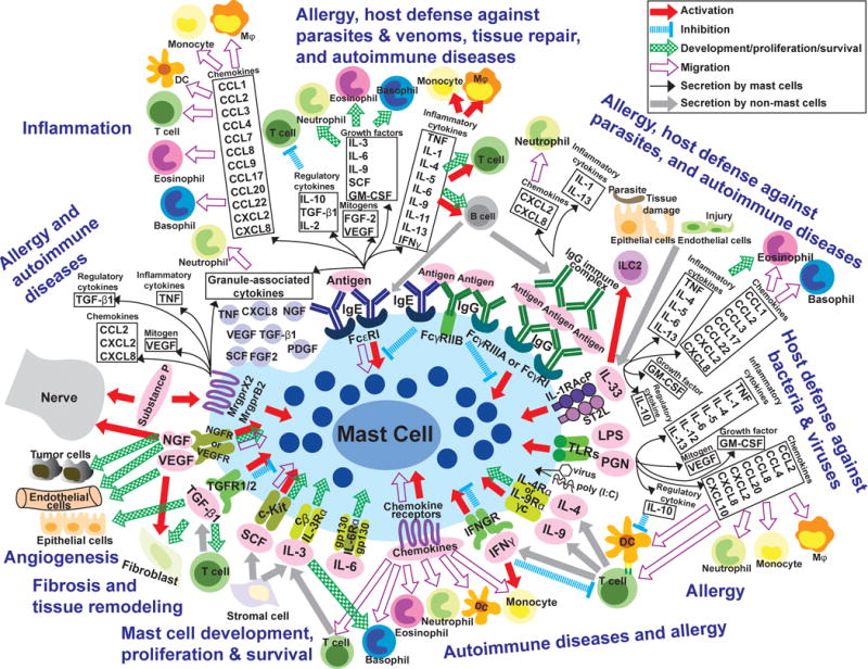
Mast cells are hematopoietic cells that reside in virtually all vascularized tissues and that represent potential sources of a wide variety of biologically active secreted products, including diverse cytokines and growth factors. There is strong evidence for important non-redundant roles of mast cells in many types of innate or adaptive immune responses, including making important contributions to immediate and chronic IgE-associated allergic disorders and enhancing host resistance to certain venoms and parasites. However, mast cells have been proposed to influence many other biological processes, including responses to bacteria and virus, angiogenesis, wound healing, fibrosis, autoimmune and metabolic disorders, and cancer. The potential functions of mast cells in many of these settings is thought to reflect their ability to secrete, upon appropriate activation by a range of immune or non-immune stimuli, a broad spectrum of cytokines (including many chemokines) and growth factors, with potential autocrine, paracrine, local, and systemic effects. In this review, we summarize the evidence indicating which cytokines and growth factors can be produced by various populations of rodent and human mast cells in response to particular immune or non-immune stimuli, and comment on the proven or potential roles of such mast cell products in health and disease.

Keywords: chemokines; cytokines; growth factors; immunity; inflammation; mast cells.


Conflict of interest statement

CONFLICT OF INTEREST

The authors have no conflict of interest.

Figures

Figure 1
Figure 1. Highly simplified overview of the diverse stimuli and potential consequences of mast cell activation and secretion of cytokines, chemokines and growth factors
Mast cells (MCs) can be activated through various receptors when they are exposed to the corresponding ligands (e.g., in pink ovals). This can induce MC activation (red arrows), inhibition (blue dotted lines), or migration (purple open arrows), influence MC development/proliferation/survival (green patterned arrows), and/or induce MCs to secrete many cytokines, chemokines and growth factors (black arrows and related boxes). Grey arrows depict secretion of products from cells other than MCs. Depending on the type of stimulus/stimuli, as well as the type/phenotype of the MCs, such activated MCs also may secrete many other stored and/or newly synthesized mediators (not shown). In adaptive immune responses (e.g., elicited by parasites, animal venoms or allergens), MCs can be activated when IgE bound to surface FcεRI receptors is crosslinked by bi- or multivalent antigens, or when immune complexes (IgG-ICs) bind to FcγRs. In some settings, for example in mouse BMCMCs, co-ligation of FcεRI with inhibitory FcγRIIb receptors can down-regulate MC activation , . FcγRI is a high affinity receptor induced in human MCs by IFNγ stimulation in vitro or in the IFNγ enriched environment of skin MCs in the setting of psoriasis . However, this FcγRI expression is observed in humans not in mice. Upon antibody/antigen-mediated stimulation, MCs can synthesize and secrete a panel of factors as indicated in the black boxes. In turn, those factors can influence other immune and non-immune (structural) cells and contribute to pathogenesis of various types of allergic reactions and perhaps autoimmune disorders, such as some forms of arthritis, as well as to host defense against venoms or parasites. Many of the immune and structural cells depicted are comprised of functionally distinct subtypes (e.g., T cells, DCs, macrophages, fibroblasts, nerves) and the effects of particular MC products on such cells may vary importantly depending on the target cell subtype (not shown). In some settings, such MC-derived products also may contribute to tissue repair and remodeling, both through effects on structural cells and by regulating aspects of the inflammatory/immune response. Antibody/antigen-mediated stimulation also can induce MCs to secrete preformed mediators such as histamine, serotonin (in rodents, primarily), proteoglycans, and proteases (not shown), as well as certain cytokines and growth factors which can be granule-associated (black boxes and the purple granules underneath), as well as many lipid mediators including cysteinyl leukotrienes and certain prostaglandins (not shown). IL-33, which is produced by endothelial/epithelial cells in sites of tissue damage, can stimulate MCs to secrete many factors (indicated in the black boxes) with diverse potential effects on other immune and non-immune cells that can contribute to the pathogenesis of allergies and to host defense. Products of pathogens such as LPS (lipopolysaccharide) and PGN (peptidoglycan), poly (I:C), and certain viruses can directly activate MCs through TLRs (toll-like receptors), resulting in the secretion of a variety of factors (as indicated in black boxes); depending on the setting, this could contribute to host defense and/or disease (e.g., there is a well-established clinical association between certain viral infections and exacerbations of asthma). During Th2 cell-associated immune responses, IL-4 or IL-9 from T cells or from immature cells in the MC lineage can activate MCs and promote their development/proliferation. IFNγ can deliver positive or negative signals to MCs, probably depending on species of animal, MC subpopulation, and setting (such as a disease or a particular beneficial host response). MCs can migrate in response to certain chemokines, but MCs also can be activated by chemokines. IL-3 and SCF (stem cell factor) are representatives of factors which support MC development, proliferation and/or survival (others include, depending on the MCs, IL-4, IL-6, IL-9, and NGF). IL-3 can have similar effects on basophils. NGF (nerve growth factor), VEGF (vascular endothelial growth factor), FGFs (fibroblast growth factors), and TGF-β1 (transforming growth factor type-β) can contribute to the development of fibrosis or angiogenesis, and there is some evidence indicating that these factors, like TNF (tumor necrosis factor), can be constitutively stored in the granules of some MCs. These factors also can influence MCs (as indicated with arrows). Substance P is a product of certain neurons that can potently activate some types of MCs, which in turn can secrete preformed mediators that may include granule-associated cytokines (as indicated in the black boxes). Bidirectional interactions between certain nerve cells and MCs have been studied extensively, and there is considerable interest in the potential importance of such nerve-MC interactions in health and disease. Finally, it should be kept in mind that proteases released from activated MCs can degrade TNF , IL-1β , IL-18 , IL-33 , , SCF , CCL5 and CCL11 , CCL26 , and likely other factors shown in the figure, and this may represent an important mechanism by which MCs can control the intensity and duration of the biological effects of such factors. Please see Tables 1 and 2 for additional information about how variation in MC subtype may influence the extent to which these cells can produce and/or respond to the factors shown in the figure.
Figure 2
Figure 2. Longitudinal imaging of mast cell (MC) degranulation and Il10 gene activation in a model of severe cutaneous contact hypersensitivity (CHS)
Sulforhodamine 101–coupled avidin (Av.SRho; 5 μg) was injected intradermally (i.d.) into the ear pinna of mice. One week later, the mice were treated as described in to induce a severe 1-fluoro-2,4-dinitrobenzene–induced (DNFB-induced) contact hypersensitivity (CHS) reaction. (A) Longitudinal monitoring of the release of Av.SRho+ granules by dermal MCs at the site of CHS using intravital 2-photon microscopy. Representative 3D photographs of the ear pinna before DNFB challenge or at day 1, 2, or 3 after DNFB challenge. Upper panel: merged fluorescence of Av.SRho (red) and Mcpt5-EYFP (green). Lower panel: Av.SRho fluorescence (pseudocolor scale). Dashed white circles identify hair follicles. (B) Longitudinal monitoring of both the release of dermal MC Av.SRho+ granules and activation of Il10 gene transcription (IL-10-GFP, as detected by emission of GFP fluorescent signal) at the site of CHS using intravital 2-photon microscopy. Representative 3D photographs of the ear pinna before DNFB challenge or at day 1, 2, or 3 after DNFB challenge. Upper panel: merged fluorescence of Av.SRho (red) and IL-10-GFP (green). Middle panel: Av.SRho (red) fluorescence. Lower panel: IL-10-GFP (green) fluorescence. White lines identify the magnified areas and dashed white circles identify hair follicles. Scale bars: 20 μm. (C) Percentage of Mcpt5-EYFP+ cells with exteriorized Av.SRho+ structures (i.e., degranulated dermal MCs, red circles) and of Av.SRho+ IL-10-GFP+ cells (i.e., representing MCs expressing the Il10 gene, green circles) per field of view (FOV) in ear pinnae. (D) Total number of Av.SRho+ IL-10-GFP+ cells (MCs expressing the Il10 gene, green circles) per FOV in ear pinnae and total number of IL-10-GFP+ cells in ear pinnae (black circles). (E) Percentage of Av.SRho+ IL-10-GFP+ cells (i.e., representing MCs expressing the Il10 gene, green) and of Av.SRhoIL-10-GFP+ cells (i.e., representing other cell types expressing the Il10 gene, gray) among total IL-10-GFP+ cells in ear pinnae per FOV. Mean ± SEM; data (n = 3 per group) are pooled from the 3 independent experiments performed (each done with 1 mouse per group), each of which gave similar results. (This is Figure 3 from .)
Figure 3
Figure 3. Mast cell (MC) production of IL-10 limits inflammation and epidermal hyperplasia in a model of severe cutaneous contact hypersensitivity (CHS)
Mice were treated as described in to elicit a 1-fluoro-2,4-dinitrobenzene–induced (DNFB-induced) severe CHS reaction. (A) Breeding strategy to obtain Mcpt5-Cre+; Il10fl/fl (MC IL-10 deficient) mice. (B) Changes (Δ) in ear thickness over time after challenge with vehicle (squares) or DNFB (circles) in Mcpt5-Cre+; Il10fl/fl (MC IL-10 deficient, yellow) or Mcpt5-Cre; Il10fl/fl (MC IL-10 sufficient, black) mice. (C) Photomicrographs of representative H&E (upper panel) and toluidine blue (lower panel) stained sections of ear pinnae of mice sacrificed 5 days after challenge. Asterisks indicate areas shown at higher magnification (×60) in insets, arrowheads indicate MCs, and dashed white lines in insets depict epidermis. (D) Number of MCs/mm2 dermis and (E) epidermal thickness 5 days after vehicle (squares) or DNFB (circles) challenge in Mcpt5-Cre+; Il10fl/fl (MC IL-10 deficient, yellow) or Mcpt5-Cre; Il10fl/fl (MC IL-10 sufficient, black) mice. Scale bars: 200 μm. Mean ± SEM; *P < 0.05; **P < 0.01; ***P < 0.001; (B) 2-way ANOVA; (D and E) 2-tailed, unpaired t test. Data (n = 6–12 mice per group) are pooled from the 3 independent experiments performed (each done with n = 2–4 mice per group), each of which gave similar results. (This is Figure 5 from .)

References

    1. Ehrlich P. Thesis. Leipzig University; Leipzig: 1878. Beiträge zur Theorie und Praxis der histologiscen Färbung.
    1. Gordon JR, Burd PR, Galli SJ. Mast cells as a source of multifunctional cytokines. Immunol Today. 1990;11:458–464. - PubMed
    1. Chung SW, Wong PM, Shen-Ong G, et al. Production of granulocyte-macrophage colony-stimulating factor by Abelson virus-induced tumorigenic mast cell lines. Blood. 1986;68:1074–1081. - PubMed
    1. Young JD, Liu CC, Butler G, Cohn ZA, Galli SJ. Identification, purification, and characterization of a mast cell-associated cytolytic factor related to tumor necrosis factor. Proc Natl Acad Sci U S A. 1987;84:9175–9179. - PMC - PubMed
    1. Gordon JR, Galli SJ. Mast cells as a source of both preformed and immunologically inducible TNF-alpha/cachectin. Nature. 1990;346:274–276. - PubMed

Publication types