. 2018 Jun;5(3):332-342.
doi: 10.1002/ehf2.12271. Epub 2018 Feb 12.

Human cardiomyocyte calcium handling and transverse tubules in mid-stage of post-myocardial-infarction heart failure

Affiliations

Human cardiomyocyte calcium handling and transverse tubules in mid-stage of post-myocardial-infarction heart failure

Morten Andre Høydal et al. ESC Heart Fail. 2018 Jun.

Abstract

Aims: Cellular processes in the heart rely mainly on studies from experimental animal models or explanted hearts from patients with terminal end-stage heart failure (HF). To address this limitation, we provide data on excitation contraction coupling, cardiomyocyte contraction and relaxation, and Ca2+ handling in post-myocardial-infarction (MI) patients at mid-stage of HF.

Methods and results: Nine MI patients and eight control patients without MI (non-MI) were included. Biopsies were taken from the left ventricular myocardium and processed for further measurements with epifluorescence and confocal microscopy. Cardiomyocyte function was progressively impaired in MI cardiomyocytes compared with non-MI cardiomyocytes when increasing electrical stimulation towards frequencies that simulate heart rates during physical activity (2 Hz); at 3 Hz, we observed almost total breakdown of function in MI. Concurrently, we observed impaired Ca2+ handling with more spontaneous Ca2+ release events, increased diastolic Ca2+ , lower Ca2+ amplitude, and prolonged time to diastolic Ca2+ removal in MI (P < 0.01). Significantly reduced transverse-tubule density (-35%, P < 0.01) and sarcoplasmic reticulum Ca2+ adenosine triphosphatase 2a (SERCA2a) function (-26%, P < 0.01) in MI cardiomyocytes may explain the findings. Reduced protein phosphorylation of phospholamban (PLB) serine-16 and threonine-17 in MI provides further mechanisms to the reduced function.

Conclusions: Depressed cardiomyocyte contraction and relaxation were associated with impaired intracellular Ca2+ handling due to impaired SERCA2a activity caused by a combination of alteration in the PLB/SERCA2a ratio and chronic dephosphorylation of PLB as well as loss of transverse tubules, which disrupts normal intracellular Ca2+ homeostasis and handling. This is the first study that presents these mechanisms from viable and intact cardiomyocytes isolated from the left ventricle of human hearts at mid-stage of post-MI HF.

Keywords: Calcium handling; Cardiomyocytes; Excitation contraction coupling; Heart failure; Myocardial infarction; SERCA2a.


Figures

Figure 1
Figure 1
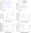
Cardiomyocyte function and Ca2+ handling. Example recordings of cardiomyocyte contraction–relaxation (A) and Ca2+ transients (B) at 2 Hz stimulation in post‐myocardial‐infarction heart failure patients (MI) (N = 9, n cells per patient: 6–10) (red lines) vs. non‐myocardial‐infarction patients (NON‐MI) (N = 8, n cells per patient: 6–10) (blue lines), reported by edge detection microscopy and Fura‐2/AM ratio (F340/380), respectively. (C) Cardiomyocyte fractional shortening. (D) Time to peak shortening. (E) Ca2+ transient amplitude. (F) Rates of Ca2+ release (time to peak Ca2 transient amplitude).
Figure 2
Figure 2
Transverse (T)‐tubule structure. T‐tubule density in post‐myocardial‐infarction heart failure patients (MI; N = 9, n cells per patient: 6–10) vs. non‐MI patients (NON‐MI; N = 8, n cells per patient: 6–10). (A) Example confocal images of T‐tubules in a di‐8‐ANEPPS‐stained cardiomyocyte from an MI patient. (B) Display of a significantly reduced T‐tubule density in MI vs. NON‐MI and (C) T‐tubule densities along relative cell length. The largest reduction in T‐tubule density of MI patients (red) compared with NON‐MI (blue) was found in the mid‐regions of the cardiomyocyte. Data are presented as mean ± standard deviation. *P < 0.01.
Figure 3
Figure 3
Cardiomyocyte diastolic function and Ca2+ handling. Cardiomyocyte diastolic function and Ca2+ handling properties at 0.5–2 Hz stimulation in patients with post‐myocardial‐infarction heart failure (N = 9, n cells per patient: 6–10) (red line) vs. non‐myocardial‐infarction patients (N = 8, n cells per patient: 6–10) (blue lines). (A) Frequency‐dependent acceleration of relaxation assessed by time to 50% diastolic re‐lengthening of the cardiomyocyte. (B) Frequency‐dependent acceleration of Ca2+ decay measured as time to 50% decay of the Ca2+ transient. (C) Diastolic cytoplasmic Ca2+ levels. Ca2+ transient tracings are reported with Fura‐2/AM ratio (F340/380).
Figure 4
Figure 4
Sarcoplasmic reticulum (SR) Ca2+ adenosine triphosphatase 2a (SERCA2a) function. SERCA2a function assessed as SR Ca2+ uptake measurements in separate biopsies of the left ventricle myocardium in post‐myocardial‐infarction heart failure patients (MI) (N = 9) vs. non‐myocardial‐infarction patients (NON‐MI) (N = 8). (A) Maximal rate of SR Ca2+ uptake was lower in MI. (B) SERCA‐2a sensitivity to cytosolic Ca2+ (Km of free Ca2+ concentration evoking half SR Ca2+ uptake rate) was significantly lower in MI; higher Ca2+ levels are needed to activate SERCA2a.
Figure 5
Figure 5
Protein expression. Western blot data from left ventricle biopsies in post‐myocardial‐infarction heart failure patients (MI) (N = 6) vs. non‐myocardial‐infarction patients (NON‐MI) (N = 5). (A) Sarcoplasmic reticulum Ca2+ adenosine triphosphatase 2a (SERCA2a) only showed a tendency of down‐regulation (P = 0.11) in MI patients. (B) Phospholamban (PLB) was not changed, but (C) the ratio PLB/SERCA2a revealed a tendency (P = 0.055) of increased levels in MI compared with NON‐MI. (D) Both the phosphorylation site of PKA at PLB serine‐16 (pPLB Ser‐16) and (E) the phosphorylation site of CaMKII at PLB theorine‐17 (pPLB Thr‐17) was significantly lower activated in MI patients. (F) Phosphorylated CaMKII at the auto‐activating site threonine‐286 (pCaMKII Thr‐286) was lower in MI. (G) Representative images of Western blot for each group. Data are presented as mean ± standard error of the mean. *P < 0.05, **P < 0.01.
Figure 6
Figure 6
Spontaneous Ca2+ release and irregular cardiomyocyte activation. Percentage of cells with spontaneous Ca2+ release events between regular electrical stimulated twitch Ca2+ releases (upper graphs) was higher in post–myocardial‐infarction heart failure patients (MI) (N = 9, n cells per patient: 6–10) compared with non‐MI patients (NON‐MI) (N = 8, n cells per patient: 6–10). Lower graphs display that the cardiomyocytes from MI had impaired ability to follow electrical stimulation when increasing the frequency from 2 to 3 Hz.

References

    1. Roger VL. Epidemiology of heart failure. Circ Res 2013; 113: 646–659. - PMC - PubMed
    1. Heinzel FR, Bito V, Biesmans L, Wu M, Detre E, von Wegner F, Claus P, Dymarkowski S, Maes F, Bogaert J, Rademakers F, D'Hooge J, Sipido K. Remodeling of T‐tubules and reduced synchrony of Ca2+ release in myocytes from chronically ischemic myocardium. Circ Res 2008; 102: 338–346. - PubMed
    1. Bers DM. Altered cardiac myocyte Ca regulation in heart failure. Physiology (Bethesda) 2006; 21: 380–387. - PubMed
    1. Schillinger W, Lehnart SE, Prestle J, Preuss M, Pieske B, Maier LS, Meyer M, Just H, Hasenfuss G. Influence of SR Ca(2+)‐ATPase and Na(+)‐Ca(2+)‐exchanger on the force–frequency relation. Basic Res Cardiol 1998; 93: 38–45. - PubMed
    1. Wachter R, Schmidt‐Schweda S, Westermann D, Post H, Edelmann F, Kasner M, Luers C, Steendijk P, Hasenfuss G, Tschope C, Pieske B. Blunted frequency‐dependent upregulation of cardiac output is related to impaired relaxation in diastolic heart failure. Eur Heart J 2009; 30: 3027–3036. - PMC - PubMed

Publication types

MeSH terms

Substances