. 2018 Apr 15;78(8):1986-1999.
doi: 10.1158/0008-5472.CAN-17-0315. Epub 2018 Feb 5.

Silencing the Snail-Dependent RNA Splice Regulator ESRP1 Drives Malignant Transformation of Human Pulmonary Epithelial Cells

Affiliations

Silencing the Snail-Dependent RNA Splice Regulator ESRP1 Drives Malignant Transformation of Human Pulmonary Epithelial Cells

Tonya C Walser et al. Cancer Res. .

Abstract

Epithelial-to-mesenchymal transition (EMT) is organized in cancer cells by a set of key transcription factors, but the significance of this process is still debated, including in non-small cell lung cancer (NSCLC). Here, we report increased expression of the EMT-inducing transcription factor Snail in premalignant pulmonary lesions, relative to histologically normal pulmonary epithelium. In immortalized human pulmonary epithelial cells and isogenic derivatives, we documented Snail-dependent anchorage-independent growth in vitro and primary tumor growth and metastatic behavior in vivo Snail-mediated transformation relied upon silencing of the tumor-suppressive RNA splicing regulatory protein ESRP1. In clinical specimens of NSCLC, ESRP1 loss was documented in Snail-expressing premalignant pulmonary lesions. Mechanistic investigations showed that Snail drives malignant progression in an ALDH+CD44+CD24- pulmonary stem cell subset in which ESRP1 and stemness-repressing microRNAs are inhibited. Collectively, our results show how ESRP1 loss is a critical event in lung carcinogenesis, and they identify new candidate directions for targeted therapy of NSCLC.Significance: This study defines a Snail-ESRP1 cancer axis that is crucial for human lung carcinogenesis, with implications for new intervention strategies and translational opportunities. Cancer Res; 78(8); 1986-99. ©2018 AACR.


Conflict of interest statement

Conflict of Interest Disclosure: At the time of publication, Dr. Ooi is a paid employee of Fluidigm Corporation, and Dr. Gomperts is an uncompensated consultant at InSpira.

Figures

Figure 1
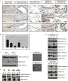
Figure 1. Snail expression is observed in human pulmonary premalignant lesions and drives EMT in an HBEC-based model of lung premalignancy
Representative fields at 400X; scale bar = 50 µm. (A) (i) Isotype control staining of a NSCLC tumor previously identified as Snail-positive (11). Snail staining of (ii) HBEC-Vector cells (negative control) and (iii) HBEC-Snail cells (positive control) grown in 3D ALI culture. Snail staining of (iv-vi) SCC-adjacent LAs and (vii-ix) ADC-adjacent SAs. Snail staining of (x-xii) SM premalignant lesions, where xi-asterisk denotes a representative focus of inflammation and xii-asterisk denotes a representative field of hyperplasia. Snail staining of (xiii-xv) AAH premalignant lesions. (B) Snail immunostaining was quantitated by assigning Intensity (I) and Positivity (P) scores (each on a scale from 0 to 4, low to high) and calculating IxP values. (C) Normal HBEC-Parental (P), -Vector (V), and -Snail (S) were evaluated for Snail and E-Cadherin expression by Western Blot (WB) analysis. (D) At-risk oncogene-modified HBEC-V and -S were evaluated for Snail and E-Cadherin. Normal HBEC-V and -S were (E) probed for a panel of epithelial and mesenchymal markers of the EMT program and (F) evaluated for morphology indicative of EMT; top = cobblestone epithelial morphology, and bottom = spindle-shaped mesenchymal morphology.
Figure 2
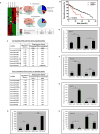
Figure 2. Genomic and proteomic profiling reveal Snail-driven cancer-associated signaling nodes operative during pulmonary premalignancy
(A) Differential gene expression was evaluated using Affymetrix U133 Plus 2.0 gene arrays and bioinformatic analyses. (i- four left columns) A heatmap of differential gene expression (> 2 FC). (i- two right columns) Color-coded log10-transformed q-values of (red) up- and (green) down-regulated genes observed in human NSCLC tumors relative to normal lung tissue. (ii- venn diagrams and pie charts) Summary of overlap between Snail-dependent genes identified in the HBEC-Snail dataset and dysregulated genes observed in previously published clinical lung cancer datasets. (ii- table) Summary of fisher exact tests justifying the reported overlap. (B) Survival analysis of Snail within the lung SCC TCGA dataset. (C) Differential miRNA expression was evaluated using Exiqon miRCURY microRNA arrays and bioinformatic analyses. Fisher exact tests were used to determine if statistically significant (p < 0.05) overlap occurred between target genes of a miRNA and differentially expressed genes. (D-H) Luminex multiplex assays were used to measure secreted proteins IL-6, MMP-3, CXCL8, CXCL5, and CXCL10.
Figure 3
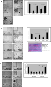
Figure 3. Snail drives key malignant phenotypes in 3D models of lung carcinogenesis
(A) Normal HBEC-V and -S were cultured in a 3D spheroid model and evaluated for morphology indicative of EMT and invasion; representative 400X fields at d3 of growth. (B-C) HBEC-Vector and -Snail were cultured in a 3D lung organotypic ALI model; representative 400X fields at d14 of growth. (B) The cultures were H&E stained and immunostained for Snail and E-Cadherin to assess EMT and invasion. (C) They were immunostained for markers of proliferation (Ki67), differentiation (Ck14 and p63), and stemness (Ck14, p63, and ALDH).
Figure 4
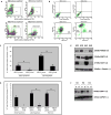
Figure 4. Snail drives stemness expansion, apoptosis resistance, and dysregulation of oncogene and tumor suppressor gene expression
(A) ALDH activity levels and (B) the CD44+CD24 cancer stem cell phenotype were evaluated in HBEC-Vector and -Snail via flow cytometry. (C-D) HBEC-Vector versus -Snail susceptibility to apoptosis in response to two distinct stimuli was assessed via flow cytometry; LR = early apoptosis, UR = late apoptosis, and UL = necrosis. (E-F) The expression patterns of select oncogenes (RAB25) and tumor suppressors (EMP3, GAS1, and ESRP1) were surveyed by WB analysis.
Figure 5
Figure 5. Snail-driven malignant transformation is augmented by driver mutations and is dependent on acquisition of stem-like traits
(A) The capacity of normal HBEC-V and -S for AIG was evaluated by seeding the cells in a miniaturized AIG assay; 1000 cells per well of a 96 well plate with 8 replicates per condition. After 2 weeks in culture, colonies were assessed by (photos) gross microscopic examination and (bar graph) biochemical assay. Each field is 20X, and each inset is 40X and centered on a representative colony in the same well. Absorbance was detected at 570 nm (600 nm reference) and is directly proportional to AIG. (B) HBEC3-V and -S harboring P53 loss (H3mP53) or the combination of P53 loss and KRAS activation (H3mP53/KRAS) were evaluated for AIG after 15 days in culture. (C) Three flow-sorted stem cell subsets, ALDH-CD44+CD24+, ALDH+CD44+CD24+, and ALDH+CD44+CD24-, evaluated for AIG after 15 days in culture.
Figure 6
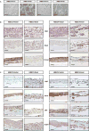
Figure 6. Snail drives malignant transformation, tumor growth, and metastasis of HBECs in vivo
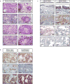
Representative fields at 400X; scale bar = 50 µm. (A-B) H&E and IHC evaluation of primary tumors and lung metastases arising from H3mP53/KRAS-Snail cells injected SC in NSG mice. (i-iv) Four primary tumors representing each of the histologies observed are shown. (v-viii) Four fields depicting the pulmonary metastatic lesion number and size. Large and well-circumscribed nodules with keratin pearls and intracellular bridges indicative of SCC tumor histology were observed in every case. (B) H3mP53/KRAS-Snail cells transformed into primary tumors and lung metastases in mice were immunostained for (top row) Snail, (middle row) ALDH, and (bottom row) CD44, all of which were required for tumor-initiating capacity in vitro. (C) Human clinical specimens containing SM-SCC or AAH-AIS-ADC were immunostained for ALDH1A3 and ALDH3A1.
Figure 7
Figure 7. Snail-dependent ESRP1 silencing drives human pulmonary epithelial cell transformation
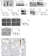
(A) Basal expression of ESRP1 was measured by qRT-PCR and normalized to B2M. (B) WB analysis performed for Snail, ESRP1, and housekeeping gene expression. (C) WB analysis performed for Snail, ESRP1, and GAPDH expression in HBEC3-V and -S and H3mutP53/KRAS-V and -S transiently transfected with ESRP1-specific cDNA constructs (and untreated control cells). (D) Normal HBEC3-V and -S with transient re-expression of ESRP1 were seeded in an accelerated AIG assay at 3000K cells/well with 10 replicates. Z-stacked photomicrographs of each well were created on d0 and d5. Each dot on the graph represents the number of colonies counted from each 3D focused image of each well. (E) At-risk HBEC3-P53/KRAS-V and -S with transient re-expression of ESRP1 were seeded in an AIG assay. (F) Normal and (G) at-risk HBEC-S with stable re-expression of ESRP1 were evaluated for Snail and ESRP1 expression by WB analysis. The (H) normal and (I) at-risk HBEC-S were then then seeded in an AIG assay and assessed after 7 days of growth. (J) Transduction with ESRP1 or control cDNA was repeated using two different approaches (CRISPR method with Hygromycin B selection or IMPDH method with MPA selection), before the normal and at-risk cells were evaluated for Snail and ESRP1 expression by WB analysis and seeded in an AIG assay. (K) Pulmonary premalignant lesions with high level Snail expression in Figure 1 (n=12) were evaluated for ESRP1 expression. Representative fields at 400X; scale bar = 50 µm.

References

    1. Albini A, DeCensi A, Cavalli F, Costa A. Cancer Prevention and Interception: A New Era for Chemopreventive Approaches. Clin Cancer Res. 2016 - PubMed
    1. Mani SA, Guo W, Liao MJ, Eaton EN, Ayyanan A, Zhou AY, et al. The epithelial-mesenchymal transition generates cells with properties of stem cells. Cell. 2008;133:704–15. - PMC - PubMed
    1. De Craene B, Berx G. Regulatory networks defining EMT during cancer initiation and progression. Nature reviews Cancer. 2013;13:97–110. - PubMed
    1. De Craene B, Denecker G, Vermassen P, Taminau J, Mauch C, Derore A, et al. Epidermal Snail expression drives skin cancer initiation and progression through enhanced cytoprotection, epidermal stem/progenitor cell expansion and enhanced metastatic potential. Cell Death Differ. 2014;21:310–20. - PMC - PubMed
    1. Reinke LM, Xu Y, Cheng C. Snail represses the splicing regulator epithelial splicing regulatory protein 1 to promote epithelial-mesenchymal transition. The Journal of biological chemistry. 2012;287:36435–42. - PMC - PubMed

Publication types