. 2018 Mar 5;215(3):827-840.
doi: 10.1084/jem.20172222. Epub 2018 Feb 6.

Caspase-1 self-cleavage is an intrinsic mechanism to terminate inflammasome activity

Affiliations

Caspase-1 self-cleavage is an intrinsic mechanism to terminate inflammasome activity

Dave Boucher et al. J Exp Med. .

Abstract

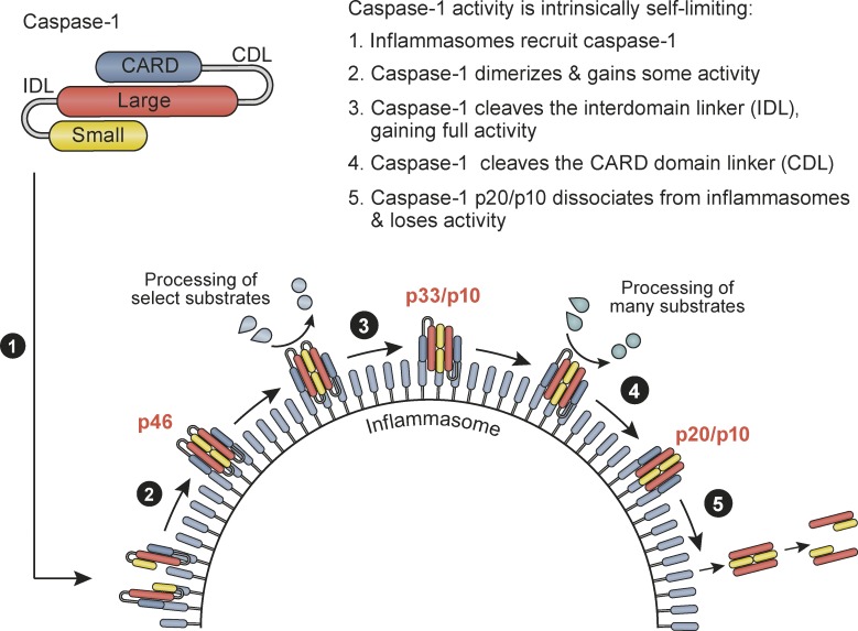
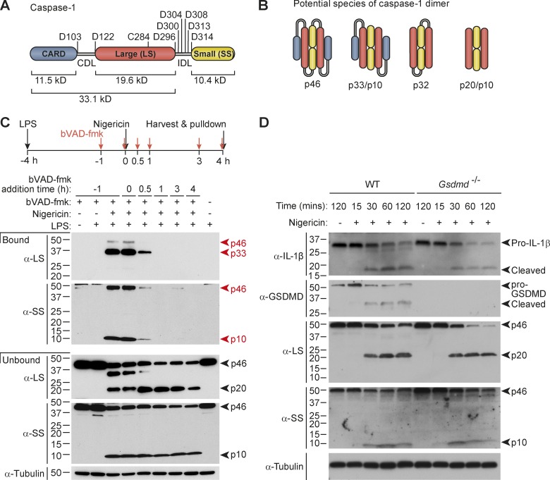
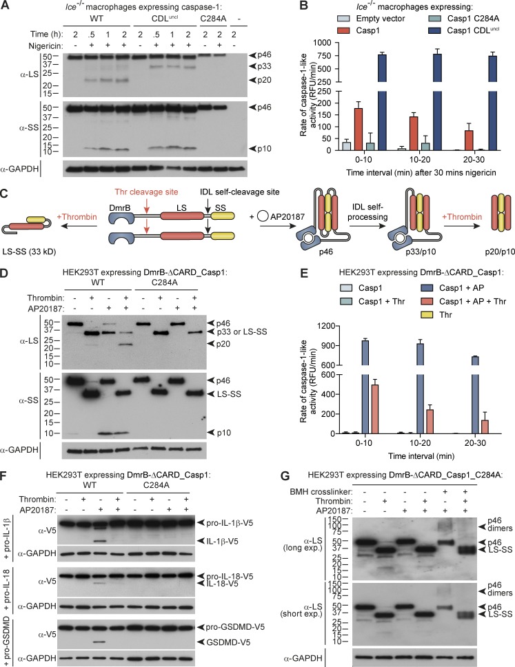
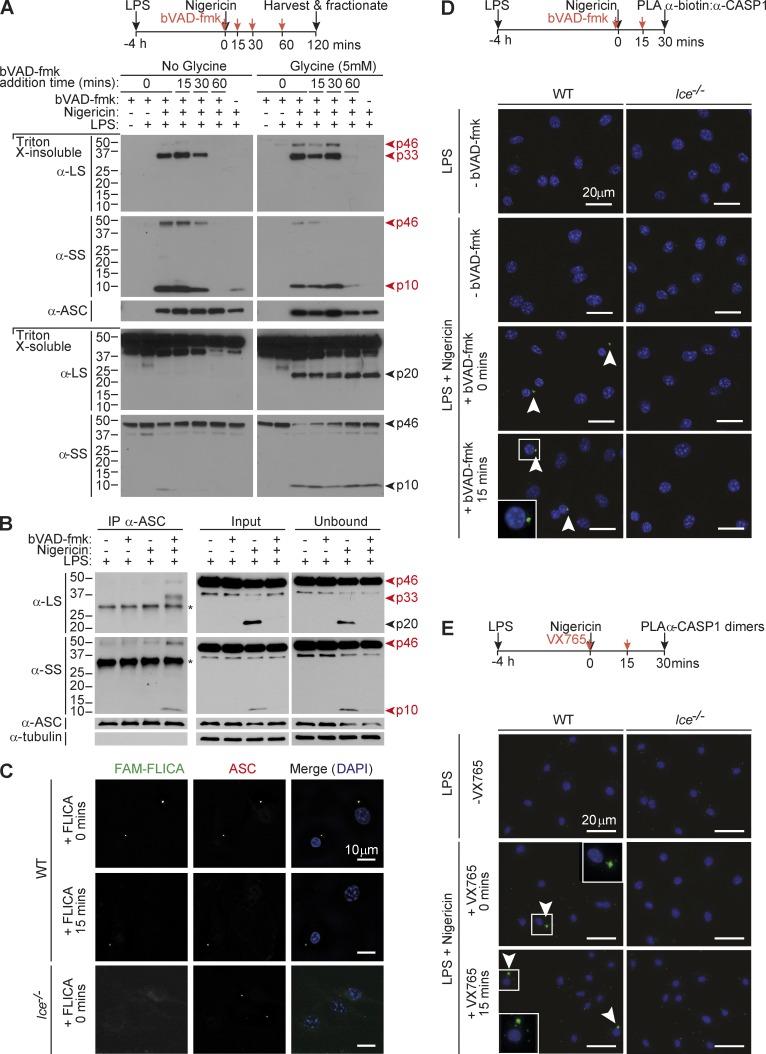
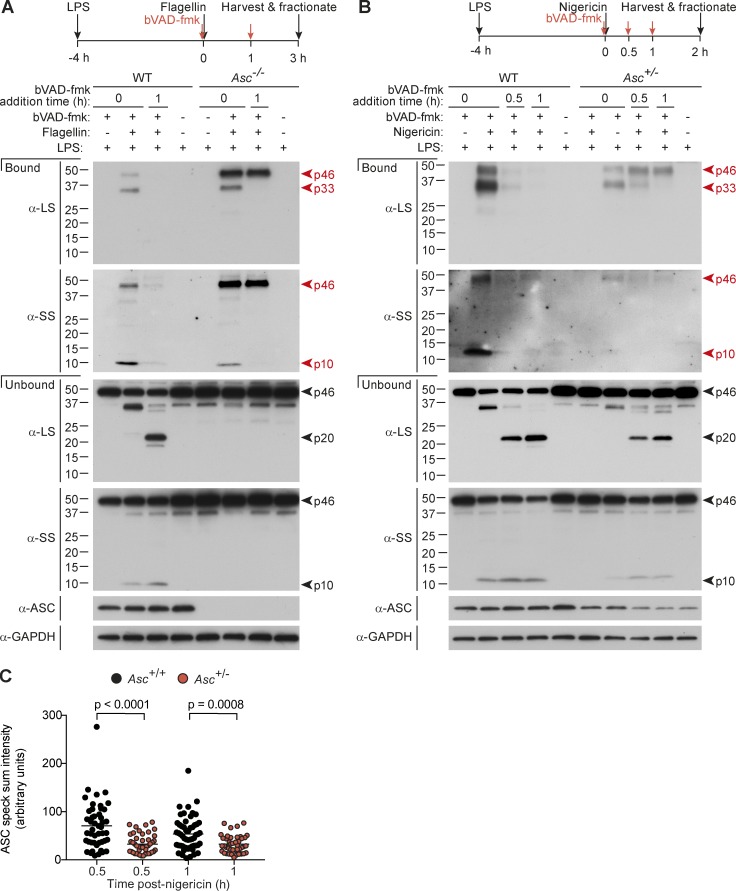
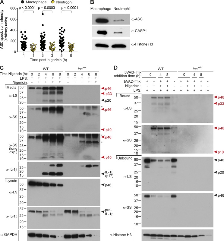
Host-protective caspase-1 activity must be tightly regulated to prevent pathology, but mechanisms controlling the duration of cellular caspase-1 activity are unknown. Caspase-1 is activated on inflammasomes, signaling platforms that facilitate caspase-1 dimerization and autoprocessing. Previous studies with recombinant protein identified a caspase-1 tetramer composed of two p20 and two p10 subunits (p20/p10) as an active species. In this study, we report that in the cell, the dominant species of active caspase-1 dimers elicited by inflammasomes are in fact full-length p46 and a transient species, p33/p10. Further p33/p10 autoprocessing occurs with kinetics specified by inflammasome size and cell type, and this releases p20/p10 from the inflammasome, whereupon the tetramer becomes unstable in cells and protease activity is terminated. The inflammasome-caspase-1 complex thus functions as a holoenzyme that directs the location of caspase-1 activity but also incorporates an intrinsic self-limiting mechanism that ensures timely caspase-1 deactivation. This intrinsic mechanism of inflammasome signal shutdown offers a molecular basis for the transient nature, and coordinated timing, of inflammasome-dependent inflammatory responses.


Figures

None
Graphical abstract
Figure 1.
Figure 1.
Active caspase-1 is predominantly a transient p33/p10 species in nigericin-stimulated macrophages. (A) Representation of potential self-processing sites within the CDL and IDL of mouse caspase-1, relative to the catalytic cysteine (C284). (B) Possible species of dimeric caspase-1 generated by CDL and/or IDL cleavage. (C) Pull-down of active caspase-1 from mouse macrophages, using the bVAD-fmk caspase activity probe. Macrophages were left untreated or primed with LPS for 4 h, and then stimulated with nigericin for a further 4 h before addition of 1% IGEPAL into the well, to lyse cells directly in their culture medium. bVAD-fmk was applied to cells 1 h before (−1 h), during (0 h), or after (0.5, 1, 3, 4 h) nigericin addition. Streptavidin-coated beads pulled down active caspase-1 bound to the biotin-labeled activity probe in mixed lysates/supernatants. Streptavidin-bound and -unbound fractions were analyzed by Western blot using antibodies directed against the caspase-1 large and small subunits (LS, SS). (D) LPS-primed WT and Gsdmd−/− macrophages were stimulated with nigericin and harvested at various time points until 2 h after nigericin stimulation. Cell supernatants were precipitated and resuspended in cell extracts, and the kinetics of caspase-1 substrate cleavage (pro-IL-1β cleavage to p17, pro-GSDMD to GSDMD p30) was examined by Western blot. All data are representative of at least three independent experiments.
Figure 2.
Figure 2.
Caspase-1 processing at the CDL destabilizes caspase-1 dimers and terminates protease activity. Bone marrow progenitors from Ice−/− (Casp1−/−/Casp11null/null) mice were retrovirally reconstituted for mouse caspase-1 (WT, C284 catalytic mutant, or CDL-uncleavable mutant) during their differentiation to macrophages. (A) Differentiated macrophages were then left untreated or LPS-treated for 6 h, or were LPS-primed for 4 h before stimulation with nigericin for 0.5, 1, or 2 h. Cell extracts and cell-free supernatants were mixed and analyzed by immunoblot. (B) Differentiated, transduced macrophages were LPS-primed for 4 h, before stimulation with nigericin for 30 min. Cell culture medium was then replaced with caspase assay buffer containing the caspase-1 fluorogenic substrate, YVAD-afc, and caspase-1–like activity was monitored over the next 30 min. (C) An engineered caspase-1 system allowed controlled caspase-1 dimerization by AP20187 (AP) and CDL cleavage by thrombin (thr). 1 µM AP20187 was added to HEK293T-expressing caspase-1 constructs (WT vs. catalytic mutant, C284A) for 10 min. Cell culture medium was replaced for caspase activity buffer supplemented with digitonin to lyse cells for activity assays (± 20 U/ml thrombin). (D) Immunoblot analysis of caspase-1 cleavage by thrombin after incubation for 1 h. (E) Caspase activity assay, in which thrombin was added to cell lysates at the same time as the YVAD-afc fluorogenic substrate. (F) Cleavage assay in which thrombin was added to caspase-1–expressing cell lysates at the same time as natural substrates (lysates from HEK293T ectopically expressing V5-tagged substrates) and incubated for 0.5 h. (G) Thrombin was added to caspase-1_C284A–expressing cell lysates and incubated for 1 h. Bismaleimidohexane was then added to lysates for 1 h to cross-link caspase-1 dimers. Graphs are mean + SD of triplicate wells from a single experiment. All data are representative of at least two (G) or three (A–F) independent experiments.
Figure 3.
Figure 3.
Caspase-1 activity localizes to the inflammasome. (A) Mouse macrophages were either left untreated or LPS-primed for 4 h before nigericin stimulation for a further 2 h. bVAD-fmk was applied to cells at various times after nigericin (0, 15, 30, 60 min). Cell supernatants were removed and subjected to streptavidin pull-down, which failed to detect active caspase-1 species in this fraction (see Fig. S3 A for pull-down analysis of cell culture medium). Cells were harvested and fractionated. The ASC speck-containing (Triton X-100–insoluble) fraction and the Triton X-100–soluble fraction were analyzed for caspase-1 species by Western blot. Cells were treated with 5 mM glycine 30 min before nigericin to delay cell rupture (right). (B) Macrophages were LPS-primed (4 h) and stimulated with nigericin for 30 min (with and without bVAD-fmk application immediately before nigericin). ASC was immunoprecipitated, and coimmunoprecipitated caspase-1 species were examined by immunoblot. *, Nonspecific band. (C) Macrophages were LPS-primed (4 h) and stimulated with nigericin for 30 min. The FAM-FLICA caspase-1 activity probe was added at the indicated times after nigericin stimulation to localize caspase-1 activity relative to ASC. (D and E) LPS-primed WT or Casp1-deficient Ice−/− macrophages were stimulated with nigericin. Caspase inhibitors (bVAD-fmk caspase activity probe, VX765) were applied at the indicated times after nigericin stimulation, with (D) or without (E) 5 mM glycine in the cell culture media. 30 min after nigericin stimulation, cells were fixed and PLA was performed to detect active caspase-1 (α-biotin/α-caspase-1 LS; (D) or caspase-1 dimers (α-caspase-1 LS:PLUS/α-caspase-1 LS:MINUS; E). DAPI labeled the nucleus. All data are representative of at least three (A–D) or two (E) independent experiments. Arrowheads indicate foci of caspase-1 activity or dimers.
Figure 4.
Figure 4.
Inflammasome size specifies active caspase-1 species and activity duration. (A–C) LPS-primed macrophages were transfected with flagellin (A) or stimulated with nigericin (B and C). bVAD-fmk was applied at the indicated time points (A and B). (A and B) Pull-down of active caspase-1 from macrophages. 1% IGEPAL was added to macrophages at 3 h after flagellin and 2 h after nigericin stimulation, to lyse cells directly in their culture medium. Streptavidin-coated beads pulled down active caspase-1 bound to the biotin-labeled activity probe in mixed lysates/supernatants. (C) ASC speck intensity of Asc+/+ and Asc+/− macrophages (4 h LPS + nigericin, with VX765 added 1 h before nigericin to prevent cell death), quantified from microscopy images. Each circle is one speck (n = 40–55), and significance between samples was assessed by nonparametric, unpaired Mann–Whitney test. All data are representative of at least three (A and B) or two (C) independent experiments.
Figure 5.
Figure 5.
Cell type specifies caspase-1 activity duration. (A–D) LPS-primed (A and B) macrophages or (A–D) neutrophils were left untreated or stimulated with nigericin. (A) ASC speck intensity of Ice−/− macrophages and neutrophils (4 h LPS + nigericin), quantified from microscopy images. Each circle is one speck (n = 20–55), and significance between samples was assessed by nonparametric, unpaired Mann–Whitney test. (B) Immunoblot for inflammasome component expression in whole-cell extracts (4 h LPS). (C) Cell lysates and cell-free supernatants were harvested at various times after nigericin stimulation (0, 2, 4, 8 h), and analyzed by Western blot. *, Nonspecific band. (D) bVAD-fmk was applied at the indicated time points. Supernatant was removed, and cells were lysed 8 h after nigericin stimulation, and active caspase-1 was pulled down using streptavidin beads. Streptavidin-bound and -unbound fractions from mixed supernatants/extracts were analyzed by Western blot. All data are representative of at least three (A, B, C [WT], D [WT]) or two (C [Ice−/−], D [Ice−/−]) independent experiments.
Figure 6.
Figure 6.
Summary model for mechanisms by which inflammasomes direct the active caspase-1 species and duration of caspase-1 activity. See Discussion for details.

Comment in

  • Defusing inflammasomes.
    Sandstrom A, Vance RE. Sandstrom A, et al. J Exp Med. 2018 Mar 5;215(3):723-724. doi: 10.1084/jem.20180241. Epub 2018 Feb 12. J Exp Med. 2018. PMID: 29440361 Free PMC article.

References

    1. Agard N.J., Maltby D., and Wells J.A.. 2010. Inflammatory stimuli regulate caspase substrate profiles. Mol. Cell. Proteomics. 9:880–893. 10.1074/mcp.M900528-MCP200 - DOI - PMC - PubMed
    1. Boatright K.M., Renatus M., Scott F.L., Sperandio S., Shin H., Pedersen I.M., Ricci J.E., Edris W.A., Sutherlin D.P., Green D.R., and Salvesen G.S.. 2003. A unified model for apical caspase activation. Mol. Cell. 11:529–541. 10.1016/S1097-2765(03)00051-0 - DOI - PubMed
    1. Boucher D., Duclos C., and Denault J.B.. 2014. General in vitro caspase assay procedures. Methods Mol. Biol. 1133:3–39. 10.1007/978-1-4939-0357-3_1 - DOI - PubMed
    1. Broz P., von Moltke J., Jones J.W., Vance R.E., and Monack D.M.. 2010. Differential requirement for Caspase-1 autoproteolysis in pathogen-induced cell death and cytokine processing. Cell Host Microbe. 8:471–483. 10.1016/j.chom.2010.11.007 - DOI - PMC - PubMed
    1. Chen K.W., Groß C.J., Sotomayor F.V., Stacey K.J., Tschopp J., Sweet M.J., and Schroder K.. 2014. The neutrophil NLRC4 inflammasome selectively promotes IL-1β maturation without pyroptosis during acute Salmonella challenge. Cell Reports. 8:570–582. 10.1016/j.celrep.2014.06.028 - DOI - PubMed

Publication types